Shocking Dentistry: Modern Clinical Applications for an Old Dental Device

Abstract
In the late 1800s, a new high-energy electrical device was introduced as a cure for a multitude of diseases and conditions that afflicted Americans. Dental pain and disease were among those treatable items. The treatments were applied by direct contact of the body with a hand-held, high voltage Tesla coil and Geissler tube, which together created heat, ozone and ultraviolet (UV) light. The device was termed “The Violet Ray.” However, it was considered mostly a fraud and was banned by the FDA in 1950. Recent experiments as to the oral antibacterial properties of the Violet Ray demonstrate that this device can quickly kill salivary bacteria and thus may have some valid patient treatment and instrument sterilization properties.
Victorian Technology
Nikola Tesla, an inventor and mechanical and electrical engineer, designed and constructed a series of coils that produced high-voltage, high-frequency currents during the late 1880s.1 Named after him, the Tesla coil was a series of two nested coils of electric-powered wire with an electric vibrator switch that rapidly opened and closed the circuit. This created high voltage energy (20,000 volts per cycle) at a low, safe amperage. When attached to a vacuum (Geissler) tube doped with argon, UV light and electric discharges were produced.2 This created heat (through diathermy), ozone and minor neuro-muscular contractions from the electric arching, which were considered beneficial by the Victorian society that first popularized it. The phenomenon was collectively called the Violet Ray, named for the violet color produced in the vacuum tube and from the electrical discharges left on the patient’s skin.2-7
At the turn of the 20th century, life was hard, death often came early, and medicine (including dentistry) remained primitive.8 The average life expectancy was 43 years of age, and 20% of the urban population died of tuberculosis (TB). The fearful population had a 50-50% chance of being helped or harmed by a doctor’s visit.8
Dental care was denture-oriented. Treatments were brutal, with exodontia as the chief treatment offered to patients. This care was occasionally helped through the use of hazardous cocaine or procaine local anesthetics. Vulcanite dentures often followed a long series of dental episodes with operative dentistry (crowns, inlays, amalgam-gold foil fillings, X-rays), an expensive, time-consuming and painful luxury.
People were receptive to many “quack” devices and treatments because professional healthcare was viewed as “imprecise,” if not outright fraudulent and useless.7 Two preferred methods of treatment, electric shock boxes (e.g., Violet Ray) and radium water (radium salts in water), were popular for general preventative and curative treatments of medical and dental illnesses. Both used new technology (electricity and atomic radiation) in an age where “new” was synonymous with “good.”2-7,9,10
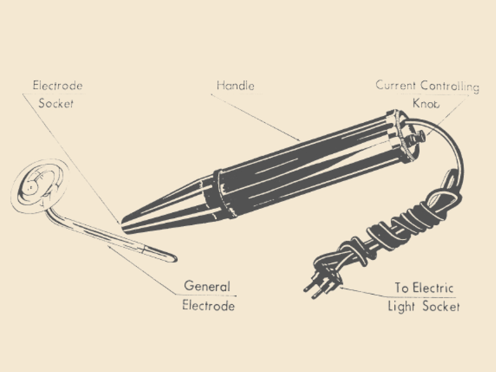
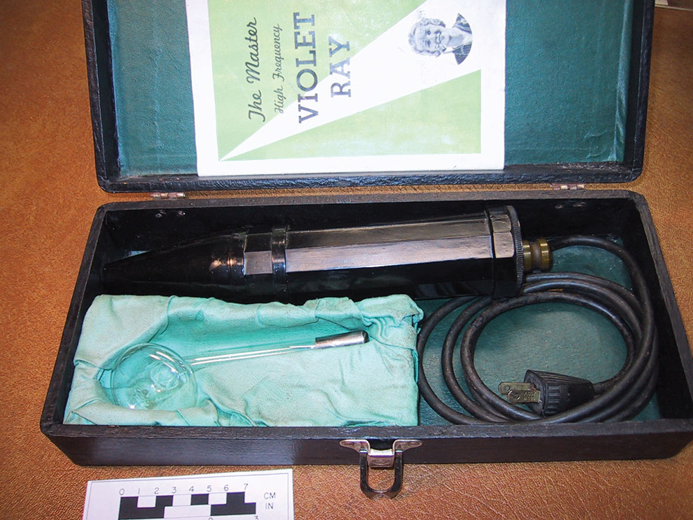
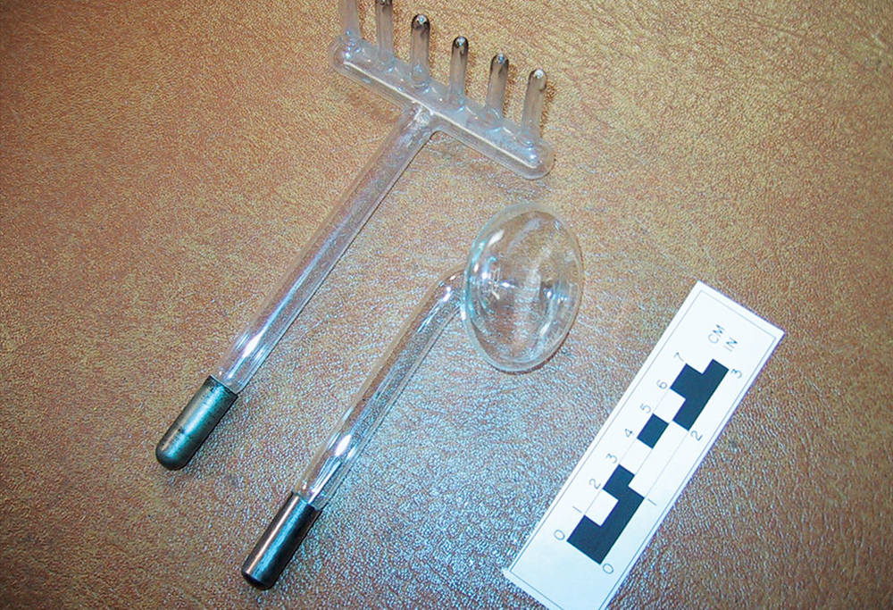
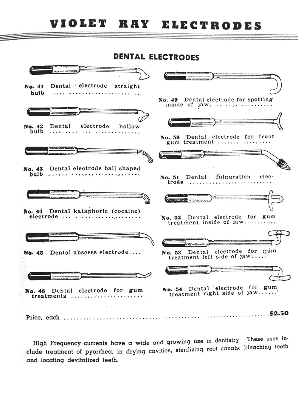
The Violet Ray generator was sold by numerous companies, in a wide variety of shapes and sizes, and retailed for as little as $5 per unit (Figs. 1, 2).3,6 Professional models for physicians and dentists were more powerful, glitzy and expensive, costing up to $45 per unit. Considering the typical worker of 1905 earned about $1 a day, these devices were affordable by the average family. There were dozens of Violet Ray “electrodes” (Geissler tubes) available, each with its own shape, use and name.3-63 There were electrodes for the left and right sides of the mouth, as well as a separate tube for the anterior teeth (Figs. 1–5). There were electrodes for numbing teeth (named the cocaine tube), for anal insertion, deep anal insertion, hair growth, etc. If there was an orifice, there was a genderized tube made to fit (Fig. 5).3-7 The Violet Ray was recommended for treatment and prevention of numerous maladies, from arthritis to zinc deficiency.2-8 The instruction manual included with purchase recommended regular treatments for diphtheria, hair loss, sore throat, consumption (TB), rectal fissures, tooth ache, pyorrhea, flat feet and lower back pain. There was little research into the efficacy of these treatments, but they remained popular for more than 50 years because of the technology used, affordability, placebo effect and some antibacterial properties of the high voltage, UV light and ozone.
The Violet Ray was “modern.” It could be administered by one’s physician, dentist, friend or oneself. It could be applied with or without undressing, an important consideration in Victorian society. It was inexpensive; it tingled, flashed, buzzed, sparked, glowed, made medicine-like smells (ozone) and relaxed muscles (overstimulation of nerve endings by the high voltage).2-8 It was safe; no one was ever reported killed. It was exactly what a fearful, medically traumatized, diseased, superstitious, populace wanted.2,7 Sales were brisk for 50+ years.
It was inexpensive; it tingled, flashed, buzzed, sparked, glowed, made medicine-like smells (ozone) and relaxed muscles (overstimulation of nerve endings by the high voltage). It was safe; no one was ever reported killed.
In 1950, the FDA sued the manufacturer of the Violet Ray for fraud and terminated all manufacture and advertising, with the exception of its use for dermatological treatments.11 And the Violet Ray became another historic, quack device, occasionally used by counter culture therapists or erotic sex devotees (the high voltage was applied to nipple rings, tongue studs, etc.).
Dental Treatments
Early Violet Ray generators came as a two-part system: the electric coils and the electrode. After 1920, a single-unit machine was sold with the coils and replaceable electrode combined in the form of a 12-inch torpedo-shaped, Bakelite unit (Figs. 1–3).2-7 It had a cord, which plugged into household power (110-220 VAC). The unit operated with an adjustable electric vibrator that created rapid voltage fluctuations, causing the two nested coils to create high voltage (20,000 VAC) with low amperage. It was tuned by ear, using a screw knob at the base of the unit, to the optimum vibration (voltage). A high-pitched audible buzz signaled maximum power. The electrode glowed a violet color and ozone was produced as the high voltage electricity and UV radiation crackled off the electrode.2-8
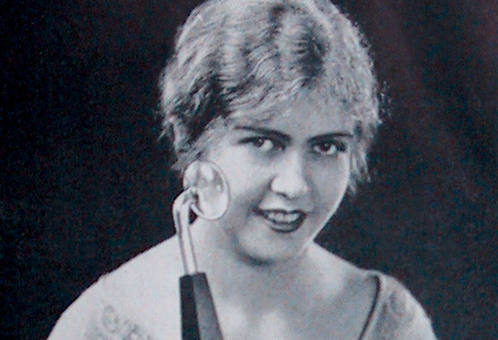
Then, the electrode was applied to the body where treatment was desired (Fig. 3). As the electrode (tube) came within 1 cm of the skin, varied electric arching occurred depending on whether the patient was electrically grounded. A snapping sound could be heard as hundreds of small bolts of electricity arched from the electrode to the skin or oral mucosa, causing a prickly sensation. There was no pain but the subject could feel unusual sensations at skin level. As the electrode touched the skin surface, the arching diminished and a warm sensation was felt. This heat gradually increased and required circular movement of the electrode in order to avoid burning the tissue by diathermy. In effect, the electrode was rubbed on the skin, creating strange lights, sensations, sparks and pungent smells.7
The recommended treatment consisted of rubbing the electrode on the skin or mucosa for five to 10 minutes, followed by rest (Fig. 5).2-7 To avoid overtreatment, the Violet Ray generator would overheat after 15 minutes of use. It then had to be unplugged in order to cool off. There was no limit to the number of treatments patients could have, but twice daily was the blanket recommendation. Contact with mucosa (oral, anal, vaginal, etc.) was initially “tingly” and comfortable treatment required the Violet Ray generator to be started after the electrode was placed on the tooth or tissue, and then moved away. This avoided the sting of the electric arching.2,7
Some recommended dental treatments lasted five minutes per session. The dental electrodes included:
- A “Straight (surfaced) bulb” (No. 41) for moderate arching.
- The “Hollow bulb” electrode (No. 42) produced greater arching.
- The “Ball shaped” electrode (No. 43) was a 4.0 mm glass ball, which produced greater heat.
- The “Dental kataphoric (cocaine)” electrode (No. 44) was a small curved straight bulb used for anesthesia. (At that time cocaine and derivatives were injected as local anesthetics.)
- The “Dental gum treatment” electrode (No. 45) was a small tipped curved tube, which was placed in the interproximal area of the teeth.
- The “Dental electrode for spotting inside the jaw” (No. 49) was a thin, curved tube, which illuminated the oral cavity.
- The “Dental electrode for front gum treatment” (No. 50) was a thin, rounded “Y” shaped tube that was placed along the anterior teeth.
- The “Dental fulguration electrode” (No. 51) was a pencil thin tube, which ended in a point causing increased heat buildup and tissue burning.
- There were a series of “Dental electrodes for gum treatment” designed to fit the lingual curvature of the anterior, right and left sides of the jaws (No. 52–54). Each electrode was priced (circa 1920s–1930s) at $2.50 per unit (Fig. 5).2-8
When more effective treatments and medications were offered to the populace, the popularity of the Violet Ray generators decreased. Why get your gums zapped when you could take antibiotics and have a scaling, which worked better in the end? The question remains, did the Violet Ray machine provide any real benefit for its users?
Modern Experiments
There is a 120+ year history of effective treatments by electricity and UV light in reputable medical and health journals.12-15 Most of the modern day UV studies show bactericidal activity and increased tissue healing on the fibroblastic level.12-15 Epithelization is enhanced by UV exposure.15 A series of preliminary tests and experiments were made to investigate the nature and effectiveness of the Violet Ray exposure in dentistry. A 1920s Montgomery Wards Violet Ray #9 generator with a “rake” and 5 cm diameter “bulb” electrodes were used (Figs. 1–4). Power was turned to the maximum voltage attainable: approximately 20,000 volts.
1. Electromagnetic Radiation: An A.F. Sperry (EMF-200A) electromagnetic force field tester was used to determine the electromagnetic field radiation (measured in mGauss) produced by the Violet Ray generator. Environmental background ranged from 0.2 to 0.3 mG. The generator produced fields of 8.0 to 184.0 mG at distances of 2 cm from the electrodes. In comparison, a standard 19-inch cathode ray tube television set produced an equivalent amount of EMF radiation (30.0–170.0 mG).
Conclusion: It appears that the Violet Ray generator and its emissions are no more therapeutic or hazardous than a standard TV set.
2. Temperature: Electronic and alcohol bulb thermometers were placed on a wooden table top, 2.0 mm distant from the electrodes and the Violet Ray generator described above. Run at maximum power for 40 seconds. The bulb electrode produced a 2-degree Celsius heat increase: from 22 to 24 degrees Celsius. No significant increase in heat was noted. The rake electrode, with its small pointed tips (3.0 mm diameter), produced a 29-degree heat increase: from 21 to 50 degrees Celsius (Fig. 4). This was a significant increase in temperature. A 60-second exposure with the bulb electrode contacting the skin surface was rapidly rubbed over premeasured 3 cm2 of the author’s forearm. The exposure produced a first-degree burn (reddening).
Conclusion: The heat generated from the Violet Ray electrodes can produce variable levels of temperature increase sufficient to burn tissue.
3. Salivary Bacteria: Ozone, heat, electric arching and UV light are produced by the Violet Ray generator, and each is a well-known antibacterial agent.8,11-15 Are the Violet Ray’s physical properties produced in amounts sufficient enough to be deemed a clinically significant antibacterial agent?
The antibacterial qualities of the Violet Ray were tested using fresh saliva as a microbe source and the STERIS bacteria identification system to determine sterility. The STERIS system consists of tubes liquid growth medium, which are incubated for three days at 37 degrees Celsius. Bacterial growth is identified by color changes (red to yellow) in the growth medium. Positive and negative controls were conducted in each experiment. Each experiment was repeated multiple times. Individually packaged, sterile cotton tipped swabs were used for sampling.
Ten experimental series with saliva covered skin and instruments (#20 Kerr endodontic files) were conducted using the rake and bulb electrodes for Violet Ray exposure (15, 30, 60 seconds). The exposures occurred from 1.0 mm to 5.0 mm of electrode-target distance. For the skin tests, a 3 cm circle was drawn on the subject’s forearm. The skin was disinfected with alcohol, allowed to dry and a negative control swab taken. A swab of fresh saliva was liberally applied. Samples were taken as positive controls. The skin was then exposed to the Violet Ray and sample swabs taken at three periods (15, 30, 60 seconds) of exposure.
The use of the rake electrode was discontinued because of the difficulty in adequately covering such a wide skin area with the pencil-thin points of the rake. The bulb electrode (3 cm diameter) proved much more convenient and accurate for this application. Results of all positive and negative controls were consistent with expected results. Contaminated control swabs (positive) incubated culture positive; swabs of sterile skin areas (negative) incubated culture negative. The samples that received 15-second exposures (of saliva contaminated skin) by the Violet Ray showed positive cultures after incubation. The 30-second exposures were culture negative in half (n=6) of the tests (n=12): a 50% disinfection rate. With a 60-second exposure, 11 of 12 cultures tested negative after incubation.
Conclusion: Violet Ray exposure (60 seconds) kills salivary bacteria.
Six additional experiments using the technique described above, but replacing skin with sterile #20 Kerr endodontic files, were conducted. In all cases, the saliva contaminated endodontic files incubated culture negative after a 60-second exposure with the bulb electrode. Violet Ray exposure (60 seconds) can effectively sterilize bacteria contaminated endodontic files.
Discussion
The Violet Ray generator appears to have more potential than a “quack” medical device. A literature search shows some legitimate therapeutic value (supported by the FDA).11-15 The preliminary experiments reported in this paper indicate that Violet Ray irradiation can kill salivary bacteria and sterilize instruments. A fast and inexpensive technique, the Violet Ray generator has significant potential for modern dentistry. Further research and studies are needed to refine the practical applications of this high voltage dental device for modern dentistry.
The Violet Ray generator appears to have more potential than a “quack” medical device. A literature search shows some legitimate therapeutic value. The preliminary experiments reported in this paper indicate that Violet Ray irradiation can kill salivary bacteria and sterilize instruments.
The Violet Ray device also teaches an important lesson: every generation has its new technology, complete with hopes and dreams that it will produce long desired cures and benefits. Is the Violet Ray generator, a product of last century’s discovery of functional electricity, different in its promises from the new products of today (e.g., lasers, magnetized water, cone beam radiography, etc.)? We have seen and heard this all before. Barring new technology, bells and whistles (or in this case, buzzing, flashing and arching), has dentistry really changed in the last 100 years? Aren’t we doing the same old thing, the same old way but with some new twists and shiny buttons? Think about it.
To contact Dr. Ellis Neiburger, call 847-244-0292 or visit drneiburger.com.
References
- ^ Oljaca M. Alternating Current. The Industrial Physicist. 2003;9(4):4-5.
- ^ Bakken Library. Treatment by high frequency electricity (1892 on). Bakken Museum of Electricity (MN). 2000;1:1-9.
- ^ Renulife Electric Co. Renulife Violet Ray: 1919 Manual. Detroit. 1919:1-32.
- ^ Haillwell-Shelton Electric Co. Hailwell-Shelton Violet Ray Manual. NY. 1936:1-20.
- ^ Montgomery Ward & Co. The Master Highfrequency Violet Ray Introduction Manual. Montgomery Ward, Chicago. 1936:1-4.
- ^ Master Electric Co. The Master High Frequency Violet Ray Manual. Chicago. 1928:1-30.
- ^ McCoy B. Quack!: Tales of medical fraud from the museum of questionable medical devices. Santa Monica Press: Santa Monica; 2000. p. 49-71.
- ^ Greer JH. A physician in the house for family and individual consultation. Greer & Co. Publishers: Chicago; 1915. p. 48, 750-7.
- ^ Brown C. The use of electricity by the general practitioner. JAMA. 1898;31(17):968-9.
- ^ Editorial. A plea for static electricity. JAMA. 1892;18:190-1.
- ^ Federal Drug Administration. Notice of Judgment. 1950. 3458 Misbranding of violet ray device U/S v 2 cases 30801, 3858.
- ^ Schwetz B. New treatment for eczema. JAMA. 2001;285(6):724.
- ^ Schultz B, Roenigk H Jr. Uremic puritis treated with ultraviolet light. JAMA. 1980;243(18):1836-7.
- ^ Rapini R. Uses of ultraviolet light. JAMA. 1981;245(19):1909.
- ^ Morykwas M, Mark M. Effects of ultraviolet light on fibroblast fibronectin production and lattice contraction. Wounds. 1998;10(4):111-7.
Written by Ellis Neiburger for Chairside® magazine. Copyright ©2009 Ellis Neiburger. All rights reserved.

