Photo Essay: The Importance of Connective Tissue

November 4, 2010
 image
Daniel Melker, DDS

It is important to determine whether the connective tissue present is attached or unattached at the start of a case. If tissue is unattached, it provides no protection for the underlying periodontal foundation.

Fibers of the connective tissue must either be attached to the root surface or bone in order to act as a barrier to bacterial infiltration, or to prevent trauma from retraction cords or impressions. In this author’s opinion, a biologic width in which only 1 mm of attached connective tissue is present is a weak attachment. When undertaking restorative procedures, 3 mm or more of attached connective tissue is ideal and acts as a strong protection barrier for the periodontal foundation.

Case Presentation

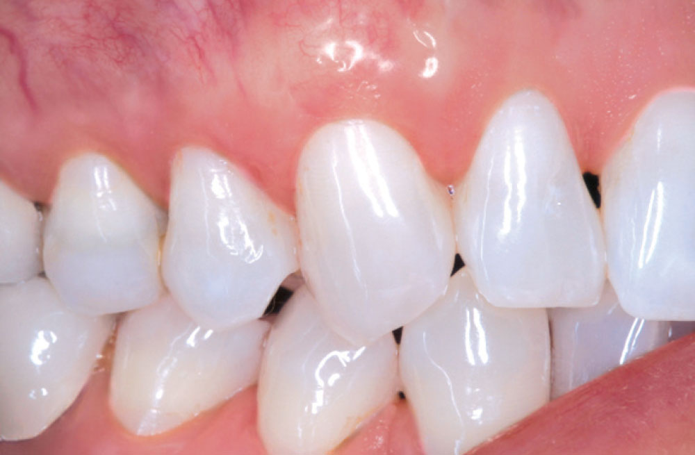
The benefits of connective tissue in protecting the underlying periodontal foundation cannot be overstated. Connective tissue, by its tenacious consistency and vascular makeup, can be extremely beneficial in its resistance to bacterial infiltration and protection against retraction cord and impression taking. The limited ability of bacteria to infiltrate connective tissue allows for better long-term stability of the periodontium. The first case was followed for 10 years and shows the importance of connective tissue in maintaining the underlying supporting tissues of the tooth.

Case 1

Q: Can we stop recession once it has started?

Yes, if we add tissue that can protect the underlying periodontal foundation. 

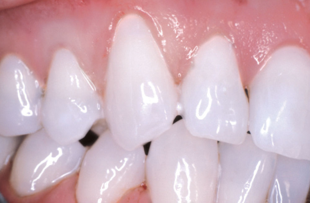
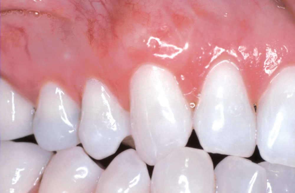
Case 1 Figure 1

Figure 1: Recession was noted on tooth #6 with no attached gingiva for underlying protection.

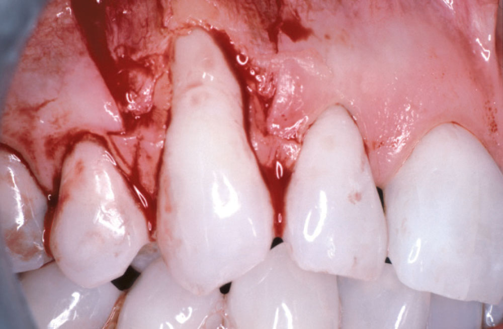
Case 1 Figure 2

Figure 2: Upon examination of the soft tissue, a large dehiscence was noted. A fenestration was also present. In total, 14 mm of bone was lost.

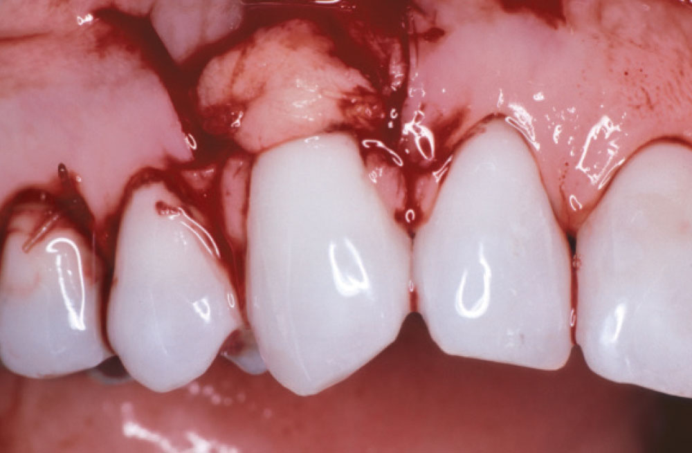
Case 1 Figure 3

Figure 3: A subepithelial connective tissue graft was placed.

Case 1 Figure 4

Figure 4: A one-year follow-up on tooth #6 showed a significant amount of connective tissue present with no probing noted.

Case 1 Figure 5

Figure 5: A 10-year follow-up showed the tissue moving in a coronal direction. Minimal probing was noted. A stable periodontal environment was also noted.

Case 2

Q: Can AlloDerm® (LifeCell Corp.; Branchburg, N.J.) be used for cases in which restorative treatment is to be undertaken?

Yes. Though AlloDerm may not always convert to keratinized tissue, it does create a thick band of connective tissue.

Case 2 Figure 1

Figure 1: Teeth #3−5 were to be restored. Minimal attached gingiva was present. The teeth were to be treated with subepithelial connective tissue grafts and biologic shaping for stability. Connective tissue was placed in order to protect the periodontium against bacterial infiltration, retraction cord placement and impressions. An extra benefit: The root surface was covered, providing a better cosmetic result.

Case 2 Figure 2

Figure 2: Cores and provisionals were placed for biologic shaping and addition of connective tissue (AlloDerm® [LifeCell Corporation; Branchburg, N.J.]).

Case 2 Figure 3

Figure 3: Biologic shaping was undertaken prior to placement of connective tissue.

Case 2 Figure 4

Figure 4: A one-year follow-up on tooth #6 showed a significant amount of connective tissue present with no probing noted.

Case 2 Figure 5

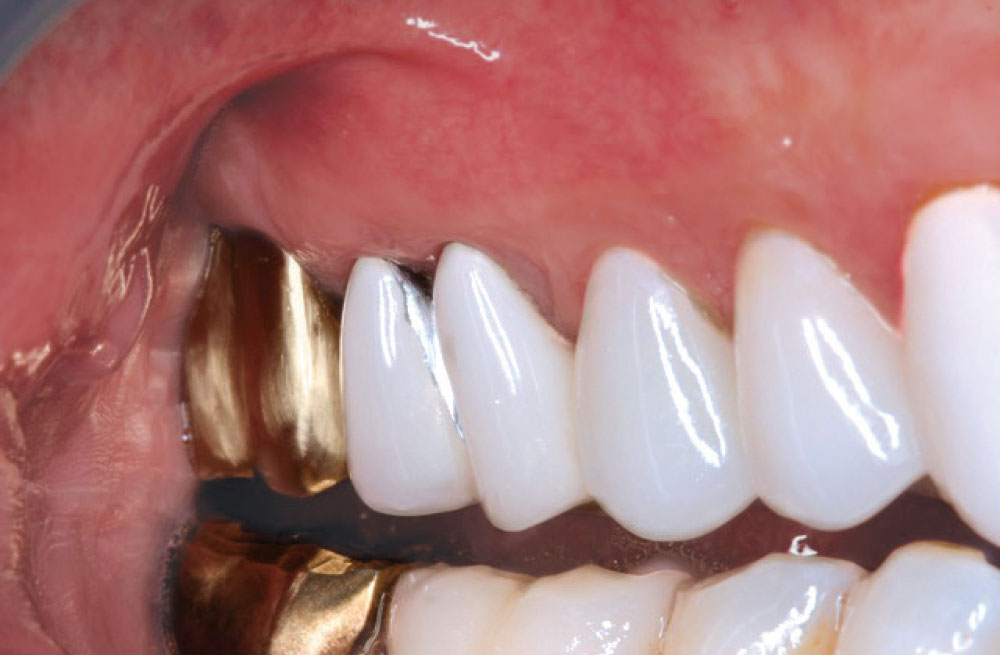
Figure 5: Final restoration placed on a sound periodontal foundation. A significant amount of connective tissue is now present to support the restoration placed. All crowns are supragingival.

Case 3

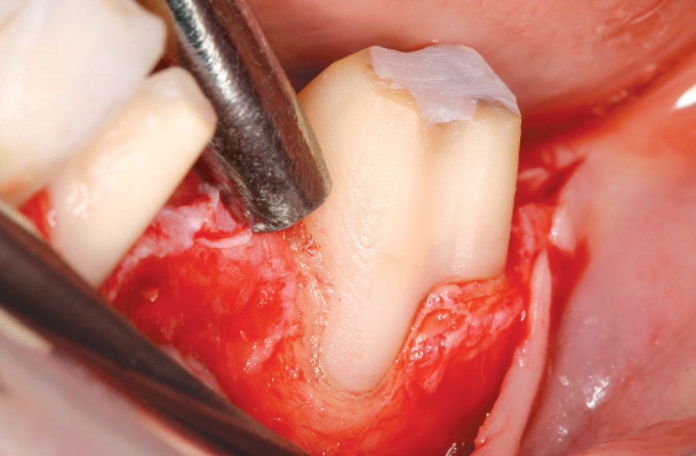
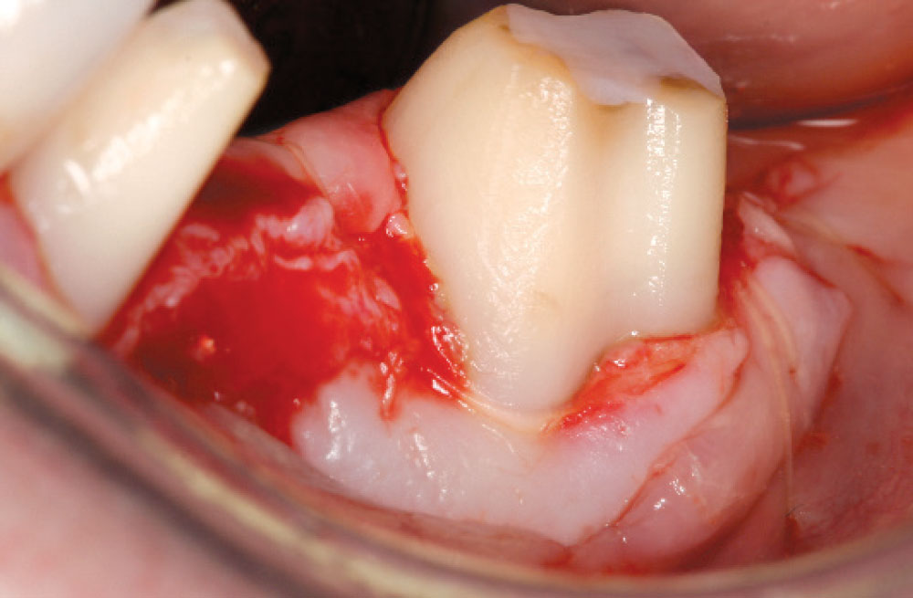
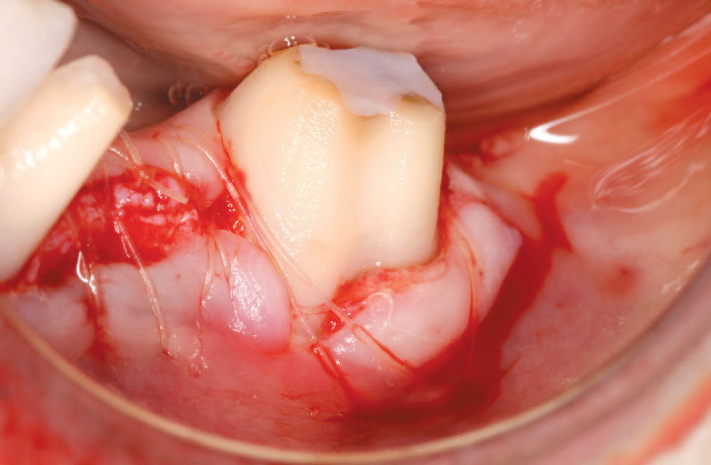
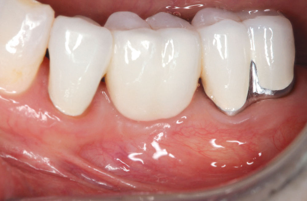
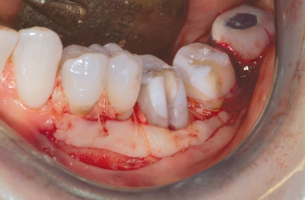
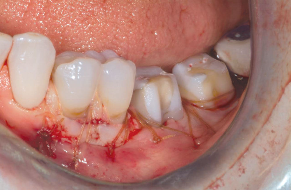
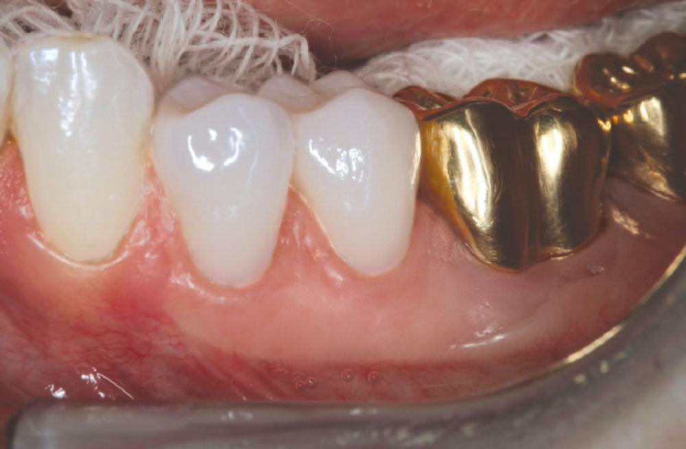
Q: What happens when a surgery is being carried out and a large dehiscence is found?

Placing a large piece of connective tissue over the root will serve as a barrier to bacterial infiltration. It also provides a stable environment for impressions to be taken without damage to the underlying support.

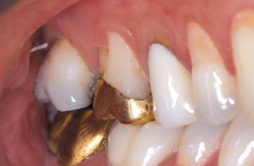
Case 3 Figure 1

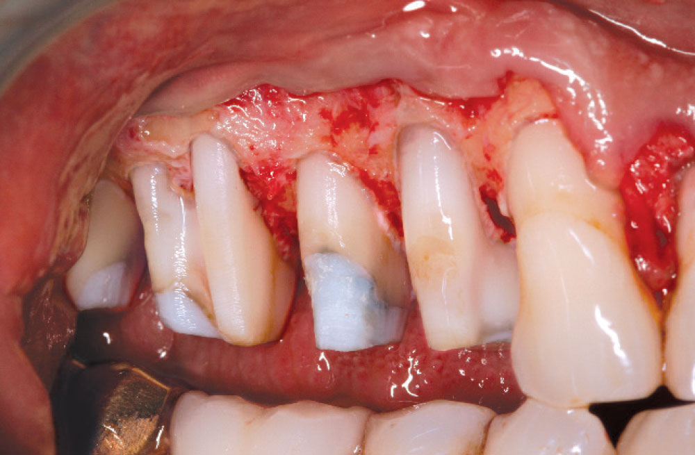
Figure 1: A new bridge was to be placed on teeth #18−20. Provisionals were placed. Notice the mucosal attachment with a lack of connective tissue attachment on the mesiobuccal root of tooth #18.

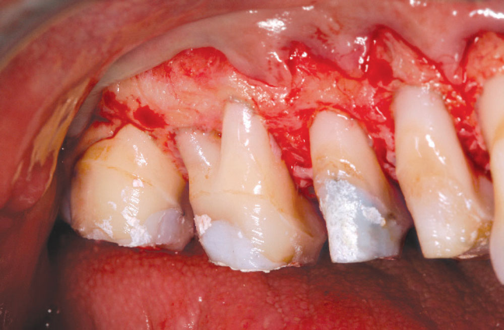
Case 3 Figure 2

Figure 2: A significant dehiscence was noted on tooth #18. Biologic shaping was performed, removing the furcation and smoothing the mesiobuccal root.

Case 3 Figure 3

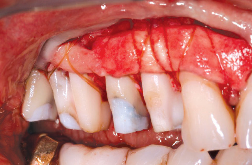
Figure 3: A large subepithelial connective tissue graft was placed.

Case 3 Figure 4

Figure 4: Tissue covered with a small portion of connective tissue exposed.

Case 3 Figure 5

Figure 5: Final restorations placed with margins placed supragingivally. The mesiobuccal root of tooth #18 was protected by connective tissue.

Case 4

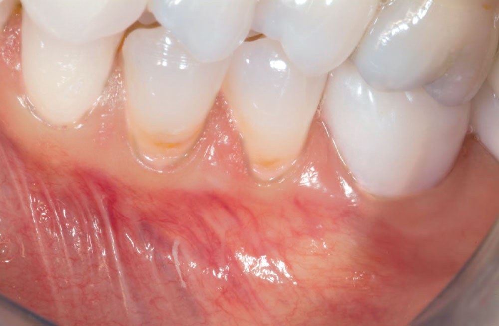
Q: What is the best way to protect an exposed root from future bone loss?

Placing dense, thick vascular connective tissue over the defect will help prevent further breakdown and stabilize the area for the future.

Case 4 Figure 1

Figure 1: Teeth #18−21 were to be restored. Minimal attached gingiva was present. Cores and provisionals were placed to allow for biologic shaping and to increase connective tissue attachment.

Case 4 Figure 2

Figure 2: A large connective tissue graft was placed to increase connective tissue attachment.

Case 4 Figure 3

Figure 3: Marginal grafts were placed on teeth #20 and #21 with a subepithelial graft placed on #19.

Case 4 Figure 4

Figure 4: Final restoration placed with supragingival margins. A large area of connective tissue is present for long-term maintenance.

Case 5

Q: Can we remove old Class V restorations and replace them with new ones?

Class V restorations can be removed and connective tissue placed in these areas to preserve the integrity of the foundation. In some cases, submarginal grafts can be placed to add the same stability.

Case 5 Figure 1

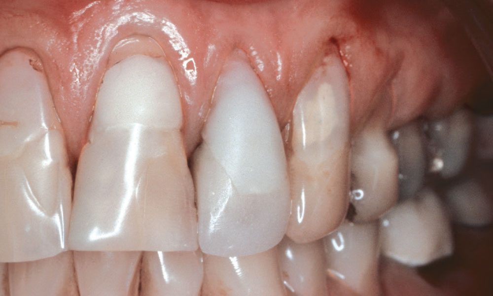
Figure 1: Preoperative photo (1989) shows large composites to be replaced with anterior crowns. The surgeon had been doing subepithelial connective grafts for just two years and was uncomfortable doing them when the restorative was placed.

Case 5 Figure 1

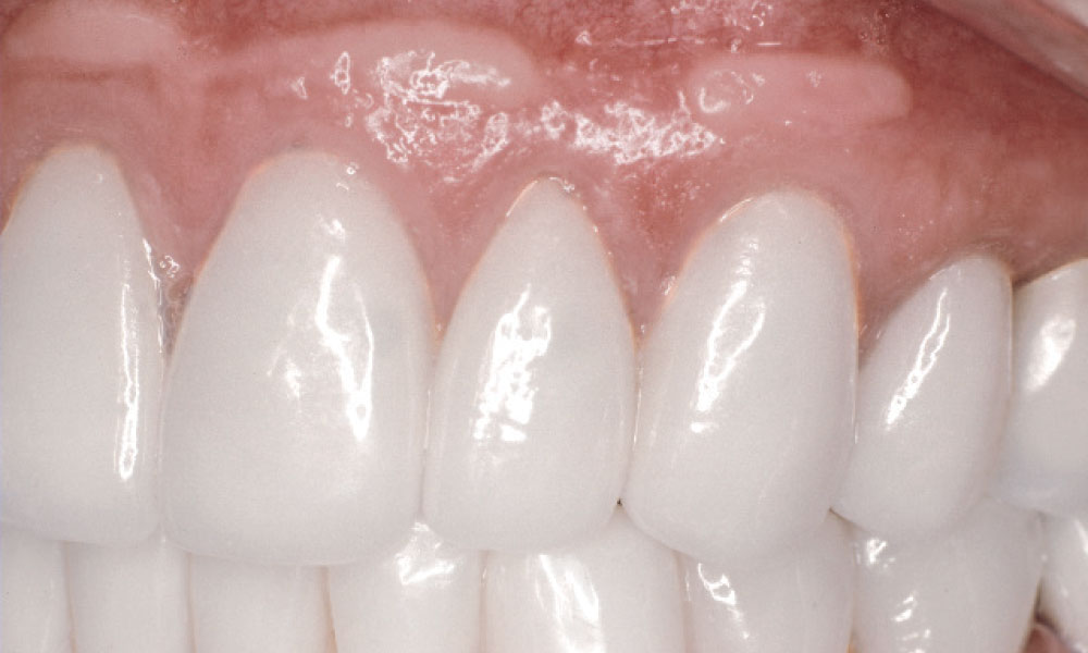
Figure 2: An 18-year follow-up to removed composites and coronally positioned flap placed along with submarginal grafts. Optimum health and periodontal stability were noted.

Case 6

Q: Can problems occur when using weak tissue in areas where restorative is to be placed?

There is a chance of recession and bone loss when weak tissue is present; strong, dense connective tissue is preferred. Trauma from impressions or the seating of restorations can cause soft tissue breakdown.

Case 6 Figure 1

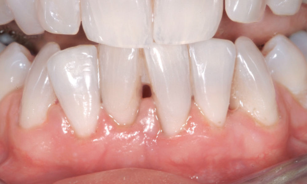
Figure 1: Mandibular anterior teeth to be restored. The surgeon elected to use only the existing tissue prior to restorative completion.

Case 6 Figure 2

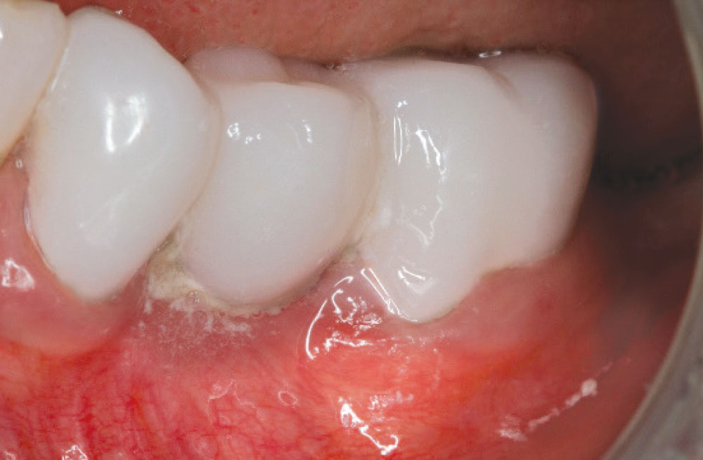
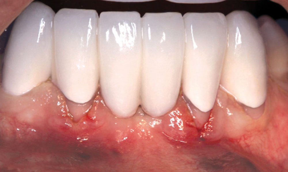
Figure 2: Final restorative presents with severe recession and a poor cosmetic result due to poor surgical technique and a lack of connective tissue.

Dr. Daniel Melker is in private practice and can be reached at 727-725-0100.