And the Impy Award Goes to … — Case of the Week: Episode 82

June 3, 2015
 image
Michael DiTolla, DDS, FAGD

This Case of the Week is not a regular Case of the Week. I looked around and noticed that a lot of people are doing award shows and stuff like that, and I wanted to have our own little award. So now we have the Impression of the Year, or the Impy Award in shorthand. But it’s not for the best one of the year; it’s for the most questionable one. Let’s take a look at our nominations now.

Figure 1

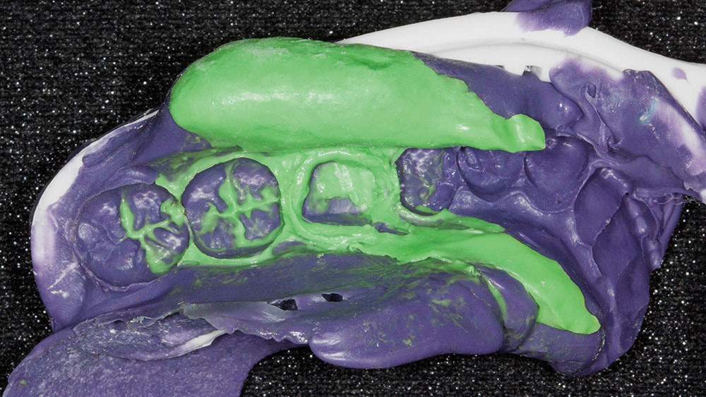
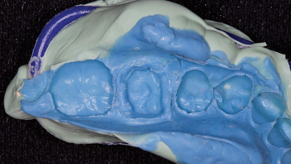
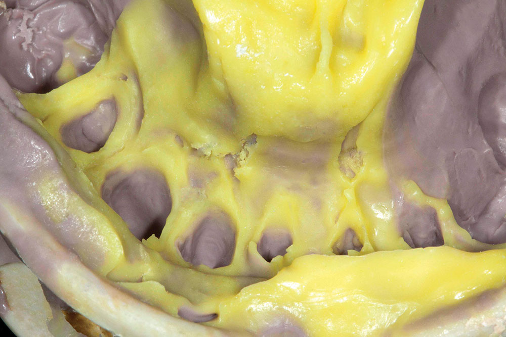
Figure 1: First, we have Dr. F. from Cincinnati, Ohio. When you look at this impression, at first glance, you can see the patient has a wisdom tooth. Dr. F. did manage to get a wisdom tooth in the impression tray, which is nice, but it looks like there is a big pull on the lingual.

Figure 2

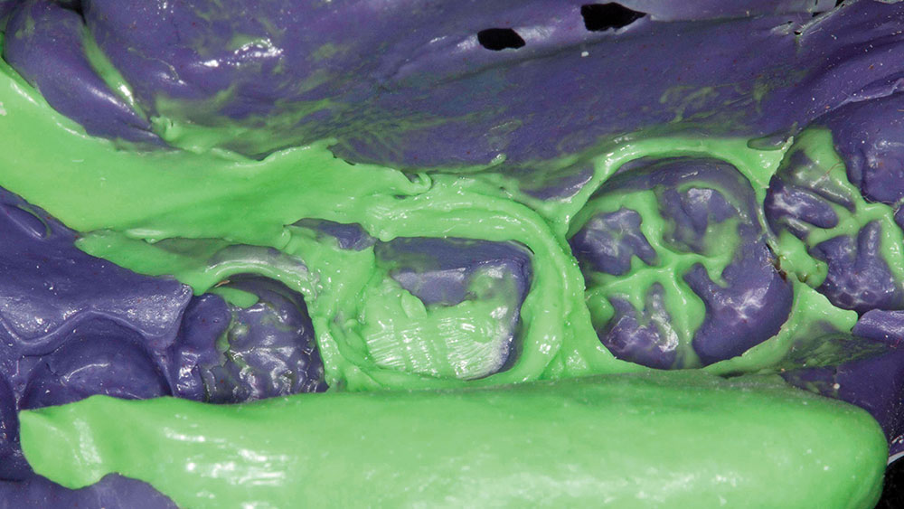
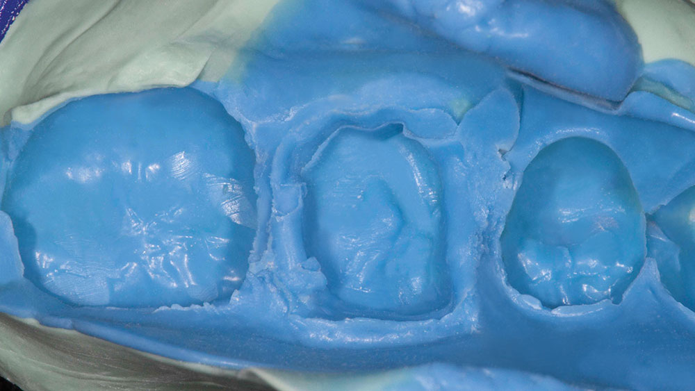
Figure 2: As we look closer, we can see that there is somewhat of a margin, and then everything just stops. There seems to have been a huge channel of either saliva or blood. I’m going to guess blood came rushing out of here because I just don’t see why there’d be that much saliva there. It looks like a dam broke and we just have water spilling out.

Figure 3

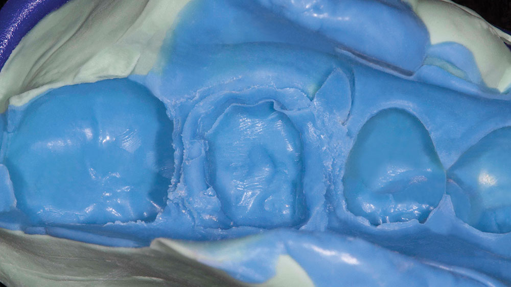
Figure 3: It’s always amazing to me just how good our impression materials are. I mean, look at the detail on the tooth posterior to the preparation there; it looks like a mountain ridge or the Sierra Nevada. These impression materials worked so well on every tooth except the one that was prepped. That is one of the consequences when you have gingiva that’s bleeding. So that’s nominee No. 1.

Figure 4

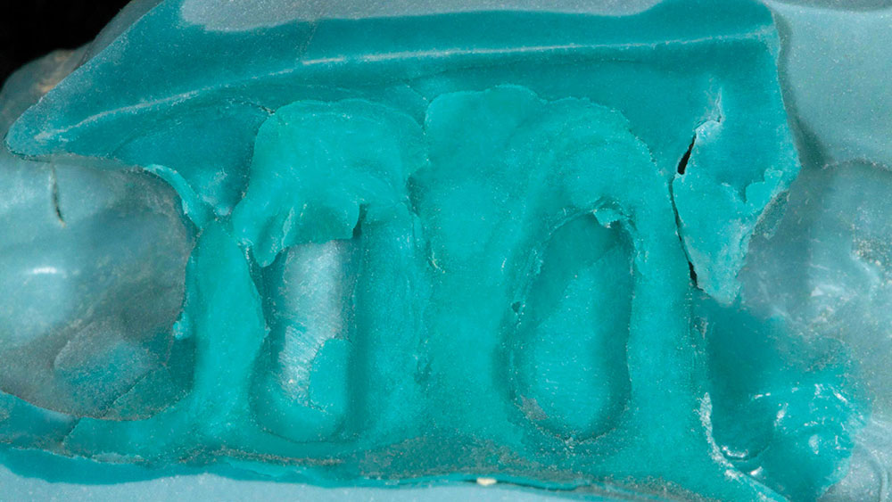
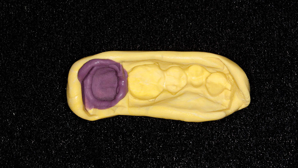
Figure 4: The second nominee (we’ve seen his work before) is Dr. H. from Miami, Florida. I believe he won Most Questionable Partial Denture last year. It looks like he’s back at it again. Here’s an impression where we have a wisdom tooth and it’s not contained in the impression; it’s actually biting down on the tray itself. That makes it a little difficult for this impression to work.

Figure 5

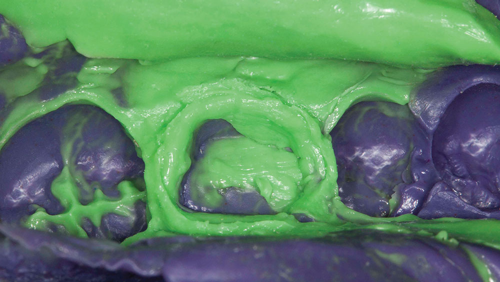
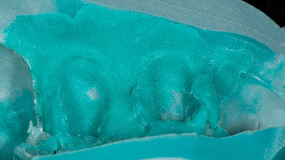
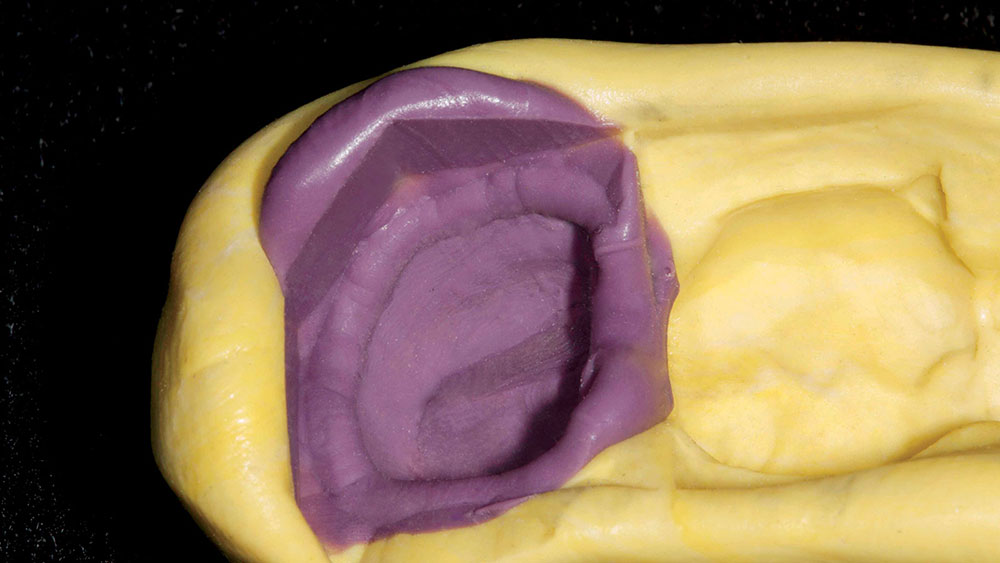
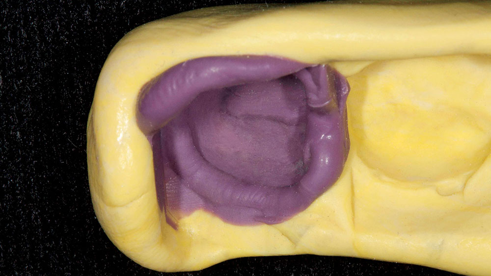
Figure 5: When we look a little bit closer, we can see extra material hanging off, unclear margins, big divots in the margin itself, and just a huge void right at the margin.

Figure 6

Figure 6: I know I say this a lot, but if you don’t take care of the tissue and it starts to bleed, these impression materials will not work. They will not set well. They’ll get pushed out of the way by the blood. This impression is a really good example of that.

Figure 7

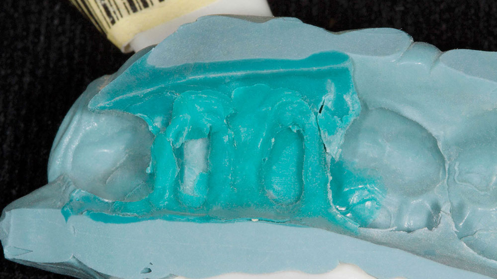
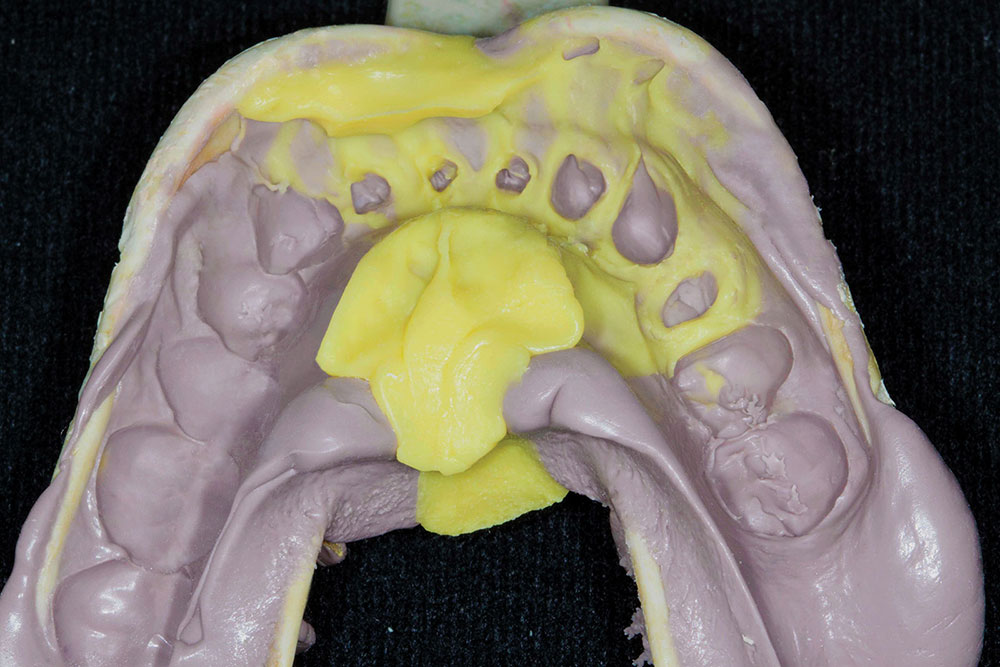
Figure 7: The third nominee is Dr. B. from sunny San Diego, California, America’s finest city. Not America’s finest impression, however. When you look at this, you can see two bicuspids prepped in this double-arch tray. Both look a little indistinct.

Figure 8

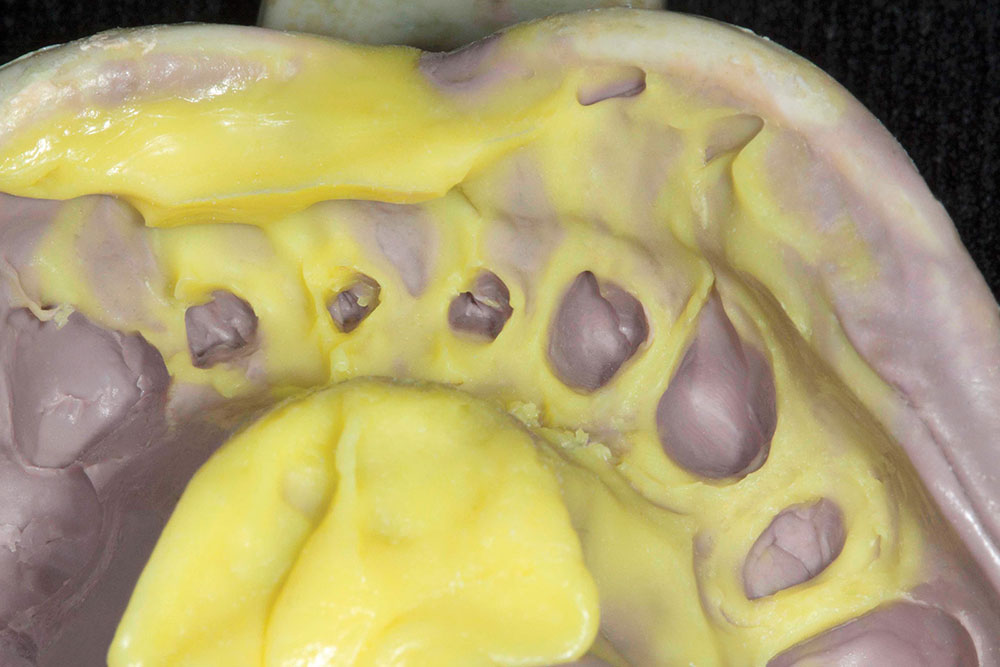
Figure 8: When we get a little closer, you can see where it looks like extra material was just added on at the end. We’ve got a little tear as well; not the end of the world, but again, just overall not a lot of detail.

Figure 9

Figure 9: You can really see what a mess this impression is toward the front. We have some white speckles here. It almost looks like it was coated with confectioner’s sugar at the end (maybe to tell the technician to be sweet with this impression). I don’t know what this is. Maybe it’s latex. It’s difficult to see. This is going to be a really hard one.

Figure 10

Figure 10: All right, we’ve got our fourth nominee: Dr. K. from Chicago, Illinois. Let’s take a look at what Dr. K. did. These are some anterior preps as you can see on the front, and they look pretty indistinct. You can see some rough outlines of what appear to be teeth; some kind of round, preplike shapes.

Figure 11

Figure 11: Looking a little bit closer, I don’t think there was any retraction cord or diode laser used here. I’m not sure what was used. In fact, I’m not even sure the teeth were necessarily prepped. It just looks like there was an impression of some tiny teeth.

Figure 12

Figure 12: If we flip the impression over and look at it toward the lingual, you can see that we really don’t have much definition there either. But at least we can tell they’re preps because we see some of the diamond marks in there. This impression had to be sent back pretty quickly. There’s one tooth toward the posterior that you can almost make out the margins and where the tooth is, but the rest of it is just kind of sloppy.

Figure 13

Figure 13: Our fifth nominee is Dr. P. from Atlanta, Georgia. This one is really confusing because when you look at this impression, it’s got some really sharp angles on it, and you can tell there’s no patient whose mouth actually looks like this.

Figure 14

Figure 14: Based on this 90-degree angle and how smooth it is, it looks like a polyvinyl impression of a model that has a base formed on it. This causes me to say, “Wait a minute: This wasn’t taken in a human mouth, yet it appears to be an impression of a human molar.” It has this big band around it that kind of looks like the margin, but it’s not. The margin is that thin line down below it.

Figure 15

Figure 15: As you look at it from multiple angles, it’s really tough to figure out what happened. Apparently, the doctor took an impression of a model, and then he relined it with light-body material. Frankly, it’s really confusing, and I’m not sure what to make of it. It’s really just kind of crazy when you look at it and see exactly what’s going on.

So congratulations to all the nominees. There was a bumper crop of questionable impressions from which to choose: A difficult choice, but I’m going to have to go with Dr. K. from Chicago. Of course, we picked impressions that were on the far end of the not-looking-very-good bell curve; but I have to say that, going through the year’s impressions, a lot of the ones that we looked at were actually taken very well. And as a laboratory, when you look at our remake rate — the rate of dentists sending stuff back to us — the annual figure went down. And it continues to get lower as time goes on due to monolithic restorations like IPS e.max® (Ivoclar Vivadent; Amherst, N.Y.) and BruxZir® Solid Zirconia. Both of these materials have lower remakes than PFMs ever did. As more dentists hop on board with these monolithic restorations, we see the remake rates go down. Any time we can do something to reduce the remake rate — whether it’s a technique, prepping, impressions or switching to a monolithic material — it is always a huge benefit for the patient.