Too Much Tooth, Not Enough Tooth: Making Decisions About Anterior Tooth Position

The restoration or creation of an esthetic smile is always the result of focused observation, thoughtful evaluation, and a systematic approach to planning and sequencing treatment. Restorative and esthetic dentistry approached through this process will incorporate the five critical elements that contribute to the beauty of a natural smile and result in a successful outcome for both patient and dentist. These five essential considerations are tooth position, gingival levels, arrangement, contour and color.
Although each of these is important in the final result, the first step is the most important — and in the esthetic process, the starting point for tooth position always is the incisal edge of the maxillary central incisor.1,2 As in denture prosthetics, this step is critical not only in the esthetic plan, but also in developing the functional treatment plan — because it determines the appropriate positions of all the maxillary teeth and subsequently, beginning with the lower incisors, determines the positions of the mandibular teeth.
Lip Mobility as a Factor in Tooth Display
In this article, I review the elements used in determining the correct position of the incisal edge of the maxillary central incisor, step No. 1 in the diagnostic and treatment planning sequence called Esthetics—Function—Structure—Biology, used in the Spear Education program. The practitioner must evaluate three aspects to ensure correct placement of that edge, and I will describe them here.
As a general rule in my practice, with the patient’s lip at rest, I always ensure that at least the edges of both central incisors are visible so that the patient does not appear to be edentulous.
The Elements of Determining Incisal Edge Placement
The three factors the dentist must evaluate for correct placement of the maxillary central incisor’s incisal edge are tooth display and lip mobility; the position of the incisal edge relative to the position of the other teeth in the maxillary arch; and phonetic considerations.
Tooth display and lip mobility. The first consideration is a combination of two elements: the amount of tooth displayed at rest and lip mobility. Lip mobility is the amount of lip movement that occurs as the patient smiles.3 The majority of observers will select as an ideal esthetic smile one that displays the full central incisor and includes a slight amount of gingiva apical to the tooth.4 The amount of tooth that shows at rest will vary depending on the amount of lip movement during the smile. As an example, if the patient’s central incisor is 10.5 mm long (an average length) and the lip moves 6 mm from rest to full smile, assuming a display of the gingival margin during full smile, 4.5 mm of tooth will be displayed at rest. If the same patient's lip is less mobile, moving only 4 mm from rest to full smile, 6.5 mm of tooth will be displayed at rest to achieve the same esthetics. Conversely, if the patient has a highly mobile lip, with 10 mm of movement, only 0.5 mm of tooth will display at rest to meet the esthetic requirements of an ideal smile.
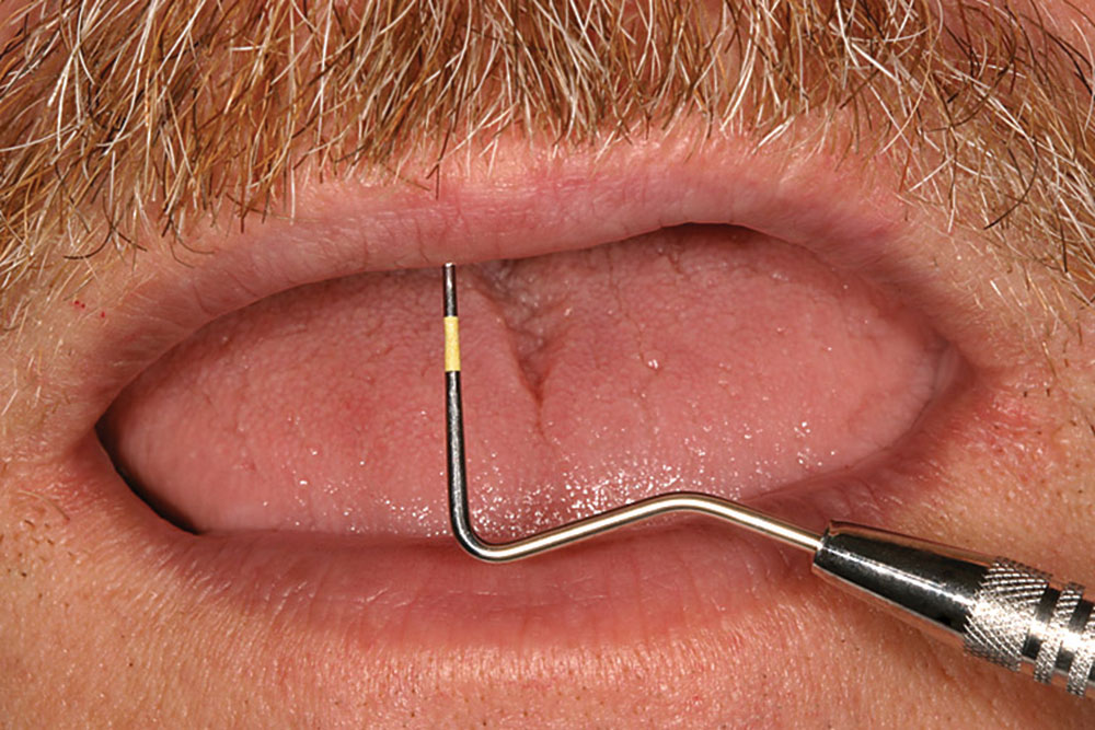
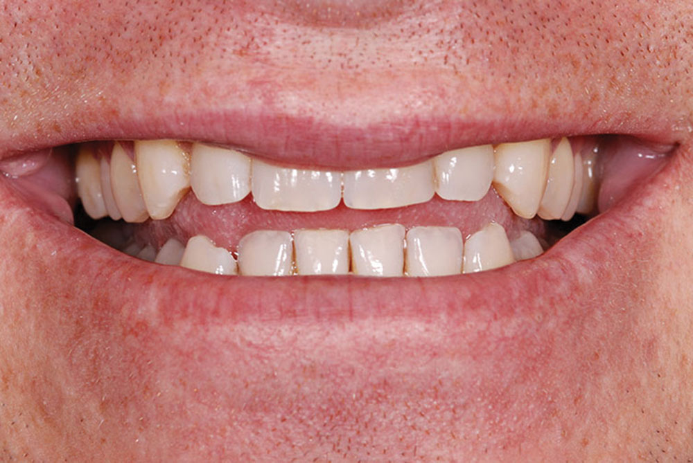
The preceding example illustrates clearly that placement of the incisal edge will be influenced dramatically by the patient's level of lip mobility and the desired appearance of the smile regarding tooth exposure and gingival display (Figs. 1a, 1b). As a general rule, the more mobile the lip, the less tooth that can be displayed with the lip at rest to create a pleasing smile; the less mobile the lip, the more tooth display at rest that will be necessary to create a pleasing, full smile. In 1978, Vig and Brundo5 examined a sample of women and determined the following averages for tooth display at rest according to age:
- Age 30, 3 mm to 3.5 mm
- Age 50, 1 mm to 1.5 mm
- Age 70, 0 mm to 0.5 mm.
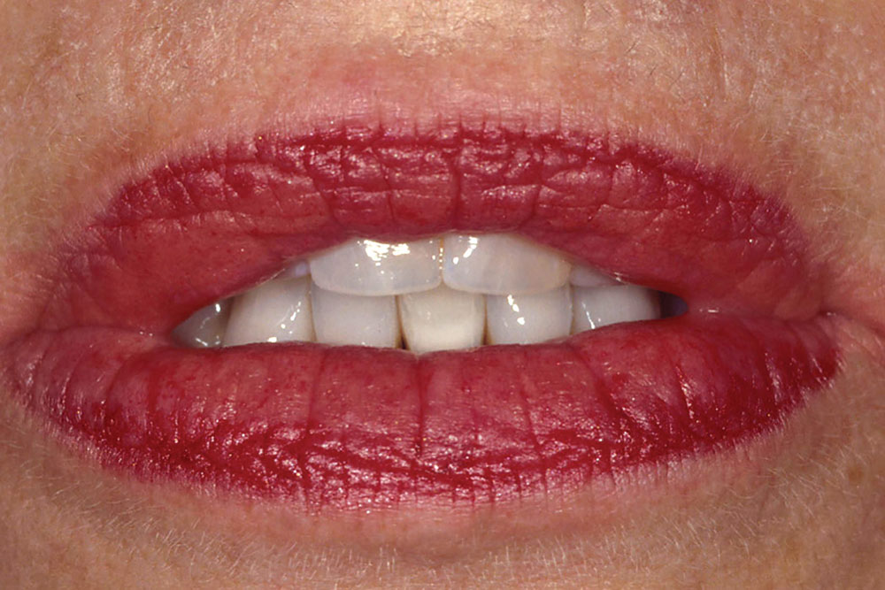
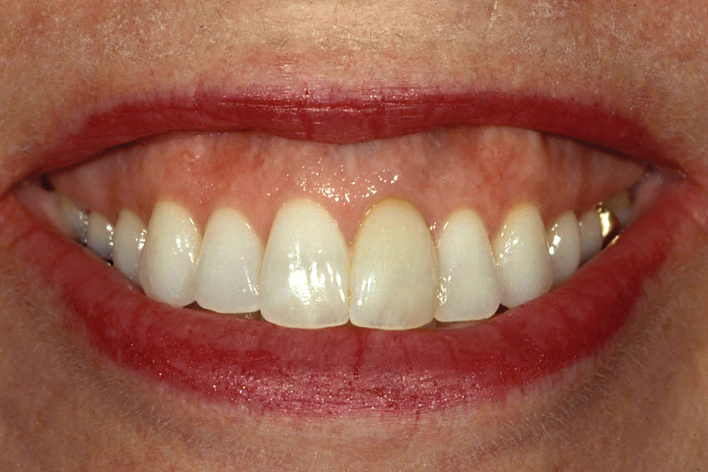
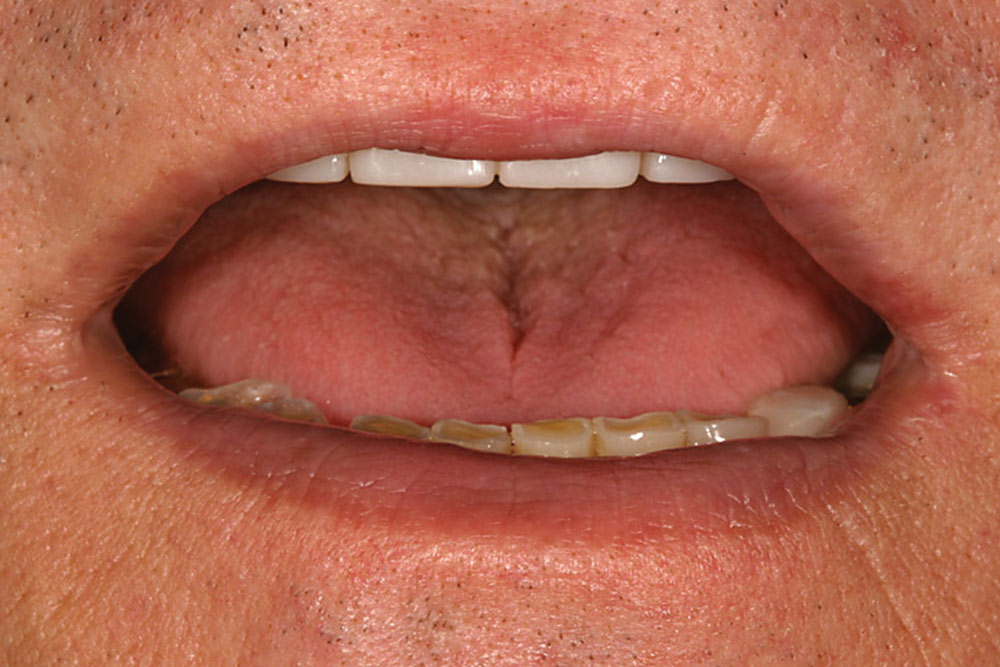
According to Vig and Brundo’s study, this change in display is less the result of tooth position than of changes in the facial tissues relative to the skeletal base. I find this information especially useful with patients who believe their teeth are too short. To begin, I evaluate how much tooth they display with the upper lip at rest. I then ask the patient to smile, and I note the amount of lip movement. If I know the amount of tooth display desired with the patient’s full smile, the patient’s lip mobility combined with the average length of a central incisor helps me determine where to begin in testing placement of the incisal edge. This is an especially useful technique with patients who exhibit extreme dental wear. Often, these patients display no tooth with the lip at rest (Fig. 2a). Using Vig and Brundo’s5 averages, I can approximate display at rest on the basis of the patient’s age and know how much to lengthen the central incisors to create an average tooth display with the lip at rest (Fig. 2b). I then can try this incisal edge position as either a composite mock-up or a provisional restoration (Fig. 2c). By asking the patient to smile fully, I can evaluate the smile and use this observation to refine the edge position. Whenever the practitioner is lengthening the incisal edge, he or she must evaluate “f” and “v” sounds and modify tooth shape and position for acceptability (see section on phonetics below).
The ultimate position of the incisal edge for patients with extreme tooth wear is a combination of tooth display at rest, lip mobility, age and functional consideration based on what the occlusion will tolerate. Vig and Brundo’s5 averages of tooth display at rest are simply useful starting points from which to make refinements to arrive at the most appropriate position for each patient. As a general rule in my practice, with the patient’s lip at rest, I always ensure that at least the edges of both central incisors are visible so that the patient does not appear to be edentulous.
Position relative to other maxillary teeth. The second consideration in establishing the correct maxillary incisor position is evaluation of the incisal edge relative to the other teeth in the maxillary arch.6,7
In a normal Class I occlusion, the incisal edge of the central incisor will be on approximately the same plane as the tips of the canines and the buccal cusp tips of the premolars and molars. When this arrangement exists, the maxillary central incisal edge position is esthetically pleasing, and the smile line exhibits symmetry with the lower lip (Fig. 3).9
When the maxillary central incisal edge is coronal to the plane of the posterior teeth, it is caused most commonly by overeruption of the teeth as a result of Class II malocclusion or of restorative dentistry completed without consideration of edge position. The teeth appear too prominent in the face, and the smile line exhibits an exaggerated curvature. Bringing the edges apically to the plane level with the posterior teeth is an excellent starting point when correcting front teeth that appear too long. After the anterior teeth are placed on the same plane as the posterior teeth, either through orthodontics or provisional restorations, the practitioner then can refine the position for the most pleasing appearance.7,8
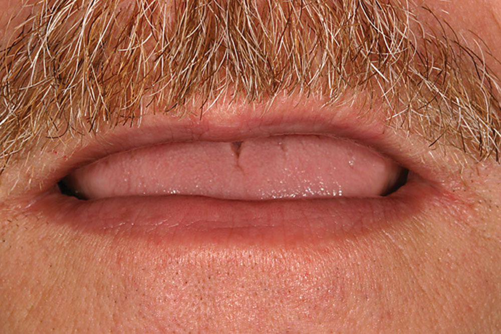
When the maxillary central incisal edge is apical to the plane of the posterior teeth, it creates a reverse smile line (Fig. 4). Common causes of this are undereruption resulting from a Class III malocclusion, ankylosis caused by trauma or a patient’s habit (such as tongue thrusting and thumb sucking). Perhaps the most common cause of this tooth position, however, is wear of the anterior teeth resulting from a protrusive bruxing habit or chemical erosion while the posterior teeth sustain minimal wear.
Placing the maxillary central incisor’s incisal edge visually on the plane of the posterior teeth, either orthodontically or restoratively, will resolve most of the esthetic problems and create a position from which the dentist then can make refinements. Although this is a useful method of starting to position central incisal edges, it cannot always be used. When posterior teeth are missing, worn away, overerupted or improperly restored, one must use the first and third considerations alone.
Given the importance of esthetic success in practice today, and the fact that every facet of treatment is affected when a dentist decides to change a patient’s incisal edge position, it is critical that dentists learn, become comfortable with and use these techniques when evaluating patients.
Phonetics. The third consideration in appropriately positioning the maxillary incisal edge is phonetics, specifically the “f” and “v” sounds, as described in classic prosthodontic texts.9,10,11 Most technique discussions mention using “s” sounds as well; however, whereas this certainly is an important consideration, the “s” sound is the result of the interaction between the maxillary and mandibular incisors.12 In the Esthetics—Function—Structure—Biology treatment planning protocol, we first position the maxillary incisors to the ideal esthetic position and then modify the mandibular incisors and the lingual aspect of the maxillary incisors to correct the “s” sound, the final position and shape being determined by the movement of the mandible during speech. Enunciation of “f” and “v” sounds creates light contact of the central incisors with the “wet-dry” line of the lower lip. Dimpling or trapping of the lower lip signals that the contact impingement by the teeth is too great and indicates teeth that are too long and must be shortened. One difficulty in using “f” and “v” sounds to evaluate length and position is that they can tell the dentist reliably whether the teeth are too long, but they do not offer much insight into whether the teeth are too short. Even when the maxillary central incisors are severely worn, formation of “f” and “v” sounds will look correct because speech is so adaptable to shortening of the maxillary incisors. Because restorative dentistry usually is involved in lengthening maxillary central incisors, using phonetics is an excellent consideration in determining whether teeth have been lengthened too much.
Conclusion
The focus of this article is the esthetic considerations of the maxillary central incisal edge as part of the Esthetics—Function—Structure—Biology process of diagnosis. Clinicians should recognize that all changes made to the position of the maxillary central incisor must address the functional etiology that placed the central incisor in a position other than one that creates the ideal smile. They also must understand clearly how the functional component, the occlusion, must be altered to produce a predictable result with the new incisal edge position.
In this article, I have presented three considerations in evaluation and positioning of the maxillary central incisal edge. Given the importance of esthetic success in practice today, and the fact that every facet of treatment is affected when a dentist decides to change a patient’s incisal edge position, it is critical that dentists learn, become comfortable with and use these techniques when evaluating patients.
Dr. Frank Spear is founder and director of Spear Education. To learn about Dr. Spear or Spear Education, visit speareducation.com or call 866-781-0072.
DISCLOSURE: Dr. Spear did not report any disclosures. The views expressed are those of the author and do not necessarily reflect the opinions or official policies of the American Dental Association.
References
- ^Boucher CO, Hickey JC, Zarb GA, editors. Boucher’s prosthodontic treatment for edentulous patients. 7th ed. St. Louis: Mosby; 1975. p. 224.
- ^Sharry JJ. Complete denture prosthodontics. 3rd ed. New York: McGraw-Hill; 1974. p. 234.
- ^Martone AL. Anatomy of facial expression and its prosthodontic significance. J Prosthet Dent. 1962;12(6):1020-42.
- ^Tjan AH, Miller GD, The JG. Some esthetic factors in a smile. J Prosthet Dent. 1984 Jan;51(1):24-8.
- ^Vig RG, Brundo GC. The kinetics of anterior tooth display. J Prosthet Dent. 1978 May;39(5):502-4.
- ^Frush JP, Fisher RD. The dynesthetic interpretation of the dentogenic concept. J Prosthet Dent. 1958;8(4):558-81.
- ^Lombardi RE. The principles of visual perception and their clinical application to denture esthetics. J Prosthet Dent. 1973 Apr;29(4):358-82.
- ^Lombardi RE. A method for the classification of errors in dental esthetics. J Prosthet Dent. 1974 Nov;32(5):501-13.
- ^Pound E. Esthetic dentures and their phonetic values. J Prosthet Dent. 1951 Jan-Mar;1(1-2):98-111.
- ^Watt DM. Tooth positions on complete dentures. J Dent. 1978 Jun;6(2):147-60.
- ^Pound E. Recapturing esthetic tooth position in the edentulous patient. J Am Dent Assoc. 1957 Aug;55(2):181-91.
- ^Rothman R. Phonetic considerations in denture prosthesis. J Prosthet Dent. 1961;11(2):215-23.
Reprinted with permission from the American Dental Association (ADA): Spear FS. Too much tooth, not enough tooth: making decisions about anterior tooth position. J Am Dent Assoc. 2010;141(1):93-6. Copyright ©2010 American Dental Association. All rights reserved. The American Dental Association makes no representation and accepts no responsibility for the accuracy, timeliness or comprehensiveness of the cover image.

