Photo Essay with Dr. Daniel Melker on the Perio-Restorative Connection

August 4, 2011
 image
Daniel Melker, DDS

Note from the Editor

I chose to include another photo essay from periodontist Dr. Daniel Melker in this issue because I have a huge amount of respect for his clinical skills and his excellent clinical photographs. About eight years ago, I begged and pleaded with Dan to let me come spend a day or two at his office to watch how he works. I finally wore him down, and he allowed me to spend some time observing him in surgery, and then going through case slides, many of which were 15 to 20 years post-op.

Dan is Dr. Bill Strupp’s periodontist — the one partially responsible for all the gorgeous tissue you’ve seen in Bill’s articles in Chairside® or at his lectures. Dan happened to be working on one of Bill’s patients the first day I spent at his office, and when he took off the temporaries, I saw those same Strupp preps I have seen so many times before. Not that there was ever any question, but it was clear Bill practices what he preaches, and the same goes for Dan.

While I was there, I got to see a couple root reshaping cases (Dan prefers biologic shaping), which at the time boggled my mind. I didn’t know of anyone else doing this at the time, and I still don’t see anyone else showing these types of cases, other than Bill and Dan. Never before had I seen anyone routinely remove the CEJ (cementoenamel junction) to improve the long-term stability of the case. I also got to see some grafting and some hard tissue crown lengthening without a gingivectomy.

You may not want to attempt surgery quite like this yourself, but I think it’s important to know that it exists, because I guarantee you have many patients who would benefit from it. Make your periodontist take you out to lunch, bring this issue of Chairside with you, and show him what you would like him to try on a couple of cases. Resist the urge to take off an old, ugly PFM with a biologic width invasion and just slap on a new IPS e.max® (Ivoclar Vivadent; Amherst, N.Y.) crown because you know it will look better. Do the right thing and let your periodontist correct the condition, and then move forward — or do it yourself, if you feel comfortable. To push on with restorative dentistry while ignoring the tissue and supporting structures is an incomplete view of what we, as dentists, are here to do.

I hope that, like me, you will look at the cases that follow and get inspired. I think seeing them will encourage you not to just turn a blind eye the next time you come across one of these clinical situations in your own practice.

Case 1

Case 1 Figure 1

Figure 1: Tooth #28 is in need of a restorative procedure due to a lack of attached gingiva. There is no way other than adding connective tissue to change the present environment.

Case 1 Figure 2

Figure 2: The tissue is reflected and a subepithelial graft is placed to add connective tissue.

Case 1 Figure 3

Figure 3: The final restoration on tooth #28 shows a significant increase in connective tissue for long-term maintenance of the periodontal foundation.

Case 2

Case 2 Figure 1

Figure 1: Severe recession and bone loss in a 22-year-old patient. The need to increase connective tissue is apparent. A soft tissue procedure is advisable to increase the connective tissue, which is needed for long-term periodontal stabilization.

Case 2 Figure 2

Figure 2: Reflection of flap shows significant bone loss.

Case 2 Figure 3

Figure 3: PerioDerm™ thick (DENTSPLY Tulsa Dental Specialties; Tulsa, Okla.) is used to increase the connective tissue.

Case 2 Figure 4

Figure 4: Flap closure is performed, covering the PerioDerm completely.

Case 2 Figure 5

Figure 5: Total root coverage is provided by the use of PerioDerm.

Case 2 Figure 6

Figure 6: Reflection of the mucosa exposes a beautiful zone of connective tissue.

Case 3

Case 3 Figure 1

Figure 1: Severe subgingival location of tooth structure following caries removal. This case requires the removal of bone and possibly tooth surface to create a space for a new biologic width to establish, so that it will not be violated by the new restorations.

Case 3 Figure 2

Figure 2: A beautiful core is placed to be used as a marker to gauge the amount of space needed for a new biologic width to form. Note: The photo is reversed because it is a mirror shot.

Case 3 Figure 3

Figure 3: A flap is reflected exposing the location of cores to bone, revealing the need for crown lengthening procedures. Note: This can only be accomplished with surgery.

Case 3 Figure 4

Figure 4: Completed crown lengthening and suturing. Removal of previous margins and a conservative amount of bone will allow for a new biologic width to establish during healing. A new crown margin will be placed just coronal to the gingival collar to prevent periodontal interference from occurring in the future.

Case 3 Figure 5

Figure 5: An occlusal view shows 360 degrees of perfect interface between tooth surface and bone for long-term maintenance.

Case 4

Case 4 Figure 1

Figure 1: A crown placed within the biologic width is causing significant inflammation. This inflammation needs to be dealt with prior to the placement of another permanent restoration.

Case 4 Figure 2

Figure 2: The existing crowns are removed and cores and provisionals are placed.

Case 4 Figure 3

Figure 3: Occlusal view.

Case 4 Figure 4

Figure 4: With the tissue reflected, severe bone destruction is clearly seen due to the biologic width invasion.

Case 4 Figure 5

Figure 5: Cratered defects are corrected, and a parabolic architecture of the bone to mimic the soft tissue is created.

Case 5

Case 5 Figure 1

Figure 1: Developmental groove is causing bone loss on tooth #9.

Case 5 Figure 2

Figure 2: A flap is laid showing visible developmental defect with small amount of blood in it.

Case 5 Figure 3

Figure 3: Removal of developmental defect creates a smooth root surface for long-term maintenance.

Case 5 Figure 4

Figure 4: Connective tissue is added for long-term periodontal stability.

Case 5 Figure 5

Figure 5: Flap is positioned to cover the connective tissue graft.

Case 5 Figure 6

Figure 6: Postoperative view showing stable environment with zero probing.

Case 6

Case 6 Figure 1

Figure 1: A recent article by Cobb and Rapley, et al.1 discusses the problems associated with CEJs and the accumulation of biofilm, plaque and calculus. When performing biologic shaping, permanent removal of the CEJ can be undertaken to produce a long-term, maintainable environment for the hygienist and patient. Note the plaque and calculus approximating the CEJs.

Case 6 Figure 2

Figure 2: Removal of all CEJs with a handpiece to allow for long-term maintenance.

Case 7

Case 7 Figure 1

Figure 1: A patient with an excessive display of gingiva and no interest in orthognathic surgery. Crown lengthening will be necessary. A strong biologic background is necessary to properly treat this case for long-term stability. A clinician with a limited background biologically might consider doing a gingivectomy, which could present major difficulty.

Case 7 Figure 2

Figure 2: Upon reflection of the tissue, massive tori or exostoses are noted. The cause of the problem is tissue having to extend over the excess bone and up onto the enamel.

Case 7 Figure 3

Figure 3: Bone is removed to create a biologic environment between the bone and soft tissue.

Case 7 Figure 4

Figure 4: Simple interrupted sutures are placed using 5-0 chromic gut material. Because of primary closure, the patient will have minimal discomfort.

Case 8

Case 8 Figure 1

Figure 1: Patient with excessive soft tissue on enamel. Once again, the surgeon needs a strong background in biology to offer this patient the proper surgical approach for a long-lasting result.

Case 8 Figure 2

Figure 2: Severe bleeding is noted with minimal probing.

Case 8 Figure 3

Figure 3: Upon reflection of the soft tissue, it is noted that the CEJ and bone are approximating each other, thus creating a condition called “altered active eruption.”

Case 8 Figure 4

Figure 4: Space created for the biologic width through removal of bone. This procedure could never be undertaken without surgery.

Case 8 Figure 5

Figure 5: Flap is sutured back into place just coronal to the CEJs with 5-0 chromic suture material.

Case 9

Case 9 Figure 1

Figure 1: Teeth #18 and #19 with existing crowns to be replaced. Cores and provisionals will be undertaken so ideal periodontal therapy can be performed.

Case 9 Figure 2

Figure 2: The buccal margin of tooth #18 approximates the furcation area and furcation involvement is evident. Tooth #19 also has furcation issues and calculus is located just apical to the CEJ on the mesial.

Case 9 Figure 3

Figure 3: Definitive biologic shaping removes all irregularities on the tooth surface and eliminates the furcation. This treatment is a definitive approach to comprehensive periodontal and restorative treatment that can only be accomplished surgically.

Case 9 Figure 4

Figure 4: Perfectly contoured restorations following the prepared tooth surface with margins placed just coronal to the gingival collar. Furcations are barreled into the occlusal surface, allowing for long-term maintenance.

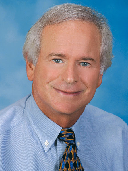
Dr. Daniel Melker is in private practice in Clearwater, Florida, and lectures nationwide. Contact him at 727-725-0100.

References

  1. Satheesh K, MacNeill SR, Rapley JW, Cobb CM. The CEJ: a biofilm and calculus trap. Compend Contin Educ Dent. 2011 Mar;32(2):30,32-7.