Photo Essay: BruxZir® Solid Zirconia Anterior Esthetic Challenge

February 16, 2012
 image
Michael DiTolla, DDS, FAGD
pointing at teeth

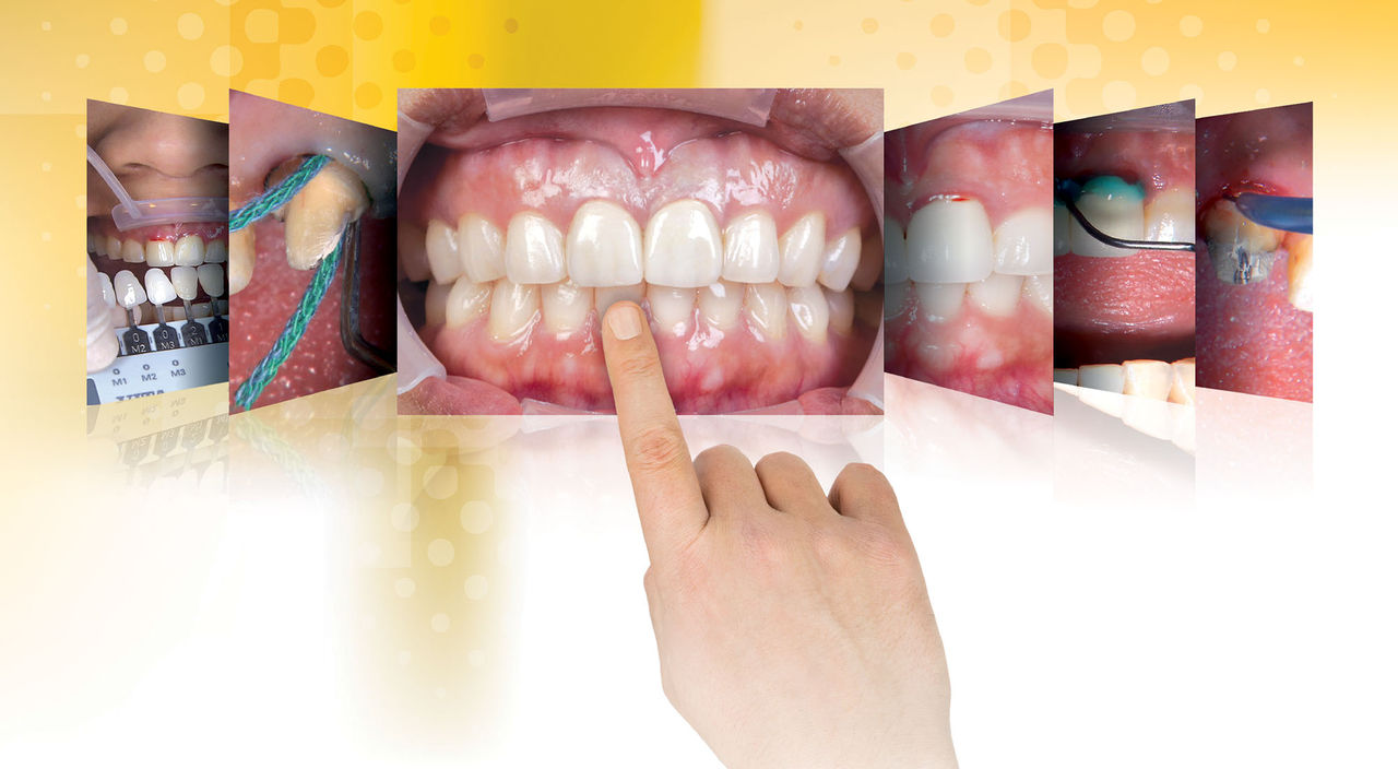
This photo essay illustrates our laboratory’s recent advancements in improving the esthetic properties of BruxZir® Solid Zirconia. Since the launch of the crown & bridge material in 2009, we have talked about BruxZir Solid Zirconia being “More Brawn Than Beauty.” As our R&D department refines our processes, improving the material’s translucency, the esthetics have continued to improve dramatically. What better esthetic challenge could there be for a material than using it to replace old crowns on teeth #8 and #9? BruxZir Solid Zirconia rises to the challenge in this case, but keep in mind, I have the advantage of in-house dental technicians, which always makes it easier to deliver great restorations. High-quality digital photographs can result in the same high-quality restorations almost as easily. After this case, we decided to upgrade the BruxZir Solid Zirconia motto to “More Brawn and Improving Beauty.” Continue reading to see if you agree!

With the margins now clearly exposed, I use an 856-025 bur (Axis Dental) with the water off and my KaVo ELECTROtorque handpiece set to 4000 rpm to slowly drop the margins to the new gingival level.
figure 1

Figure 1: First Appointment — We are going to replace the PFM crowns on teeth #8 and #9 with BruxZir Solid Zirconia crowns. This will be a good test for our newest translucent formulation. You can see how inflamed the gingiva is with the old crowns in place, which could be an allergic reaction to the existing base-metal PFMs.

figure 2

Figure 2: The first step of any restorative procedure in the anterior should be to take the shade before the teeth become dehydrated. This is especially true when using lip and cheek retractors, as we are doing here (SeeMORE [Discus Dental; Los Angeles, Calif.]).

figure 3

Figure 3: I am using the VITA Easyshade® Compact (Vident; Brea, Calif.) in the middle third of the tooth, with the tip flush against the tooth. I will shoot the shade in three spots in the middle third, in case I land on any shade anomalies.

figure 4

Figure 4: The VITA Easyshade Compact displays the shade in both VITA® Classical shades and VITA 3D-Master® shades (Vident). Having used both shade guides for many years, I strongly prefer the 3D-Master shade guide because of how well the shade tabs match natural teeth.

figure 5

Figure 5: After taking the shade, I hold the selected 2M1 3D-Master shade tab to the tooth, along with the 1M1 3D-Master shade tab for contrast. Of the spots I checked with the VITA Easyshade Compact, two were 2M1 and the third was 1M1, so I want to see how both shades look in the mouth.

figure 6

Figure 6: Now we photograph the shade tabs in the mouth. This is probably the most important part of communicating shade to the technician, so he or she can see how the natural teeth look compared to the selected shade guides. Rarely are they an exact match.

figure 7

Figure 7: I then use an Ultradent syringe to place PFG gel (Steven’s Pharmacy; Costa Mesa, Calif.) into the sulcus of teeth #8 and #9. The gingiva is so irritated that it starts to bleed just because I bumped into it with a soft brush tip. This is never a good sign.

figure 8

Figure 8: Next, I use my STA® (Single Tooth Anesthesia) System device (Milestone Scientific; Livingston, N.J.) to individually anesthetize teeth #8 and #9. Infiltrations of the maxillary central incisors are some of the most painful injections we give as dentists, and there are some patients who really hate them. This was one of those patients.

figure 9

Figure 9: The STA has a pressure sensor that lets me know if I am in the PDL during these injections, which helps me determine whether I have profound anesthesia. I used to give these types of injections by hand, but I never knew if I was giving an effective injection.

figure 10

Figure 10: The Razor® Carbide bur (Axis Dental; Coppell, Texas) is an aggressive carbide bur that easily cuts through porcelain and metal substructures. When used in combination with my KaVo ELECTROtorque handpiece (KaVo Dental; Charlotte, N.C.), it is simple to cut through an existing PFM in almost one continuous cut.

figure 11

Figure 11: Here, I am torquing the crown with a Christensen Crown Remover (Hu-Friedy; Chicago, Ill.). As we continue to use more and more high-strength, all-ceramic crowns that are more difficult to remove, there will come a day when we will reminisce about how fun it was to remove PFM crowns.

figure 12

Figure 12: We will be removing some of the unhealthy tissue to improve esthetics and gingival health, so I use a periodontal probe to sound to bone, ensuring I have enough biologic width to safely remove some tissue. To eliminate the chronic inflammation, we will need a minimum of 3 mm from the free margin of the gingiva to the crest of the bone.

figure 13

Figure 13: I use my NV MicroLaser™ (Discus Dental) to remove 1.5 mm of tissue. In addition to removing the unhealthy tissue, the diode laser helps me expose the crown margins that were buried subgingivally. This almost certainly contributed to the unhealthy gingiva that surrounded these two crowns.

figure 14

Figure 14: With the margins now clearly exposed, I use an 856-025 bur (Axis Dental) with the water off and my KaVo ELECTROtorque handpiece set to 4000 rpm to slowly drop the margins to the new gingival level. We will finish the preps at the next appointment.

figure 15

Figure 15: There is really no way to take an impression today after our gingival recontouring and still have the crown margins in the right place, so my assistant is relining BioTemps® Provisionals (Glidewell Laboratories) on teeth #8 and #9 with Luxatemp provisional material (DMG America; Englewood, N.J.), to help the tissues heal over the next two weeks.

figure 16

Figure 16: Using a thin, perforated diamond disc (Axis Dental), my assistant opens the gingival embrasures between the temps to avoid blunting the interproximal papilla. She also makes sure the gingival margins aren’t overextended and the emergence profile is flat.

figure 17

Figure 17: We use TempBond® Clear™ (Kerr Corp.; Orange, Calif.) to cement the BioTemps and avoid cement show-through in thinner temps. A word of caution with TempBond Clear: Use loupes to inspect around the temps and in the gingival embrasures to ensure no excess cement is left in place. This is an easy mistake to make with this clear cement.

figure 18

Figure 18: Second Appointment — After two weeks, we remove the temps and clean the preps with a KaVo SONICflex scaler. I know of no better way to ensure all the temporary cement is removed from the preps than by using this scaler, especially for cases where we have used Durelon™ (3M™ ESPE™; St. Paul, Minn.) as our temporary cement for its retentive properties.

figure 19

Figure 19: There is still minor irritation around the gingival margin, so I do a little trimming with the diode laser right at the gingival margin prior to placing the first retraction cord. I was worried there would be bleeding during the cord placement if I didn’t take care of this now.

figure 20

Figure 20: With the irritated tissue gone, I can now place my first cord, Ultrapak® #00 cord (Ultradent; South Jordan, Utah). Because this cord is hollow, it goes into place quite easily. I use this cord without solution (contains no epinephrine and has not been dipped in a hemostatic solution), as it could be in place for up to 45 minutes.

figure 21

Figure 21: I cut the cord on the lingual with curved scissors, while my assistant removes the cut end with cotton pliers. I cut the cord intraorally to make sure the two ends can be positioned flush to each other and do not overlap. This ensures there will be room for the second (top) cord.

figure 22

Figure 22: Because the placement of the first cord did not make the margin visually obvious, I place a second cord prior to refining the preparation. This top cord is an Ultrapak #2E cord (Ultradent). “E” refers to the epinephrine contained in the cord to help prevent bleeding.

figure 23

Figure 23: As I pack the #2E cord on tooth #8, you can see how this second cord has exposed the margin on tooth #9. Once each top cord is in place and the margins are exposed, we can begin the final finishing of the preps, which should take about 60 to 90 seconds per tooth.

figure 24

Figure 24: Now that I can finally see the margins, I use the same size bur I used before, but with a different grit (a fine grit 856-025 bur [Axis Dental], as indicated by the red stripe around the shank). The 30-micron diamond particles will smooth the prep, especially on the margins where our coarse bur broke off chunks of tooth.

figure 25

Figure 25: Two ROEKO Comprecap Anatomic compression caps (Coltène/Whaledent; Cuyahoga Falls, Ohio) are moistened internally and placed on the preps. The patient is instructed to bite with medium pressure for eight to 10 minutes. The Comprecaps ensure that the patient does not disrupt the cords with their tongue, and the pressure on the marginal gingiva provides added protection against bleeding.

figure 26

Figure 26: After my assistant removes the Comprecaps and pulls the top cord from tooth #9, I syringe medium body impression material around the preparation. Note the wide-open sulcus on the mesial of the tooth, which makes it almost impossible to miss this impression. I use medium body for my syringe material to prevent the material from tearing in the sulcus.

figure 27

Figure 27: For me, an ideal impression needs to have the prep margin clearly visible 360 degrees around the tooth, as well as 1 mm of impression material beyond the margin. This extra 1 mm of impression material beyond the margin represents an impression of the root surface, leading to ideal margin placement and optimal emergence profiles.

figure 28

Figure 28: Here, you can see how my assistant has placed the bite registration material exactly where it should be, covering the incisal third of the prepared teeth and the incisal third of the opposing teeth. Ideally, there should be no bite registration between the unprepared teeth and no contact with any soft tissue. The temporaries are then replaced, and the patient is asked to come back in two weeks for the try-in.

figure 29

Figure 29: Third Appointment — It’s been two weeks, the temps are off, the BruxZir Solid Zirconia crowns have been tried in and approved, and we are now placing a layer of desensitizer on the teeth (G5™ All-Purpose Desensitizer [Clinician’s Choice; New Milford, Conn.]). Dr. Gordon Christensen’s research shows that two coats of this glutaraldehyde/HEMA solution actually increases the bond strength of adhesive cements.

figure 30

Figure 30: I use a Warm Air Tooth Dryer (A-dec; Newberg, Ore.) for 10 seconds after applying both coats of the G5. Meanwhile, my assistant places Z-PRIME™ Plus (Bisco; Schaumburg, Ill.) inside the BruxZir crowns, and then we air-thin that for 10 seconds. Z-PRIME Plus is a zirconia adhesive that helps strengthen the bond of the cement to the crown.

figure 31

Figure 31: After my assistant loads the BruxZir crowns with a resin-modified glass ionomer cement (RelyX™ Luting Plus Automix [3M ESPE]) and the crowns are seated, I use a pinewood stick (Almore International; Portland, Ore.) to make sure they are fully seated. I then turn the stick sideways and hold it against the two incisal edges to verify they are the same length.

figure 32

Figure 32: One of the advantages of the new RelyX Luting Plus Automix is that you can tack cure the cement for five seconds with your light and then clean up the excess immediately, or you can do what you did in the past and wait two minutes for it to self-cure. It’s the only RMGI with a tack cure option available today.

I use a Warm Air Tooth Dryer (A-dec; Newberg, Ore.) for 10 seconds after applying both coats of the G5. Meanwhile, my assistant places Z-PRIME™ Plus (Bisco; Schaumburg, Ill.) inside the BruxZir crowns, and then we air-thin that for 10 seconds. Z-PRIME Plus is a zirconia adhesive that helps strengthen the bond of the cement to the crown.
figure 33

Figure 33: Here is an immediate, nonretracted shot of the BruxZir crowns on teeth #8 and #9 with the lips at rest. This is probably the easiest shot to take for crowns to look good because we are looking only at the incisal half, where reduction is nearly always adequate. The gingival third is where crown & bridge tends to look fake.

figure 34

Figure 34: A retracted view of the BruxZir crowns on teeth #8 and #9. I used to always under-reduce in the gingival third before I started doing the Reverse Preparation Technique, which ensures 1 mm of reduction in this area. Thanks to this technique, these crowns look decent even in the retracted view.

figure 35a
figure 35b
figure 35c

Figures 35a–35c: Looking at this series of “after” pictures, the most amazing part is that there is not any porcelain on these BruxZir crowns; they are solid zirconia. This is why they have superior strength and are stronger than all other restorative materials, with the exception of cast gold. The other amazing thing I notice is the facial anatomy that you see on the crowns in the lateral views. That flat facial profile in three planes is what makes a tooth look real. Because that anatomy is built into the CAD/CAM database, we are able to deliver it every time — provided the doctor gives us enough reduction. The promise of CAD/CAM dentistry is being able to deliver predictable esthetics because the restoration contours are based on a library of ideal teeth, not on a technician’s skill level or whether he or she is having a good day. As BruxZir Solid Zirconia has become more translucent, I find myself more willing to use it for challenging esthetic cases like this one. While I’m not suggesting that you suddenly switch all of your anterior restorations to BruxZir crowns immediately, you may want to consider using it for patients with parafunctional habits, or patients with old PFMs like the ones in this case, where an esthetic improvement is essentially guaranteed.

The other amazing thing I notice is the facial anatomy that you see on the crowns in the lateral views. That flat facial profile in three planes is what makes a tooth look real. Because that anatomy is built into the CAD/CAM database, we are able to deliver it every time — provided the doctor gives us enough reduction.