Photo Essay: Porcelain Veneers for Tetracycline Using Blockout Method

May 8, 2012
 image
Michael B. Miller, DDS, FAGD, FAACD
Photo Essay: Porcelain Veneers for Tetracycline Using Blockout Method Cover Image

NOTE FROM THE EDITOR


I don’t know how many dental books you own, but I have a couple of cabinets full of them. Most of them start gathering dust after my initial read, some come out a couple times per year (especially my favorite local anesthesia book), but only one has its own permanent parking spot on my desk: REALITY Publishing’s “The Techniques: Volume 1.” This publication, based on everyday applications of research and clinical experience, is the greatest gift young dentists could receive to help them achieve success in esthetic dentistry. It is also a useful reference manual for us older dentists. Dr. Michael Miller put this volume together and has been generous enough to share excerpts of it in Chairside®. In this installment, Dr. Miller shares an esthetic challenge we have all faced: placing porcelain veneers on tetracycline-stained teeth. Visit realityesthetics.com for more information on REALITY’s various publications.

Figure 1
Figure 1
Figure 2
Figure 2
Figure 3
Figure 3
Figure 4
Figure 4
Figure 5
Figure 5

Figures 1–5: Patient, with recently completed orthodontics and maxillary all-ceramic crowns on incisors and porcelain veneers on canines and premolars, wants mandibular veneers to mask tetracycline stains, despite not showing his mandibular teeth in a full smile. This type of color change can be done with an extended regimen of home bleaching instead of the expense and invasive nature of veneers, but patient wants immediate improvement and is not concerned about the upkeep necessary or cost for veneers.

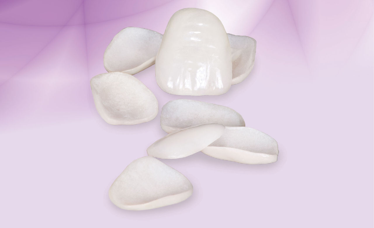
Note that the tetracycline stains are in the incisal half of each tooth, except for the central incisors, where the stains extend almost to the gingival crest. Masking stains in the more incisal portions of the teeth is much easier than when the stains are at the gingival margins. Because the stains are more incisal, the gingival color is quite normal.

Figure 6

Figure 6: Cord is placed prior to the preparation to accelerate the procedure.

Figure 7

Figure 7: Finished preparations. Note that tetracycline teeth usually become darker when they are prepared. The bonded lingual retainer obviously eliminates interproximal extensions.

Figure 8

Figure 8: Areas of preparations not to be masked with opaquer are covered with resin blockout material. Only dark-stained areas need to be masked. Resin blockout material keeps etchant and adhesive off stained areas.

Figure 9

Figure 9: Areas of preparations not to be masked with opaquer are covered with resin blockout material. Only dark-stained areas need to be masked. Resin blockout material keeps etchant and adhesive off stained areas.

Figure 10

Figure 10: Opaquer has been applied to dark-stained areas after etching and adhesive application. Even though the preparations are relatively aggressive, mandibular teeth cannot be reduced to the extent possible for larger maxillary siblings. Therefore, the opaquer must be kept quite thin.

Figure 11

Figure 11: Resin blockout has been removed. Note that only dark-stained areas are covered by the opaquer.

Figure 12

Figure 12: Veneers returned from the lab. Note that, despite their polychromatic buildup, the veneers have built-in masking ability. This built-in masking can eliminate the need to apply opaquer directly on the teeth in all but the darkest cases, assuming the veneers are at least 1 mm thick.

Figure 13

Figure 13: Mandibular anterior teeth at luting appointment two weeks after preparations. No provisionals were placed, but tissue is still inflamed.

Figure 14
Figure 14
Figure 15
Figure 15
Figure 16
Figure 16
Figure 17
Figure 17

Figures 14–17: Postoperative views two months after seating veneers. Maxillary and mandibular restorations match perfectly. There is no evidence of dark stains and tissue health has improved. Patient has been using an electronic interproximal cleaning device, which may have caused minor recession of papillae.

Dr. Michael Miller is the co-founder, president and editor-in-chief of REALITY. He maintains a dental practice in Houston, Texas. Contact him at mm@realityesthetics.com.

Reprinted by permission of REALITY Publishing. REALITY: The Information Source for Esthetic Dentistry, The Techniques, Volume 1, 2003, REALITY Publishing Company, pp. 272–74.