Achieving Reliable Denture Stability: The Need for Implant-Retained Overdentures to Increase

Poor denture stability and the challenges found in finishing restorative materials (composite, acrylic, porcelain and metal) are common and widespread problems. It is the author’s belief that these problems will significantly increase with the aging of the population.
The objective of this article is to provide the clinician with some straightforward solutions that can be employed when faced with the challenges associated with the correction of denture instability.
MINIMALLY INVASIVE DENTAL REHABILITATION
Attaching an overdenture to implants or resilient retained roots can improve denture stability. This is no different than earthquake-resistant anchoring of a building to a foundation that allows for a small amount of movement, while at the same time retaining the building in place. There are various attachments to connect the osseous anchorage and the removable prosthesis. Some attachments can be used on both natural and man-made roots (e.g., ERA® Attachment System [Sterngold; Attleboro, Mass.] and Locator® Implant Attachment System [Zest Anchors; Escondido, Calif.]). Other attachments are permanently incorporated into implants (e.g., Micro ERA® [Sterngold], Atlas® Implants [Dentatus; New York, N.Y.] and MDI Mini Dental Implants [3M™ ESPE™; St. Paul, Minn.]).
Ideal overdenture attachment characteristics include predictable placement, wear resistance, quick repair, low profile, good retention, patient-friendly use, reasonable cost and various diameters to fit differing ridge widths.
CASE REPORT
DIAGNOSIS AND TREATMENT PLANNING
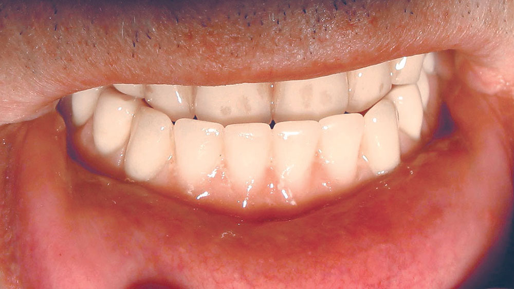
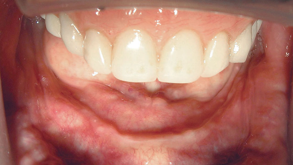
An 80-year-old male patient (retired physician) presented complaining of a mobile mandibular denture and associated pain. In addition, the patient had secondary complaints of speech difficulties, poor mastication, traumatized buccal mucosa, denture sores, poor esthetics, a prominent mandible and poor upper lip support. A Class III denture and jaw relationship was noted (Fig. 1). This gentleman had been edentulous in the mandible in excess of 30 years, resulting in obvious problems, which included the loss of the mandibular ridge and the subsequent loss of adequate denture retention (Fig. 2).
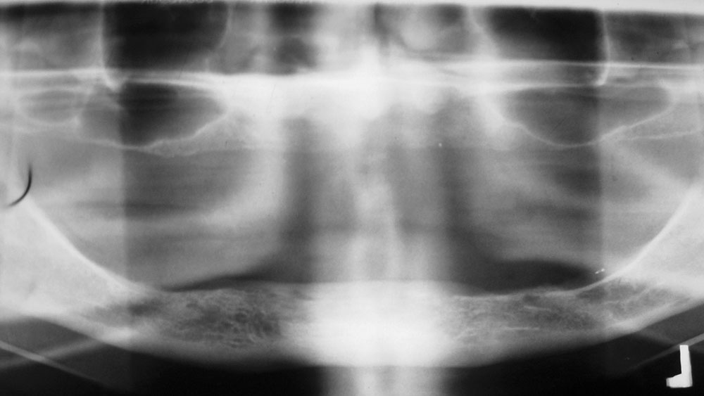
In addition to the clinical oral examination, a panoramic radiograph (Fig. 3), intraoral radiographs, dental models, photographs, complete dental history and medical histories were secured to assist in determining a treatment plan.
The patient had a long history of serious cardiac disease and hypertension that, along with possible associated complications, would cause a cautious clinician to limit the extent of any treatment and surgery. Less was better in this case.
The diagnosis included denture and skeletal Class III, significant posterior mandibular ridge and keratinized soft-tissue losses, significant medical complications, pain and speech alterations from poorly fitting dentures, diminished mastication efficiency from denture instability, poor dental esthetics and resultant decreased social interaction.
The treatment goals included stabilizing his health; minimizing any surgery and intraoral treatment; and improving denture retention, speech, esthetics, mastication and the Class III occlusion. The treatment plan would include a minimally invasive implant-retained mandibular overdenture, denture relining for stabilization, increased overbite and overjet for improved occlusion, and direct resin composites on the denture teeth for stabilization.
For this case, two ERA and two Micro ERA implants would be used to retain and stabilize the mandibular denture, both immediately and for the long-term. This system was chosen because it has: resilience, angle correction of abutments, radicular snap configuration, internal and external retention, vertical movement, hinge movement, six different retention grades, fabrication male with integrated spacer, and an optional metal receptor jacket (inside of the denture) placed by the lab or chairside. In addition, the fabrication and resilient males can be changed without bonding/processing, and there is also a micro version option.
Angle correction of the implants was anticipated due to the residual mandibular morphology. Direct composite resin veneers would be used to reshape the maxillary and mandibular dentition. A combination of soft and hard reline materials would be used to modify the soft-tissue surfaces of the existing dentures and the new denture.
MINIMALLY INVASIVE FLAPLESS IMPLANT PLACEMENT
A minimally invasive flapless implant procedure with local anesthetic was chosen to mitigate any possible unfavorable complications for this medically compromised patient.
A tissue-punch technique was used to remove soft tissue at the four designated implant sites: #27 ERA Angled Implant with 3.25 mm diameter, 10 mm length, 1 mm cuff, acid-etched; #23 ERA 0-degree Micro Head Implant with 2.2 mm diameter, 10 mm length, 1 mm cuff, acid-etched; #25 ERA 0-degree Micro Head Implant with 2.2 mm diameter, 10 mm length, 1 mm cuff, acid-etched; and #22 ERA Angled Implant with 3.25 mm diameter, 10 mm length, 1 mm cuff, acid-etched.
A latch-type #2 round bur (SS White® Burs; Lakewood, N.J.) was used with sterile saline irrigation to create a small pilot hole (1 mm) at each site. Osteotomies for the 3.25 mm diameter ERA Implants (#22 and #27) and the 2.2 mm diameter ERA Implants (#23 and #25) were completed.
For the 3.25 mm diameter implant placement sites (#22 and #27), the 1.6 mm drill (with external sterile saline irrigation) was used to create the osteotomies to 10 mm (full length of the implants). A 0-degree ERA Correct Angle Gauge (Sterngold) (metal post) was placed in the #27 first osteotomy hole to act as a guide to prepare the next site, #22. The 0-degree, 5-degree, 11-degree and 17-degree ERA Correct Angle Gauges were placed in the osteotomy sites to determine that the 0-degree for #27, 5-degree for #22, and angled female implant abutment should be used for the #22 and #27 osteotomy sites.
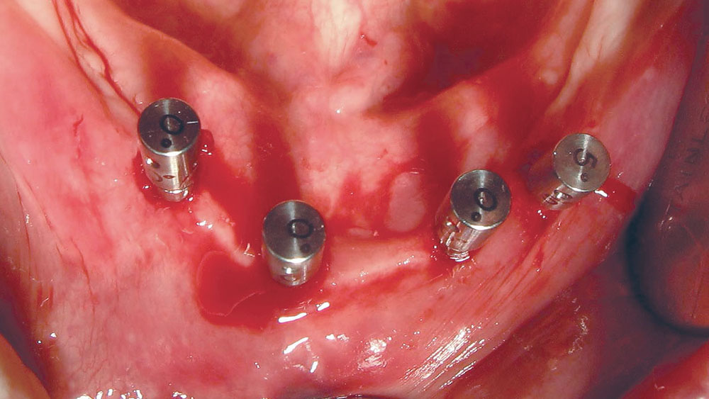
For the 2.2 mm diameter implant placement sites (#23 and #25), a 1.6 mm diameter countersink drill (with external sterile saline irrigation) created a pilot hole (as deep as the length of the untapered portion of the implant screw) with a flat area for the complete seating of the ERA female head of the implants. After the first 2.2 mm diameter 0-degree implant osteotomy, a 0-degree Correct Angle Gauge was placed in that osteotomy site. The #22 (5-degree) and #27 (0-degree) ERA Correct Angle Gauges were also in their respective osteotomy sites to allow for alignment of the last 2.2 mm diameter 0-degree implant. The ERA Correct Angle Gauges were used to determine if a straight 2.2 mm diameter 0-degree implant would align within the required 7 degrees of the other 2.2 mm diameter implant. The second 2.2 mm diameter 0-degree implant osteotomy was undertaken with a 1.6 mm diameter countersink drill (with external sterile saline irrigation). A 0-degree ERA Correct Angle Gauge was placed in this osteotomy site to verify alignment of all four implants (Fig. 4).
An ERA 3.25 mm combination countersink drill was used (with sterile saline irrigation) to expand the diameter of each 3.25 mm implant osteotomy using the appropriate 10 mm length, and to create proper contours to allow the ERA attachment heads to sit on the bone surface. The bone was not dense enough to require the tapping of the osteotomies with a bone tap and sterile saline irrigation.
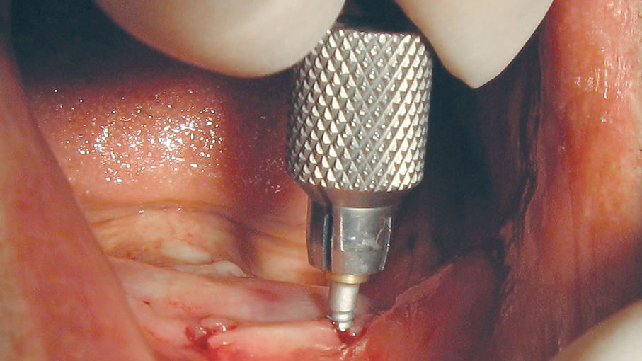
The #23 and #25 ERA straight implant packages (containing 2.2 mm diameter, 10 mm length, 1 mm cuff, acid-etched sterile implants) were opened, and the titanium tubes that hold the implants were placed in the plastic package’s round depressions. The implant insertion tool was inserted into the implant and used to carry the implant to the osteotomy. The self-tapping implant was slowly screwed into the osteotomy site with the implant insertion tool (with external sterile saline irrigation). Sometimes the implant can be fully seated using the insertion tool with downward pressure (Fig. 5).
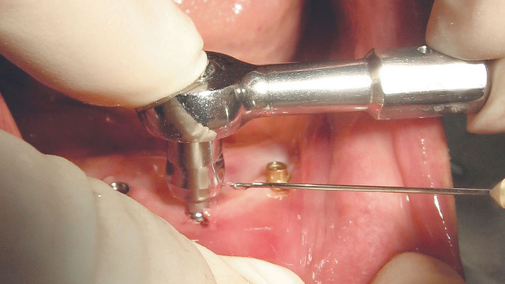
The implant insertion tool is gently tipped to disconnect it from the implant. An ERA socket with a short-handled ratchet wrench was used with downward pressure to slowly complete the seating of the implant into the osteotomy site with external sterile saline irrigation (Fig. 6).
Fractures or failures can occur if friction creates thermal trauma, fast movements with the ratchet wrench heat the implant, excessive torque (especially in cortical bone) is applied, or the ratchet is tilted away from a 90-degree angle to the implant. These actions will damage bone. Failures and fractures can be avoided in dense bone by using a ratchet wrench to slowly seat the implant with a cooling sterile saline irrigation. This is done by taking seven seconds for each quarter turn (with the thumb or forefinger of the opposite hand applying downward pressure to the head of the ratchet wrench), and then waiting for a period of five to 10 seconds or more between turns to allow bone displacement/expansion. Implants are fully seated only when all blasted threads are engaged in bone and the apical tip of each implant is stabilized by cortical bone.
The implant insertion tool, ERA socket and ratchet were used to place the 3.25 mm ERA implants (with external sterile saline irrigation) in the same manner as the 2.2 mm implants.
PROSTHETIC PROCEDURE
Progressive loading of the implants was chosen due to the patient’s advanced age, dystrophic edentulous ridge, thin soft tissue and the clinician’s desire to retain all four implants indefinitely. At the end of surgery, an immediate improvement in retention was accomplished with progressive loading of the implants.
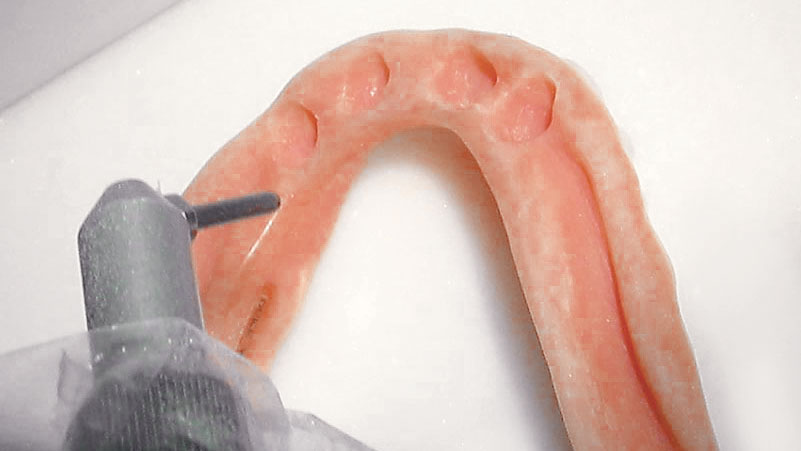
The existing mandibular denture was relieved and undercut over each of the four implants, as well as over the edentulous ridge. The tissue surface of the denture was sandblasted and cleaned (Fig. 7). Then, reline adhesive was placed on the tissue surface of the denture and thoroughly dried. Next, a soft silicone denture reline material (SOFRELINER TOUGH® [Tokuyama Dental America Inc.; Encinitas, Calif.]) was placed on the tissue surface of the denture, and it was passively seated in the mouth. Any excess silicone was trimmed and buffed.
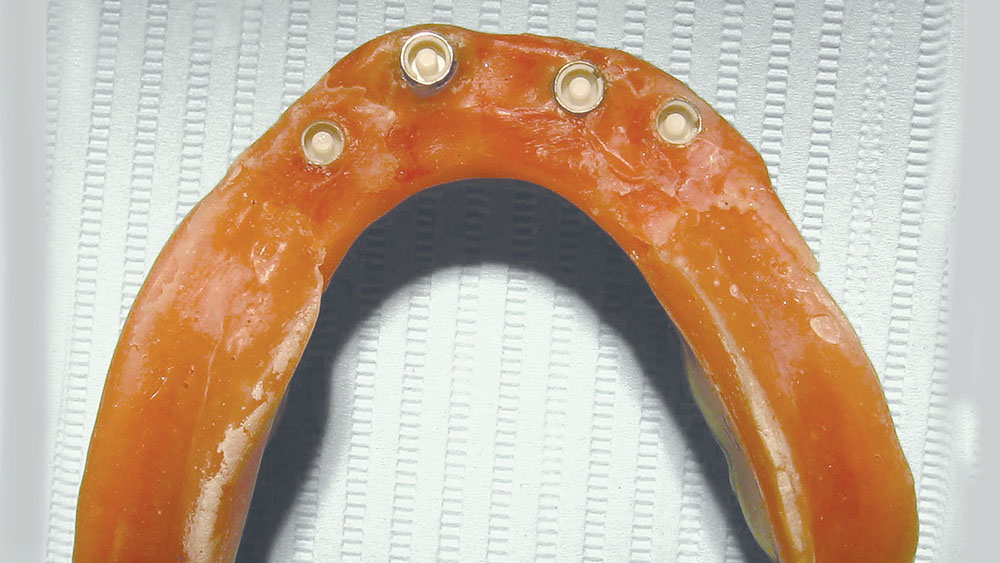
About one month post-op, the metal housing for the polymer males was placed in the mandibular denture. The silicone in the denture’s relieved wells over #23 and #25 was removed. The wells were then sandblasted and cleaned. Rubber dam covers were placed over the #23 and #25 surgical sites for protection and to block out undercuts, and the metal housings with black processing males were placed on the implants. Hard reliner (REBASE II [Tokuyama Dental America Inc.]) was then mixed and placed in the denture wells, and the mandibular denture was passively seated on the implants and edentulous ridge until the reliner set (Fig. 8).
The metal jackets with black fabrication males have built-in spacers. These were used for immediate loading and retention of the denture. The denture was placed into a solution of TOKUSO RESIN HARDENER II (Tokuyama Dental America Inc.) for a minimum of three minutes. The denture was then rinsed, dried and polished as necessary with one-step polishers (Jazz® Supreme [SS White Burs, Inc.]). These versatile, diamond-impregnated, soft-polymer polishing instruments trim, smooth and polish in one action by varying the applied force: Heavy force provides trimming, and light force generates fine polishing that quickly provides a high luster. These one-step polishers can be used on composite resins, denture teeth, hard denture reline materials and most denture acrylics.
The recesses over the 3.25 mm diameter ERA Implants were coated with SOFRELINER TOUGH PRIMER (Tokuyama Dental America Inc.), air-dried, then filled with Tokuyama SOFRELINER TOUGH, and the denture was seated in the mouth. The patient bit lightly in occlusion. (Note: DentuSil™ [Bosworth; Skokie, Ill.] and Light Liner® [Bosworth] are examples of alternative silicone liners that could also be used with these procedures.)
The black processing males were replaced with white retentive Micro ERA males. ERA males are black (processing), white (minimal retention), orange, blue, gray, yellow and red (maximum retention). The black processing males were removed with a Micro ERA core cutter (trephine bur) in a straight handpiece at a slow to medium speed. Short spurts of an in-and-out cutting motion were used to remove the center of the black fabrication male from the metal jacket. An explorer was used to remove the black processing male remnants. New white Micro ERA males were placed in the Micro ERA metal housings (#23 and #25) with a Micro ERA seating tool. An ERA white male creates a 0.4 mm space for resiliency and hinging.
OSSEOINTEGRATION
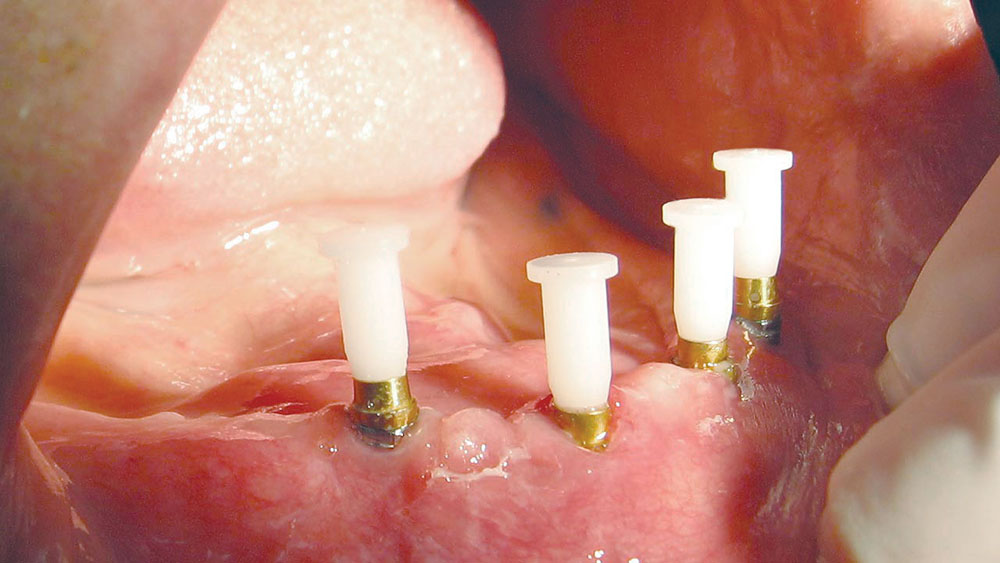
The 3.25 mm diameter ERA Implants were left undisturbed for more than the minimum of three months to allow osseointegration. The ERA female abutments were placed in the #22 and #27 positions. ERA Alignment Handles (Sterngold) were snapped into the #22 ERA Female 5-degree Angle Correction abutment (Sterngold) and the straight 0-degree #27 ERA abutment. The #22 ERA Female 5-degree Angle Correction assembly, the #27 ERA 0-degree straight assembly, and the two straight Micro ERA Alignment Handles were snapped into their respective 3.25 mm and 2.2 mm ERA Implants (Fig. 9). The #22 ERA Female 5-degree Angle Correction assembly was rotated until all of the white plastic alignment handles determined the proper path of insertion for the denture. A vertical line was marked across the implant bases and female abutments (#22 and #27) to verify correction alignment at cementation. The female abutments were removed, and a small amount of self-cure ERA Lock Cement (Sterngold) was placed into the bases and female abutments. Next, the female abutments were snapped into the implant bases and realigned by the vertical markings. Then, excess cement was removed (Fig. 10).
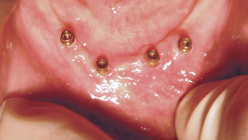
Subsequently, the 3.25 mm ERA Metal Jackets with black fabrication males and built-in spacers were placed into the mandibular denture in the same manner as they were placed into the 2.2 mm implant positions (#23 and #25). These were used for retention of the denture (Fig. 11). The 3.25 mm ERA black fabrication males were replaced with 3.25 mm ERA white retention males, using the same technique as was employed for the 2.2 mm implants but with regular ERA instrumentation (Fig. 12).
NEW MANDIBULAR DENTURE FABRICATION
The Class III occlusion was corrected using direct composite resin veneers.
The maxillary denture teeth were sandblasted, washed and dried. A one-step bonding agent (One Coat 7.0 [Coltène/Whaledent Inc.; Cuyahoga Falls, Ohio]) was applied to the denture teeth, air-thinned and light-cured. Then, flowable composite (SYNERGY® D6 Flow [Coltène/Whaledent Inc.]) was applied to the occlusal surfaces of the posterior to open the bite, allowing the shift from a Class III to a flat plane Class I occlusion. Next, hybrid composite (SYNERGY® D6 [Coltène/Whaledent Inc.]) was applied to the facial surfaces of the denture teeth, shaped into direct veneers and light-cured.
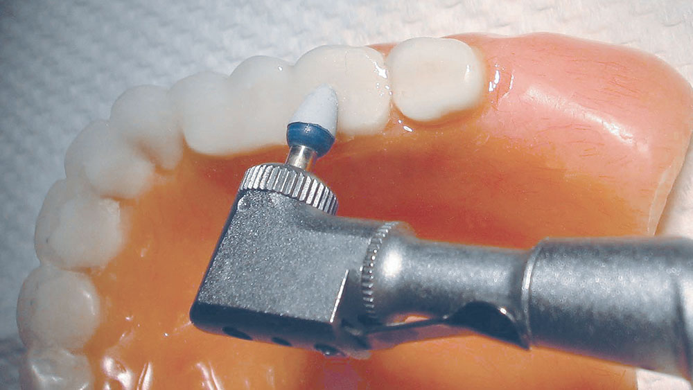
The use of safe-ended finishing burs (Safe End Composite Finishing Burs [SS White Burs]) allowed for easy trimming and shaping of the composite resin rehabilitation of the maxillary denture dentition. Then, use of the one-step polishers (Jazz Supreme) efficiently imparted high luster to the composite resin as well as to the denture acrylic surfaces adjacent to the teeth (Fig. 13).
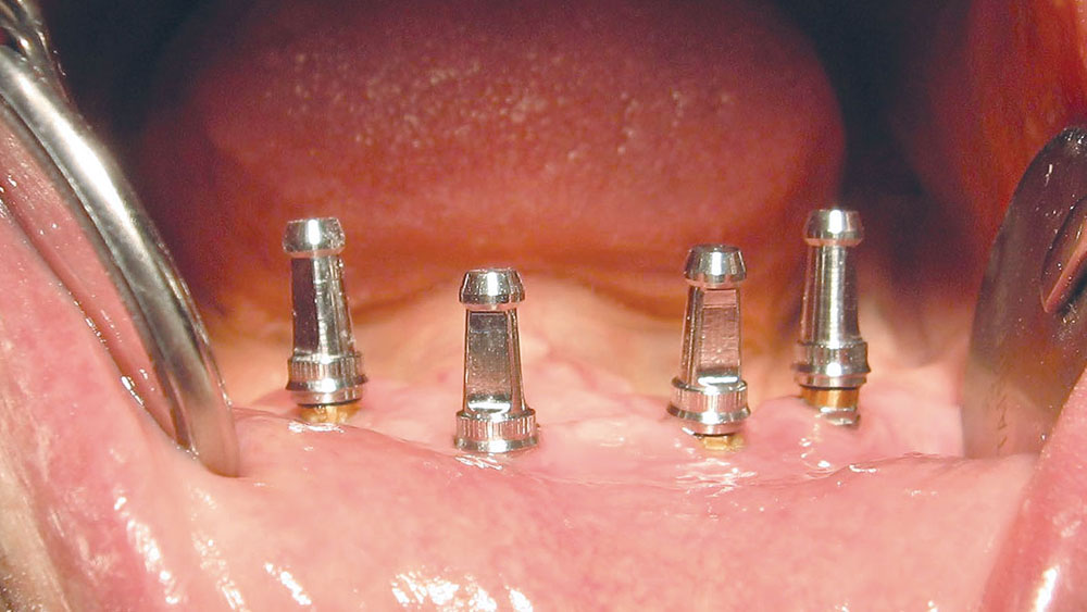
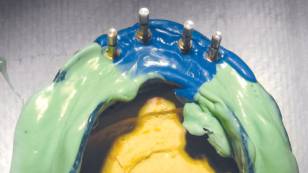
ERA Impression Copings (Sterngold) were placed on each implant female abutment (Fig. 14). An open tray was used to take a vinyl polysiloxane impression (SternVantage® Putty [Sterngold] and SternVantage® Monophase [Sterngold]) of the mandible with impression copings. ERA overdenture processing jigs were attached to the ERA Impression Copings (Fig. 15).
An experienced team at Glidewell Laboratories provided their advice and expertise regarding the ERA Implants. A wax rim was fabricated by the lab and tried in (Fig. 16). Adjustments were done and a bite registration was made. Plastic denture teeth were set in the wax rim. The teeth were tried in and minimally adjusted. The dental laboratory team packed, processed and finished the mandibular denture with integrated metal jackets for the remaining female ERA implant abutments.
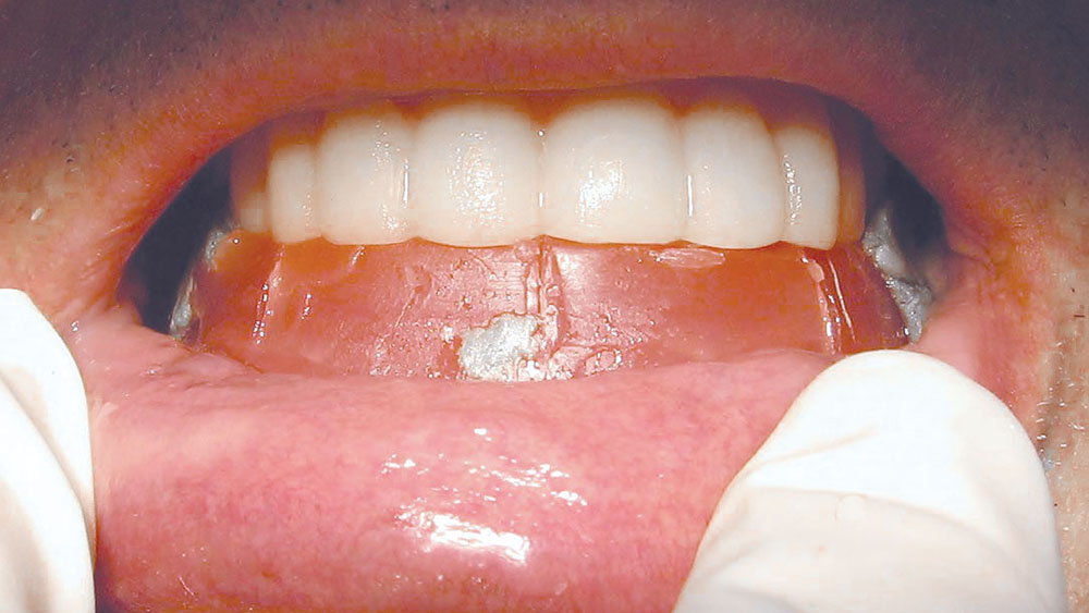
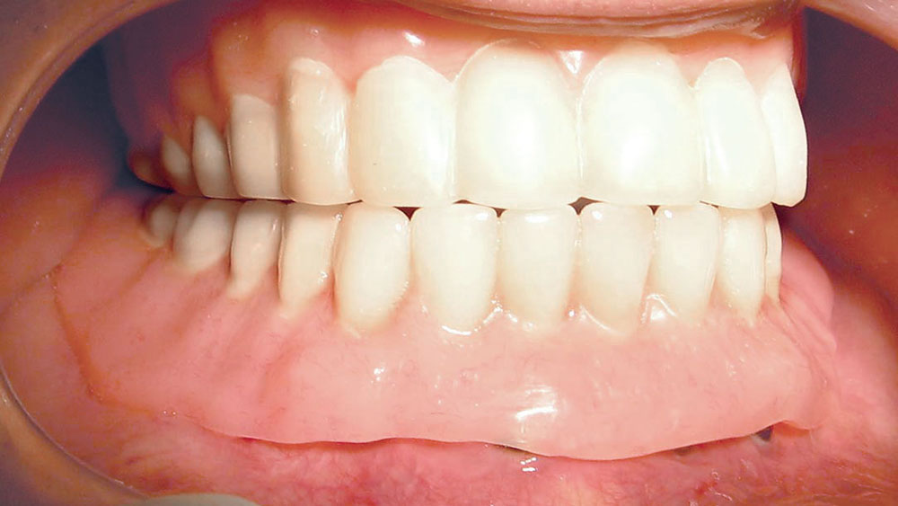
The denture was delivered (Figs. 17–19) and adjusted using an articulating foil (TrollFoil [TrollDental; Oakdale, Minn.]) that has a thin, built-in plastic frame/handle.
The black males can be replaced with white males, and then later with orange males, to increase retention as required.
BRIEF DISCUSSION OF MATERIALS
This clinical case report demonstrated simple immediate and long-term retention of overdentures with flapless placement of mini- and traditional-sized implants. This was coupled with angle-correcting, semi-precision resilient attachments that allowed for an idealized path of insertion without the demand of idealized implant alignment.
Sterngold anglecorrecting ERA abutments can be used on almost any brand of existing traditional-sized implants. Resilient ERA males can be utilized in almost any denture, and many partial dentures. The wide range of retention strengths of the ERA males allows the clinician alternatives for the initial placement and future retention requirements alike. The range of reline materials offered by manufacturers like Bosworth, Sterngold and Tokuyama have made it possible to provide quick, effective and economical alterations to removable prosthetics.
A high luster is necessary for the esthetics of implant-retained overdentures, direct restorations, hard relines, composite repairs of denture teeth, retrofitting of existing dentures to be overdentures and natural tooth bonding. Because chairside polishing of various restorative materials has often been a confusing and frustrating experience for many clinicians, the simple system (Jazz Supreme) was chosen for the purpose of demonstrating how the polishing process can be streamlined. The use of the one-step bonding, hybrid composite, finishing burs and one-step polishers allowed a very quick rehabilitation of the existing maxillary denture from Class III to Class I using a chairside auxiliary for part of the work.
CLOSING COMMENTS
The patient discussed herein is representative of an ever-growing group of medically compromised patients with chronic dental disabilities who seek oral rehabilitation from general clinicians. The presented technique of angle-correcting resilient implant-retained overdentures, with flap or flapless implant placement, allows an immediate cost-effective treatment plan for denture stabilization.
Economic pressure to control overhead expenses and time have accelerated the growth of the angle-correcting semi-precision resilient attachment system with mini- and traditional-sized implants that are coupled with innovative denture liners and polishing systems. Patient demands for increased comfort, esthetics and cost-effectiveness are propelling the expansion of these new technologies and techniques, as demonstrated in this case presentation.
The average restorative-turned-surgical-restorative dentist can avoid problems through proper case selection. It is possible for the average restorative dentist to learn to perform the minimally invasive implant surgery and overdenture fabrication that patients demand.
Dr. Gary Johnson is a chemist, dentist, lecturer, author and a director of the Clinical Research Institute. Contact him at smilewithus7@aol.com.
Disclosure: Dr. Johnson reports the following disclosures: Sterngold Dental supplied the dental implant materials and financial support for research to the Clinical Research Institute; SS White Burs supplied rotary and polishing instruments and financial support for research to the Clinical Research Institute; Tokuyama Dental America, Coltène/Whaledent, Harry J. Bosworth and TrollDental donated dental materials to the Clinical Research Institute for use in this case study; Glidewell Laboratories donated dental laboratory procedures used in this case study; and The Clinical Research Institute has provided dental materials to the author, as well as travel expenses and honoraria for presentations.
SUGGESTED READING
- Ambard AJ, Fanchiang JC, Mueninghoff L, Dasanayake AP. Cleansability of and patients’ satisfaction with implant-retained overdentures: a retrospective comparison of two attachment methods. J Am Dent Assoc. 2002 Sep;133(9):1237-42.
- Doundoulakis JH, Eckert SE, Lindquist CC, Jeffcoat MK. The implant-supported overdenture as an alternative to the complete mandibular denture. J Am Dent Assoc. 2003 Nov;134(11):1455-8.
- Carrick JL. Dentistry for an aging population: the ERA Implant Overdenture. Inside Dentistry. 2006 Jan;2:82.
- Rossein KD. Alternative treatment plans: implant-supported mandibular dentures. Inside Dentistry. 2006 Jul;2:42.
- Stoker GT, Wismeijer D, van Waas MA. An eight-year follow-up to a randomized clinical trial of aftercare and cost-analysis with three types of mandibular implant-retained overdentures. J Dent Res. 2007 Mar;86(3):276-80.
- Stanford CM. Application of oral implants to the general dental practice. J Am Dent Assoc. 2005 Aug;136(8):1092-100.
- Stanford CM. Dental implants. A role in geriatric dentistry for the general practice? J Am Dent Assoc. 2007 Sep;138 Suppl:34S-40S.
Reprinted by permission of Dentistry Today. ©2013 Dentistry Today.

