In this issue:
Utilizing a Digital Workflow for Provisionalization with BioTemps®
Author
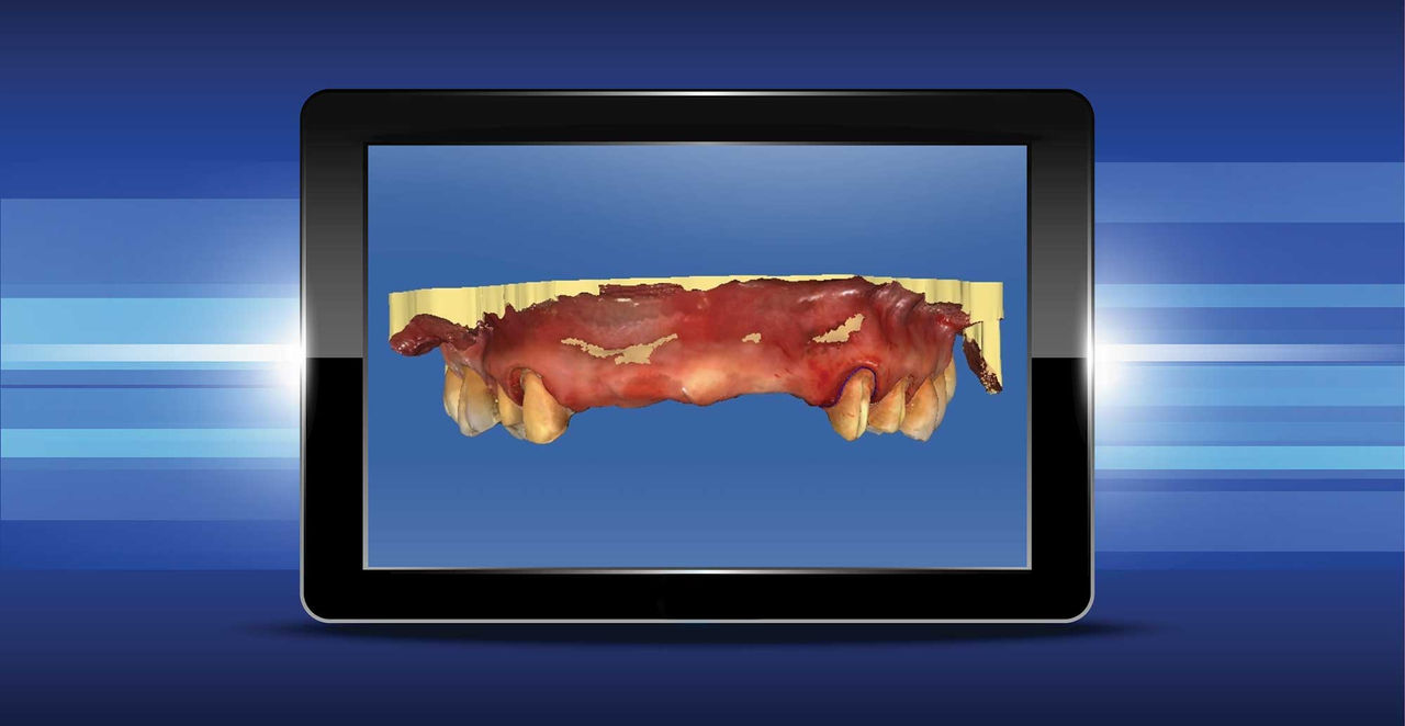
Today’s digital impression technology allows dentists to create a virtual, computer-generated replica of the hard and soft tissues in the mouth quickly and accurately using their choice of optical scanning device. As an ardent supporter of digital impressions, I make every attempt to digitize our restorative workflow. There are numerous benefits to a digital impression:
- Efficiency: It takes less time to take a digital impression than a traditional impression.
- Quicker Turnaround Time: Clinicians often forget or fail to realize the true value of this. Getting restorations back faster is better for the patient, the practice and the overall case outcome.
- Cost Savings: Have you ever calculated the cost of taking a traditional impression for a final restoration? If you add up what your office spends on impression materials, chair time and case shipping fees, you will be amazed at how much is spent on traditional methods.
CASE PRESENTATION
The female featured in this article has been a patient in our practice for nearly eight years. She has an existing PFM bridge from teeth #5 to #12 replacing missing #7 to #10. She is not terribly unhappy with the look and feel of the bridge, but the bridge has been no stranger to the big issue facing PFM restorations: the chipping of porcelain from the metal substructure. Over the past eight years, we have patched various corners and lingual surfaces.
Recently, the patient agreed to replace her long-span PFM bridge with an implant-supported bridge on #7 to #10 and individual crowns on the abutment teeth. However, she was adamant about not going a day without teeth. We advised her that this would not be an issue.


Due to the complexity of her implant surgery, immediate loading was not possible. This meant we needed a long-term esthetic provisional that would last the duration of the treatment, could be removed for surgery and was adjustable for post-surgical contouring. A BioTemps® provisional bridge (Glidewell Laboratories; Newport Beach, Calif.) was the quick and easy answer.
Traditionally, BioTemps are made prior to preparation and relined chairside. In this case, I wanted to have the BioTemps made to fit the final preparations of the abutment teeth, which would later be converted to individual restorations. As an advocate of digital impressions, I chose to follow a digital workflow.
We needed a long-term esthetic provisional that would last the duration of the treatment, could be removed for surgery and was adjustable for post-surgical contouring.


The provisional BioTemps bridge offers the following important advantages in this case:
- Trial Smile: The patient gets a "trial" of the new contours. Any modifications to length or contour can be made chairside, avoiding costly remakes and unhappy patients.
- Long-Term Durability: Due to the complexity of this case, full treatment will take well over 12 months. An acrylic provisional fabricated chairside simply won’t hold up this long.
- Removability: For implant surgery, the specialist will need the ability to remove and re-cement the provisional with relative ease.
- Adjustability: The necks of teeth #7 to #10 will need to be adjusted post-surgery to remove any pressure to the surgical sites. BioTemps are easily adjusted.
- Surgical Assistance: The contours and esthetics of the BioTemps will serve as a “guide” to the surgeon for grafting and placement of the implants.


As this case illustrates, digital impressions are not just limited to final restorations, and certainly not just to single units. It’s time for you to take a closer look at digital restorative technologies and see how they can benefit your practice and your patients.
Dr. Tarun Agarwal maintains a full-time private practice emphasizing esthetic, restorative and implant dentistry in Raleigh, North Carolina. Contact him via email at dra@raleighdentalarts.com or visit raleighdentalarts.com.