Photo Essay – Central Details: Renewing a Smile with an Esthetic Obsidian® Crown
June 24, 2016
 image
Anamaria Muresan, DMD, ME, CDT
Photo Essay – Central Details: Renewing a Smile with an Esthetic Obsidian® Crown

With a variety of dental materials available for crown fabrication, clinicians now have more options than ever to restore a patient’s smile. One challenging variable is to match the correct restorative material to the dentin shade of the preparation, especially in situations with a darker dentinal shade. In this case study, we will review the options available to help mask intrinsic staining of the natural tooth structure and provide an esthetic outcome for the patient.

Case Study

A 27-year-old male patient presented with a porcelain fused to metal crown on tooth #9, which had undergone endodontic treatment 10 years earlier. The patient explained that originally the cause of the treatment for this tooth was due to trauma at the age of nine. The patient reported that the tooth was neither sensitive nor altered in color following the trauma.

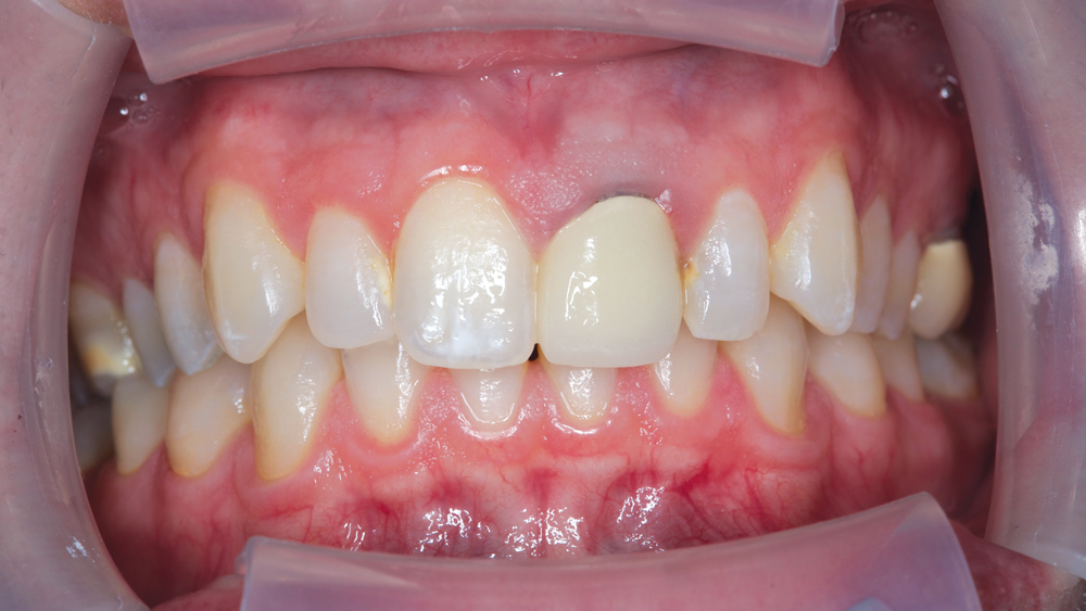
The crown required replacement due to recurrent decay. At the time of treatment, a darkened gingival margin, visible due to recession, posed a distinct challenge for this anterior case. In addition, the esthetics of the existing restoration were inadequate because of non-symmetric contours and poor shade match.

To achieve an optimal outcome and meet the patient’s expectations, two immediate goals were to match the gingival height of tooth #9 to that of tooth #8 and to whiten the patient’s smile. Choosing the right material for the new crown restoration was equally important. As the primary concern was to mask the dark gingival third of the root canal-treated tooth, Obsidian® Fused to Metal (Glidewell Laboratories; Newport Beach, Calif.) was chosen to satisfy the patient’s desire for esthetics as well as clinical standards for strength.

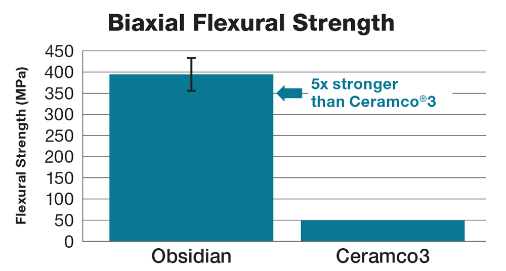
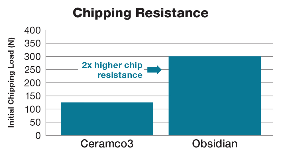
Obsidian Fused to Metal restorations put an innovative spin on PFMs, with an esthetic lithium silicate ceramic replacing the traditional porcelain. The lithium silicate ceramic maintains the translucency properties of the porcelain and is five times the strength and more than two times the chip resistance of traditional PFMs, according to recent tests (Figs. 1a, 1b).

Figure 1a
Figure 1b

Figures 1a, 1b: Obsidian ceramic takes over four times the force to fracture compared to the force necessary to fracture conventional feldspathic porcelains (PFM veneering ceramics). Tested for chip resistance, Obsidian Fused to Metal withstands more than twice the load compared to feldspathic porcelains (Data provided by the Glidewell Laboratories Research & Development department, 18651 Von Karman Ave., Irvine, CA 92612). Ceramco is a registered trademark of Dentsply Sirona Inc.

Figure 2

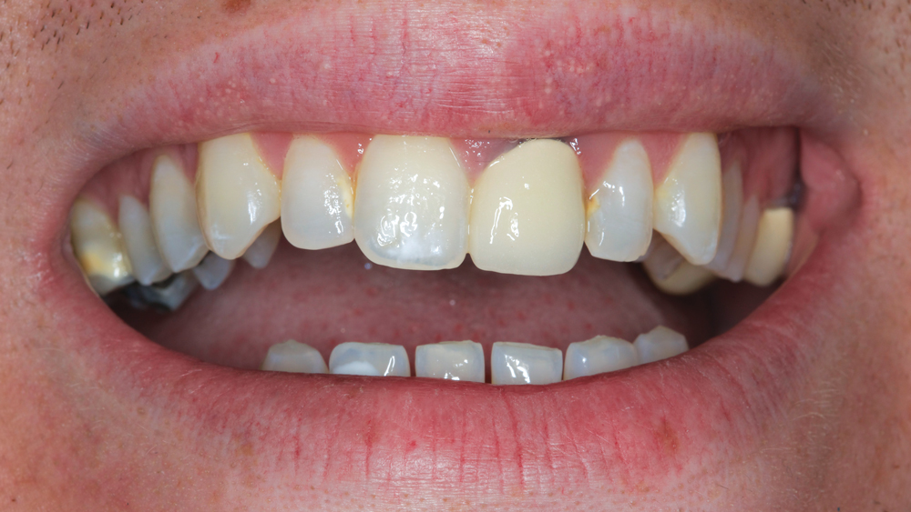
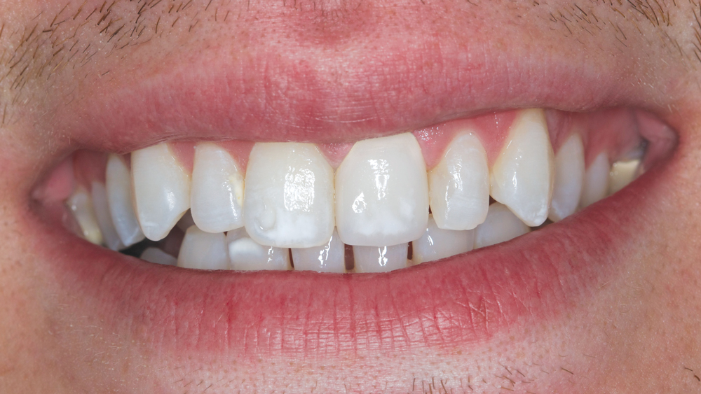
Figure 2: The PFM crown on tooth #9 fails to mirror the natural translucency and character of the adjacent teeth.

Figure 3

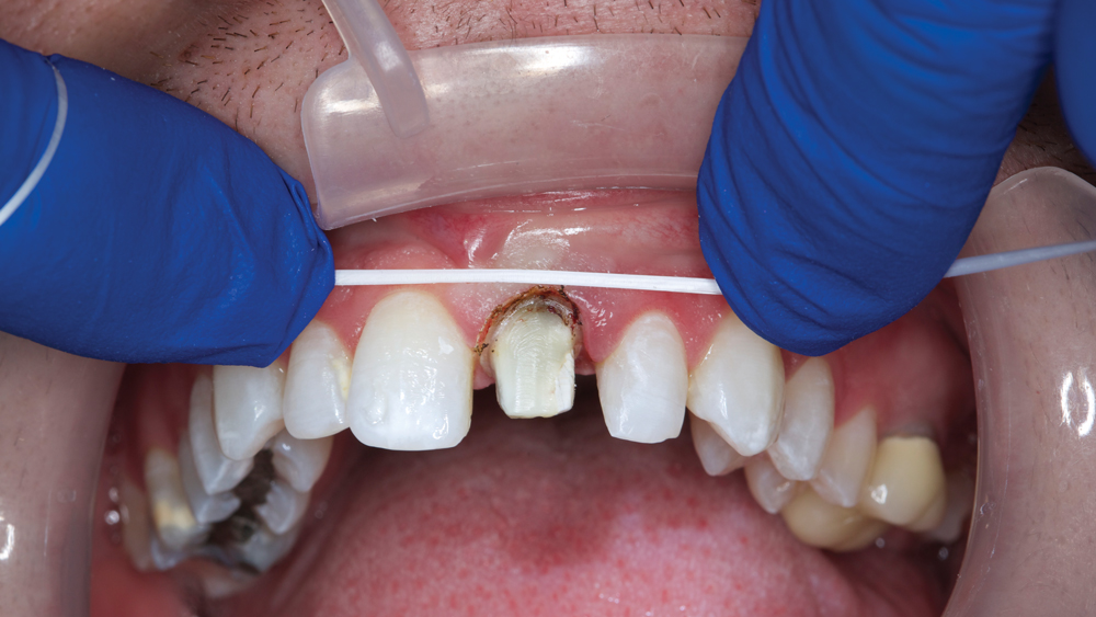
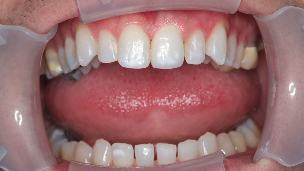
Figure 3: A retracted view reveals the darkened margin of the endontically treated tooth.

Figure 4

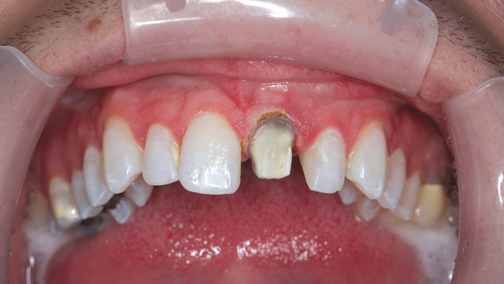
Figure 4: The gingival tissue of tooth #9 shows improved contours following gingivectomy, which was completed with a Picasso™ Lite diode laser (AMD Lasers; Indianapolis, Ind.). The patient’s thick gingival biotype, and inherently dense connective tissue, called for applying a touch-and-brush-away motion with the diode laser. Approximately 1.5 mm of tissue was removed, with strict care taken not to impinge on the biological width. Prior to the procedure, periodontal probing identified 4.5 mm on the facial and 6 mm of interproximal papilla height.

Figure 5

Figure 5: The area was cleaned with hydrogen peroxide. After the gingivectomy, the gingival height of tooth #8 & #9 are now more symmetrical. In order to approach a normal cervical contour with the provisional and the final restoration, it is important to mirror the CEJ of the adjacent central. Otherwise, the restored tooth will have a narrow form and will not look esthetic.

Figure 6

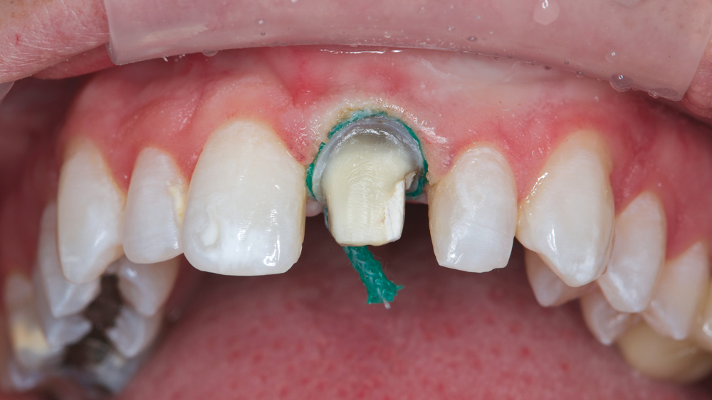
Figure 6: With size 00 and size 2 retraction cords in place, the Two-Cord Impression Technique was utilized, providing a precise view of the margin. This case features a shoulder preparation of 1 mm, to ensure enough thickness for the ceramic labial margin to block the darkness of the preparation at the gingival third.

Figure 7

Figure 7: A temporary restoration — mocked up chairside — provides a preview of how the new anterior restoration’s staining can mimic the fluorosis on tooth #8 and blend in with the overall smile. Photos of the temporary were included in the information provided to the lab technician.

Figure 8

Figure 8: After sandblasting the intaglio surface of the restoration, the Obsidian Fused to Metal crown is ready to be cemented with RelyX™ Luting Plus (3M™ ESPE™; St. Paul, Minn.), chosen for its ease of cleanup.

Figure 9

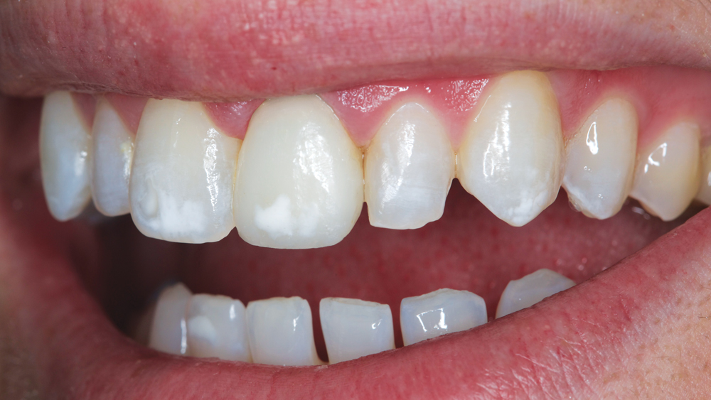
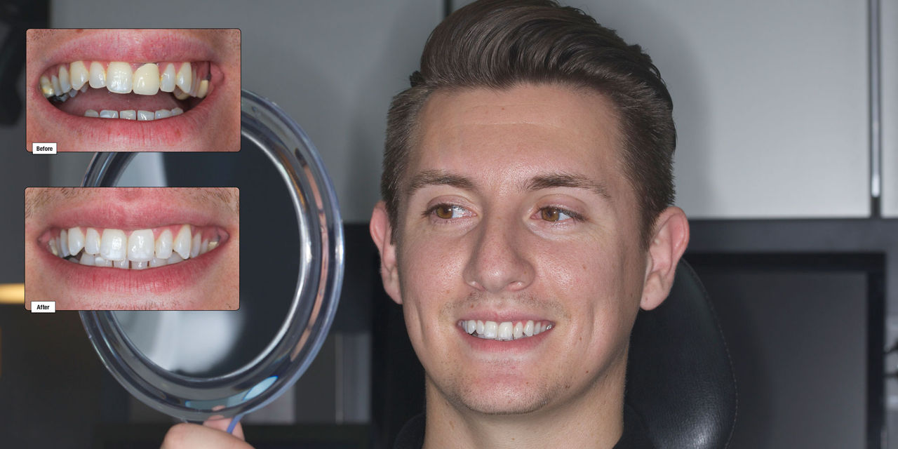
Figure 9: The Obsidian Fused to Metal crown successfully masks the darkened stump shade at the gingival third while also blending in with the overall smile, thanks to details including incisal translucency and fluorosis staining. A whiter smile was achieved with two weeks of bleaching, which took the patient from A2 to A1. The patient was pleased with the final results and even noted that his wife could tell a difference in his smile upon completion of treatment.

Conclusion

Previously, a PFM was the common restorative choice for a case involving a darkened dentinal shade. Fortunately, today’s clinicians can choose from restorative materials such as Obsidian Fused to Metal to supplant and indeed outperform traditional PFMs. In this case report, the natural esthetics and improved strength of the Obsidian Fused to Metal restoration helped achieve the proper esthetic outcome as desired by the patient.