Predictability by Design: A Full-Mouth Makeover (1 CEU)

October 1, 2026
 image
Justin Chi, DDS, CDT
Predictability by Design: A Full-Mouth Makeover

My patient Roger Torres presented with severely worn dentition throughout his mouth, resulting from a combination of attrition and erosion. The extensive loss of tooth structure was accompanied by a significant reduction in the vertical dimension of occlusion (VDO). As a consequence of advanced wear, the patient had developed an anterior edge-to-edge occlusal relationship.

The patient was also partially edentulous. All four third molars were absent, in addition to teeth #18, #19 and #30. The patient had implants placed in those sites approximately three to four months before the definitive restorative treatment began.

Tooth #31 was missing an existing crown, leaving the tooth essentially prepared for restoration. In the maxillary arch, he had existing restorations, including an amalgam restoration on tooth #2 and a crown on tooth #14. In addition, several teeth had previously been endodontically treated.

CASE REPORT

APPOINTMENT 1: INITIAL DATA

(Digital scan and diagnostic wax-up)
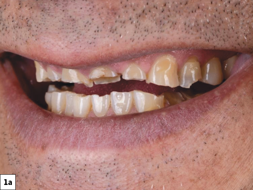
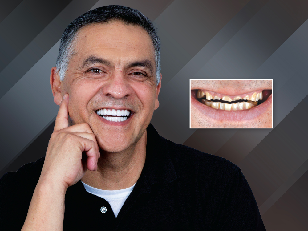
Facial view of a patient's smile showing severely worn and fractured upper and lower anterior teeth.
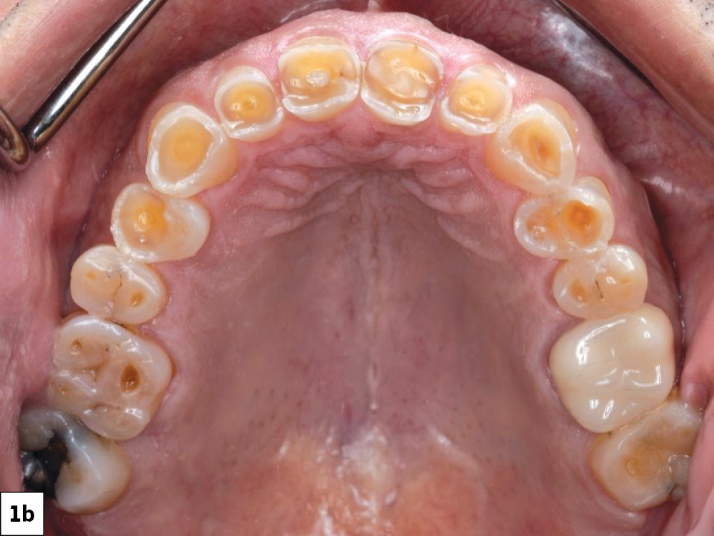
Occlusal intraoral view of the maxillary arch showing generalized severe tooth wear and decay throughout.
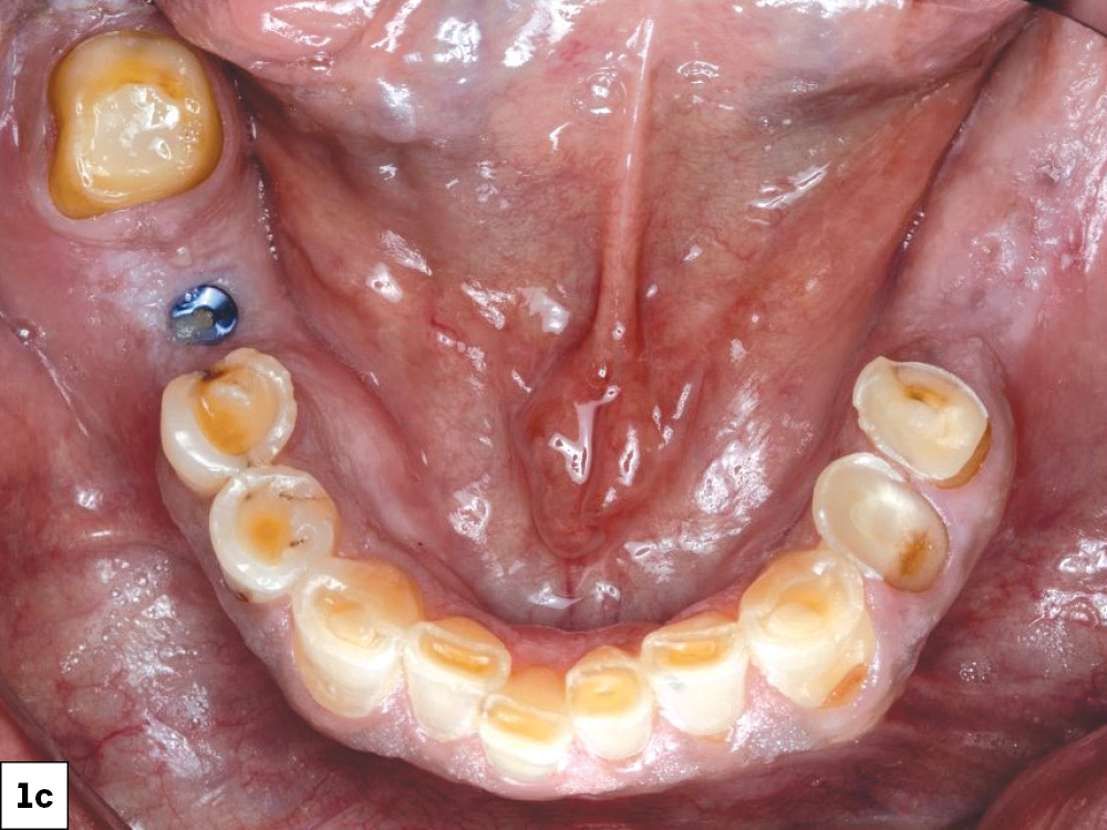
Occlusal intraoral view of the mandibular arch showing severely worn and decayed teeth with missing anterior teeth.

Figures 1a–1c: The patient’s chief complaint was esthetic. He was self-conscious about his smile and wanted to fix it. Because of the extreme wear and the amount of tooth structure that had been lost, comprehensive full-mouth restorative treatment was indicated. The treatment plan was designed to restore function, reestablish the VDO and proper tooth contours, and improve the esthetics of the patient’s smile.

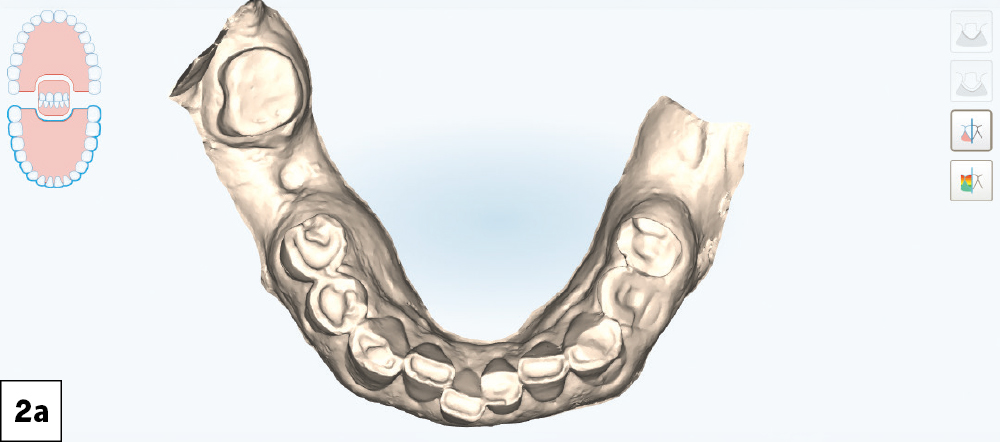
iTero intraoral scan showing the mandibular arch with severely worn dentition, buccal view.
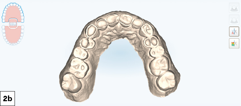
iTero intraoral scan showing the mandibular arch with severely worn dentition, occlusal view.
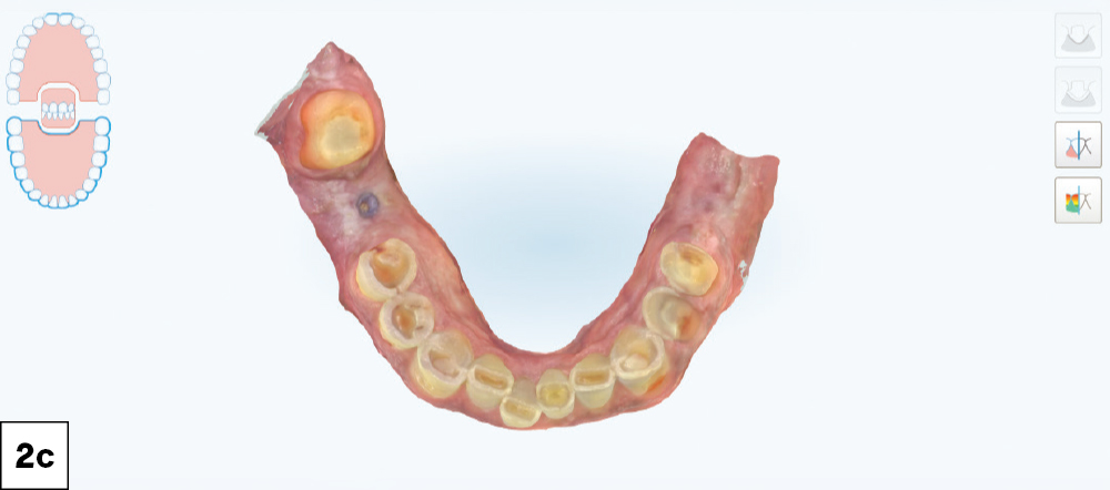
Color iTero intraoral scan of the mandibular arch showing worn and decayed teeth, occlusal view.
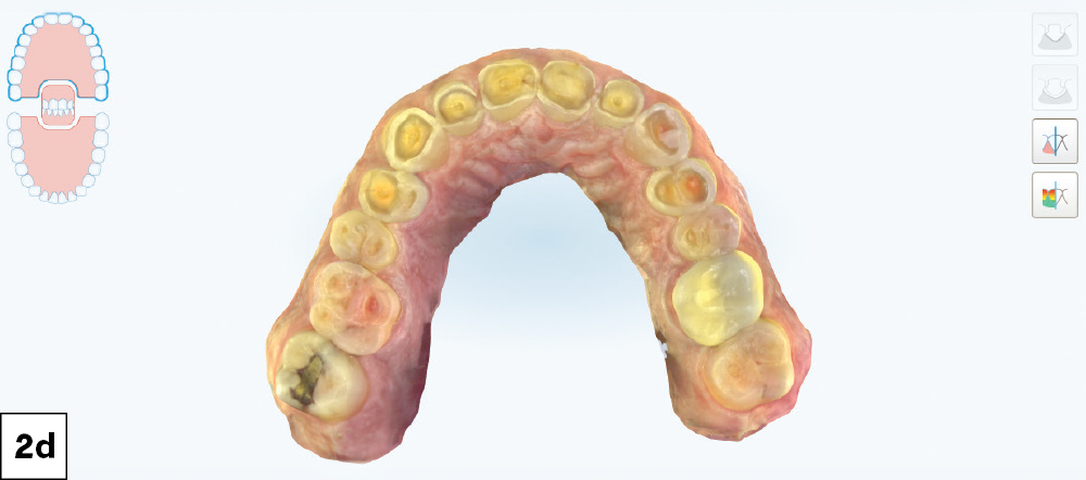
Color iTero intraoral scan of the maxillary arch showing generalized severe wear and decay, occlusal view.
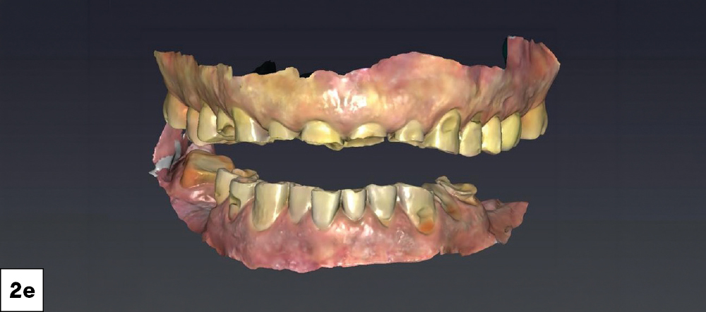
Color iTero intraoral scan showing both arches separated, revealing the extent of full-mouth wear and tooth loss.

Figures 2a–2e: A digital intraoral scan was acquired using the iTero Element® 5D Plus Intraoral Scanner (Align Technology, Inc.; Tempe, Ariz.) and submitted to Glidewell for fabrication of a diagnostic wax-up. The goal was to give the lab a clear idea of the desired VDO and the amount of occlusal opening required to achieve our restorative plan.

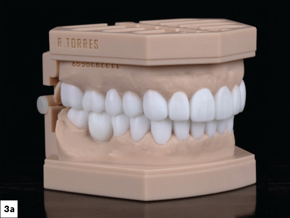
Lateral view of full-mouth diagnostic wax-up on articulated stone models showing planned tooth contours and VDO.
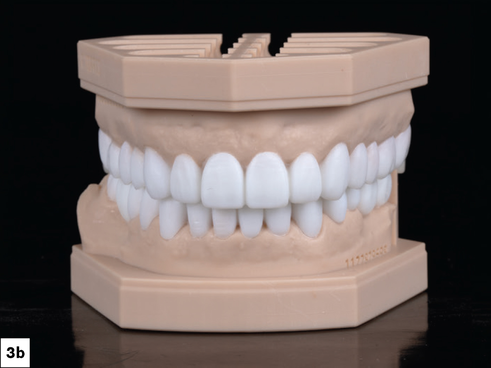
Facial view of full-mouth diagnostic wax-up on articulated stone models showing anticipated anterior esthetic outcome.
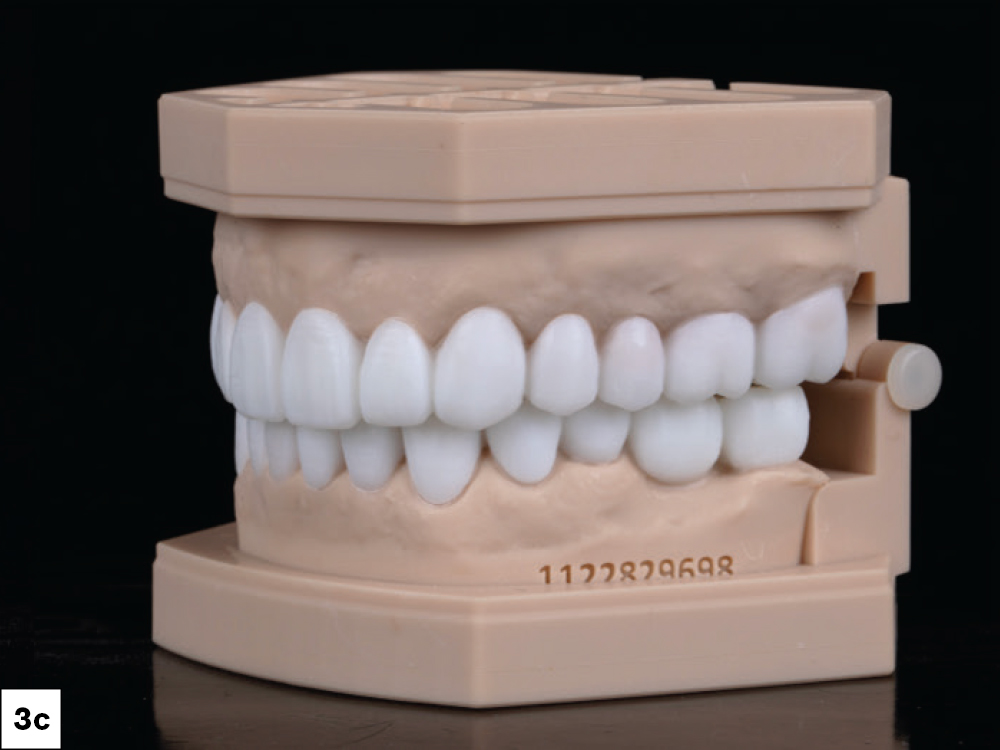
Lateral view of full-mouth diagnostic wax-up on articulated stone models showing planned occlusion from the opposite side.

Figures 3a–3c: Frontal and lateral views of the maxillary and mandibular diagnostic wax-up. By establishing proper tooth contours and function, the wax-up also provided a preview of the anticipated esthetic outcome.

APPOINTMENT 2: VERIFICATION

(Mock-up confirmation)
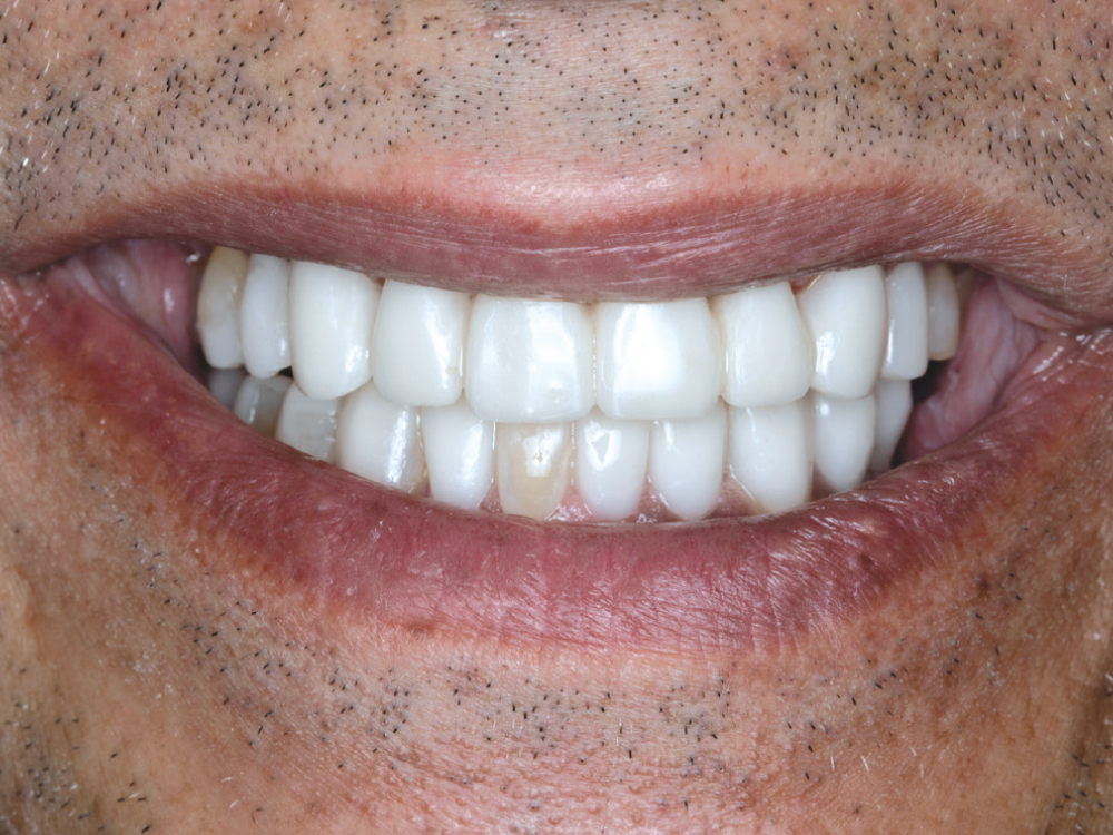
Facial view of a patient's smile with a bis-acryl mock-up in place for esthetic and functional evaluation.

Figure 4: The lab provided a wax-up and putty matrix, which I used to evaluate the proposed esthetic and functional outcome. I etched the teeth, applied 3M™ Scotchbond™ Universal Adhesive (3M; St. Paul, Minn.), and bonded the bis-acryl mock-up to his teeth so he could wear it for a test drive.

The patient wore the bonded mock-up for approximately one month as a trial period. During this time, I maintained communication with him to ensure he was comfortable with the proposed esthetics, occlusion and function. He was pleased with the esthetic result and approved proceeding with the definitive restorative phase.

MOCK-UP CARE AND MAINTENANCE

Because the mock-up was placed as one piece, all the teeth were splinted together. That meant the patient could not floss normally during the test-drive period, so I gave him detailed hygiene instructions to help keep the gingival tissues healthy.

I also explained that the bis-acryl material was softer than the definitive restorations would be. The purpose of the mock-up was primarily to confirm appearance, evaluate comfort and allow the patient to experience the proposed changes over time. 

The first part of the restorative phase was to stabilize the posterior dentition. At the next appointment, I prepared the maxillary posterior teeth for full-coverage restorations.

APPOINTMENT 3: MAXILLARY POSTERIORS & MANDIBULAR MOLARS

(Pretreatment scans, prep, scan, design, mill and deliver 12 units)

The goal of this appointment was to address the maxillary posterior teeth and mandibular molars. This approach allowed us to establish posterior support and stabilization of the occlusion before proceeding with treatment of the anterior teeth.
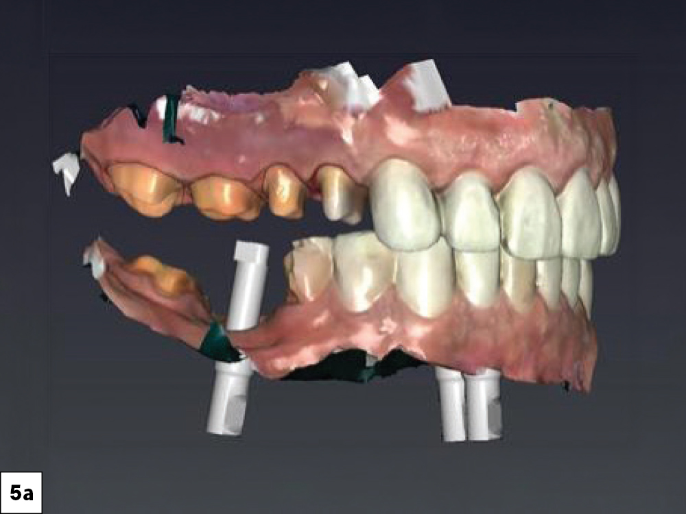
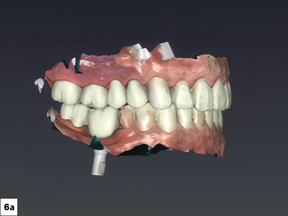
3D intraoral scan showing maxillary posterior preparations alongside scan bodies seated on mandibular implants, lateral view.
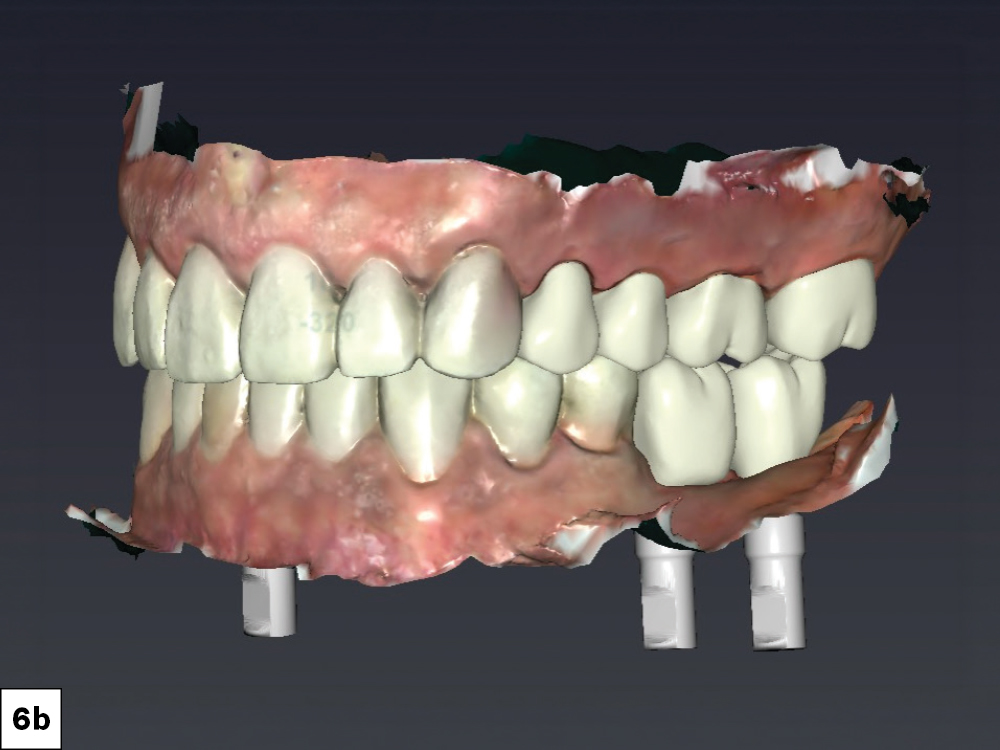
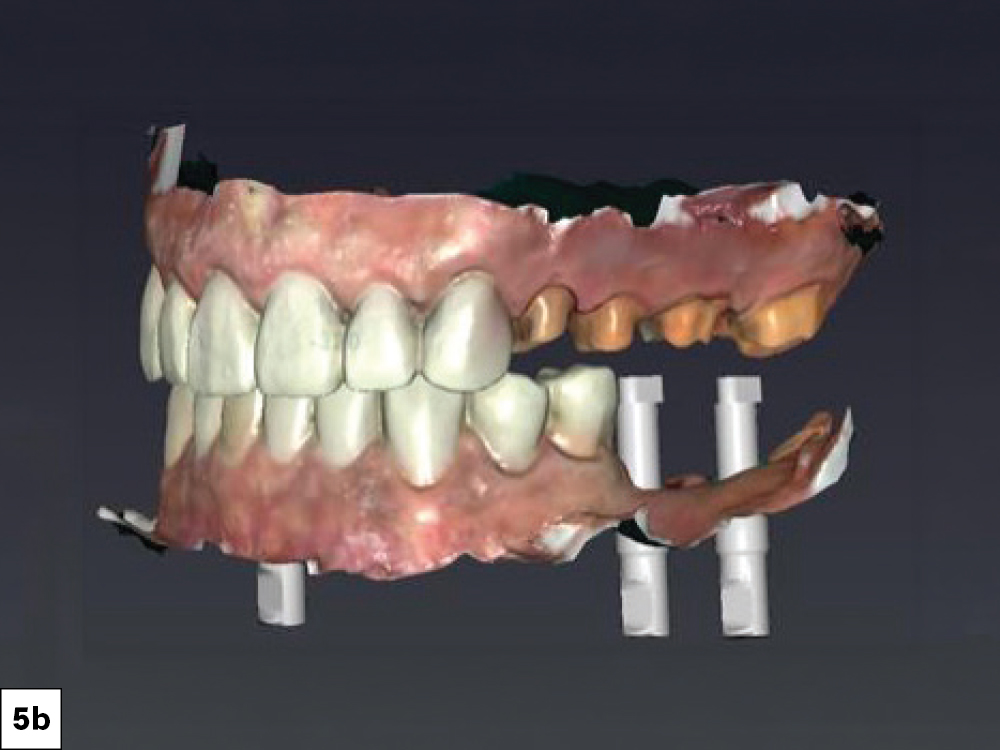
3D intraoral scan showing both arches with anterior provisionals in place and scan bodies on mandibular implants, facial view.
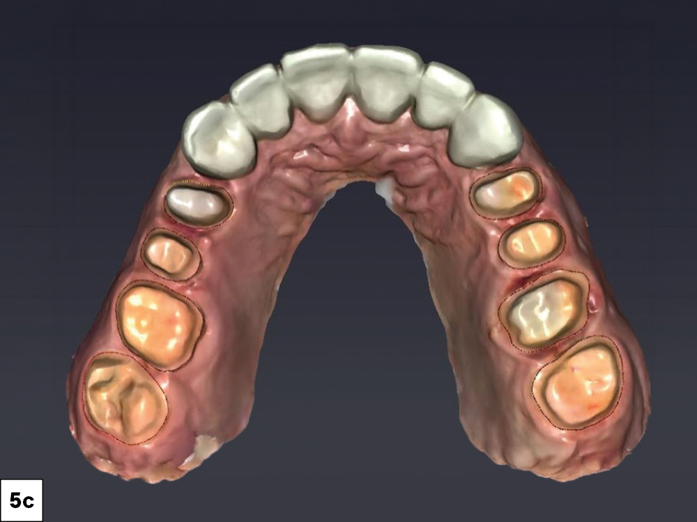
3D intraoral scan occlusal view of the maxillary arch showing posterior crown preparations with margin lines alongside seated anterior provisionals.

Figures 5a–5c: The maxillary posterior preparations included teeth #2–5 and #12–15. The preparation on tooth #31 was also refined, as the tooth had previously lost its crown. During this same phase, I completed the second stage by uncovering the implants. Scan bodies were seated, and digital impressions were acquired for the implant restorations.

3D intraoral scan showing both arches in occlusion with mandibular implant scan bodies visible, lateral view.
3D intraoral scan showing both arches in occlusion with mandibular implant scan bodies visible, facial view.

Figures 6a, 6b: All designs were done chairside, using the fastdesign.io™ Software and Design Station.

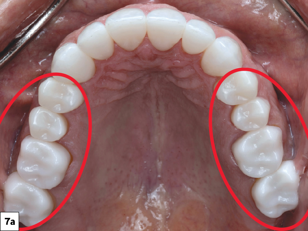
Occlusal intraoral view of the maxillary arch with bilateral posterior BruxZir crowns highlighted, showing completed restorations.
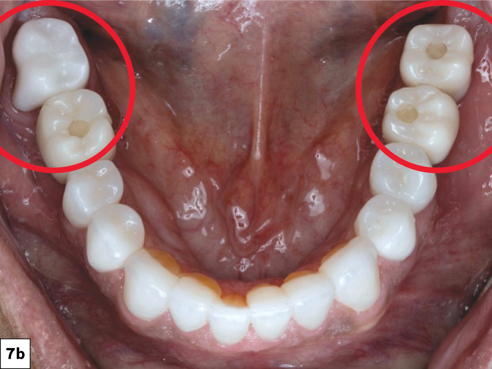
Occlusal intraoral view of the mandibular arch with bilateral posterior BruxZir crowns and implant restorations highlighted, showing completed restorations.

Figures 7a, 7b: The posterior restorations were fabricated using the fastmill.io™ In-Office Mill as follows: teeth #2–3, #14–15 and #31 were restored using BruxZir® NOW (bleach 1); teeth #4–5 and #12–13 with BruxZir Esthetic NOW (bleach 1); and implants on #18–19 and #30 were delivered using BruxZir NOW SRC (screw-retained crowns) (shade B1).

APPOINTMENT 4: MAXILLARY & MANDIBULAR ANTERIORS/PREMOLARS

(Pretreatment scans, prep, scan and temporize 16 units)

Approximately one week later, treatment progressed to the anterior teeth and mandibular premolars. The maxillary anterior teeth, #6–11, were prepared for full-coverage crowns.
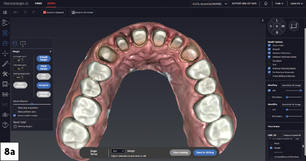
Margin detection view of maxillary posterior crown preparations in fastdesign.io, occlusal view.
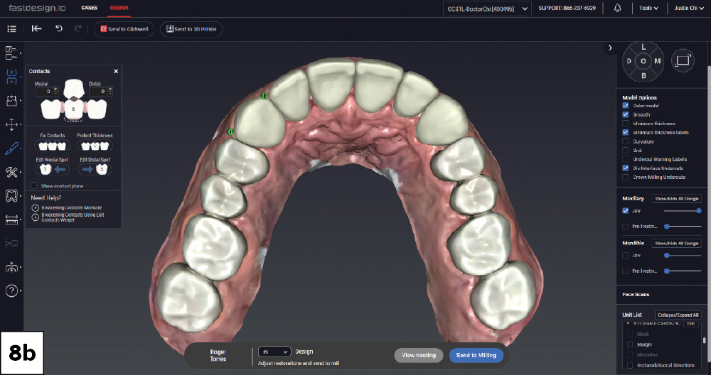
Contact adjustment view of maxillary crown designs in fastdesign.io, occlusal view.
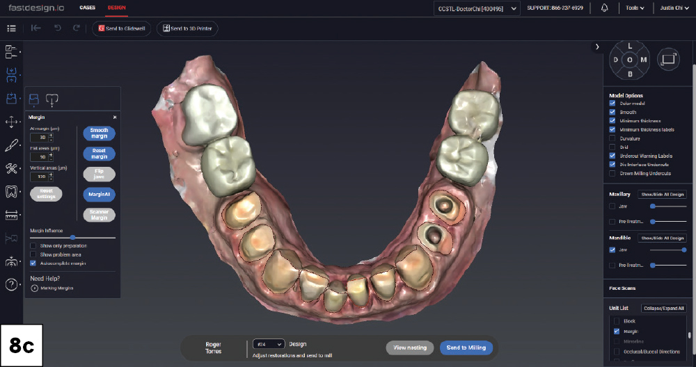
Margin detection view of mandibular posterior crown and veneer preparations in fastdesign.io, occlusal view.
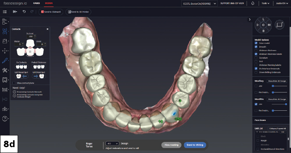
Contact adjustment view of mandibular crown and veneer designs in fastdesign.io, occlusal view.
Both arches displayed open in fastdesign.io showing full-mouth crown and veneer designs, facial view.
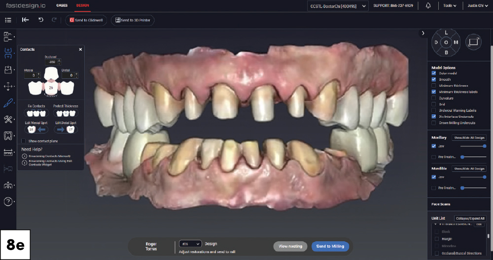
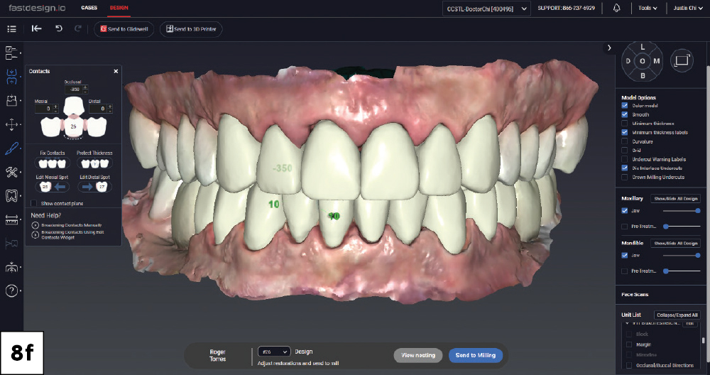
Both arches in occlusion in fastdesign.io showing completed full-mouth crown and veneer designs with contact data, facial view.

Figures 8a–8f: Margin marking and final designs were done on the fastdesign.io system. In the mandibular arch, teeth #22–27 were prepared for veneers. Teeth #28 and #29 were prepared more conservatively as overlay-style restorations with supragingival lingual margins. Teeth #20 and #21 were endodontically treated, so I restored those with endocrowns. Anteriors were temporized until the final appointment the next day.

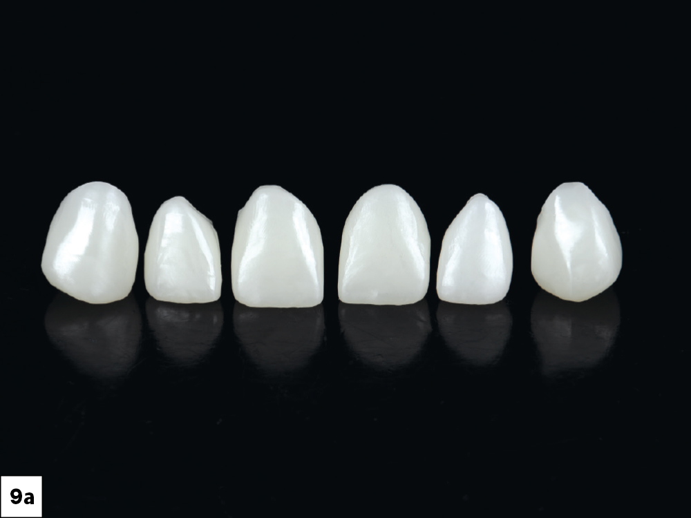
Six milled BruxZir Esthetic anterior crowns displayed in a row against a black background.
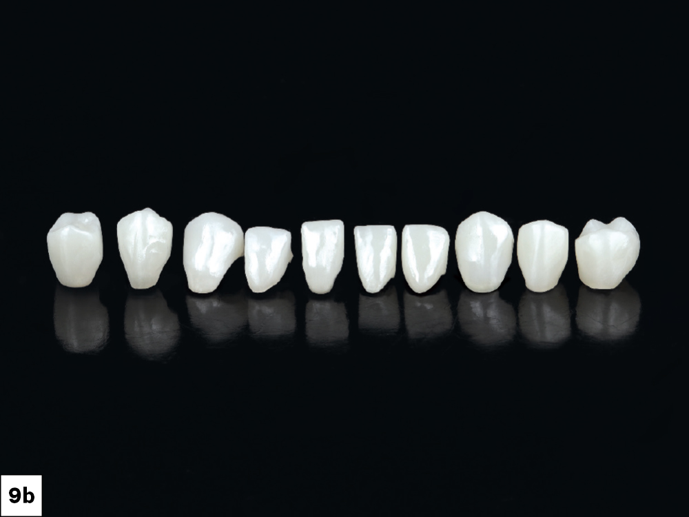
Ten milled BruxZir Esthetic crowns, veneers, and endocrowns displayed in a row against a black background.

Figures 9a, 9b: Teeth #6–11 were restored with BruxZir Esthetic NOW (bleach 1); teeth #20–21 were delivered as BruxZir Esthetic NOW (bleach 1) endocrowns; teeth #22–27 as BruxZir Esthetic NOW (bleach 1) veneers; and teeth #28–29 using BruxZir NOW (bleach 1).

Bonding Protocol

Restoration Preparation: After confirming fit, contacts and occlusion, I sandblasted with 50 µm aluminum oxide to properly clean and roughen the intaglio surface of the BruxZir NOW and BruxZir Esthetic NOW restorations. After sandblasting, I applied Monobond® Plus (Ivoclar Vivadent, Inc.; Amherst, N.Y.), which contains the MDP primer required for bonding zirconia. Isolation was maintained until final seating.

Tooth Preparation and Cementation: The prepped teeth were etched with phosphoric acid, rinsed, cleaned and isolated. After etching, I applied Gluma® Desensitizer (Kulzer GmbH; Hanau, Germany), followed by 3M Scotchbond Universal Adhesive.

I cemented the maxillary posterior restorations using BruxZir Dual Cure Resin Cement (Glidewell Direct; Irvine, Calif.). For the maxillary anterior crowns, mandibular veneers and lower premolar restorations, I used NX3 Nexus™ Third Generation Universal Adhesive Resin Dental Cement (Kerr Corporation; Brea, Calif.). Once the restorations were fully seated, excess cement was removed, the bonded restorations were light-cured, and final occlusal adjustments were completed.

APPOINTMENT 5: FINAL DELIVERY

(Remove temps, deliver anteriors, scan for nightguard and 3D print in-office)
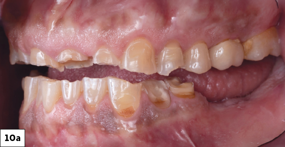
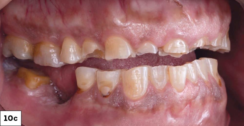
Pretreatment lateral intraoral view showing severely worn and damaged dentition on both arches.
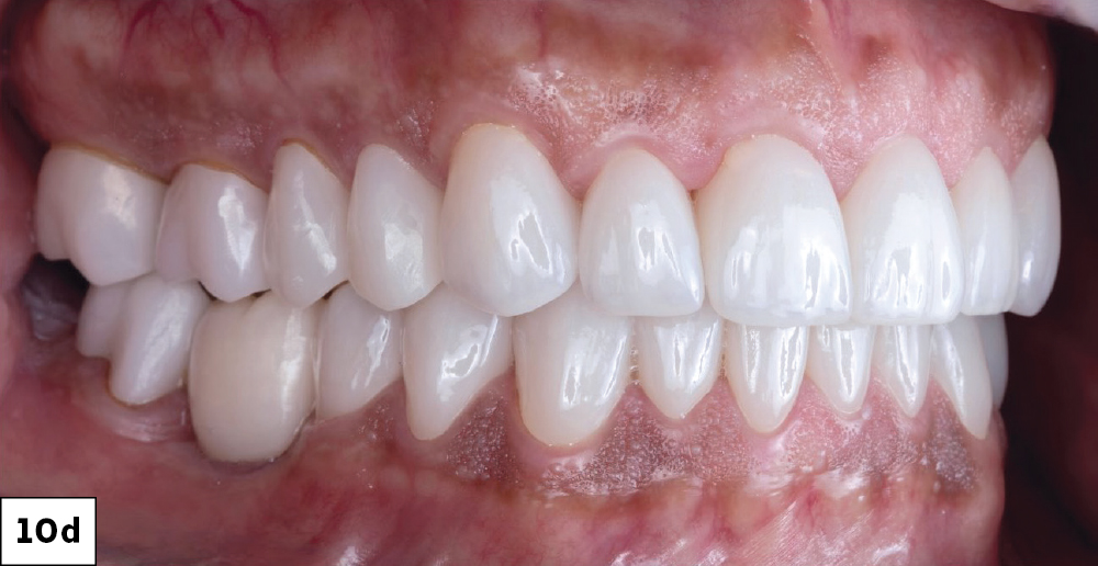
Post-treatment lateral intraoral view showing completed full-mouth BruxZir restorations with restored occlusion, left side.
Pretreatment lateral intraoral view showing severely worn dentition on the right side.
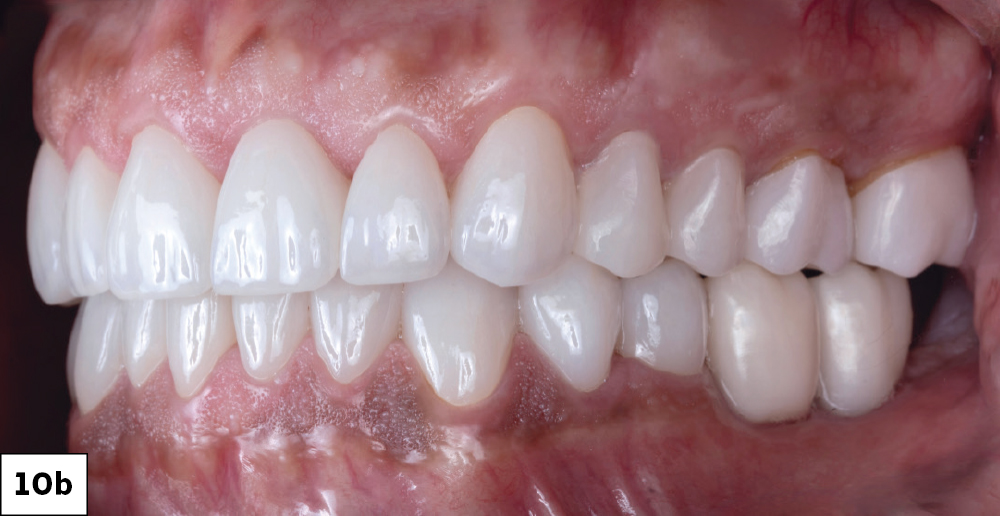
Post-treatment lateral intraoral view showing completed full-mouth BruxZir restorations with restored occlusion, right side.

Figures 10a–10d: The final delivery intraoral photographs show the dramatic improvement in tooth form and overall esthetics. Restoring occlusion helped create a more functional relationship between the arches while also improving the smile.

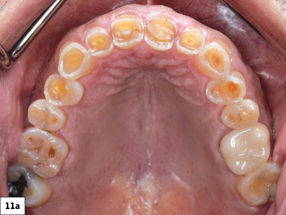
Pretreatment occlusal view of the maxillary arch showing generalized severe wear and decay.
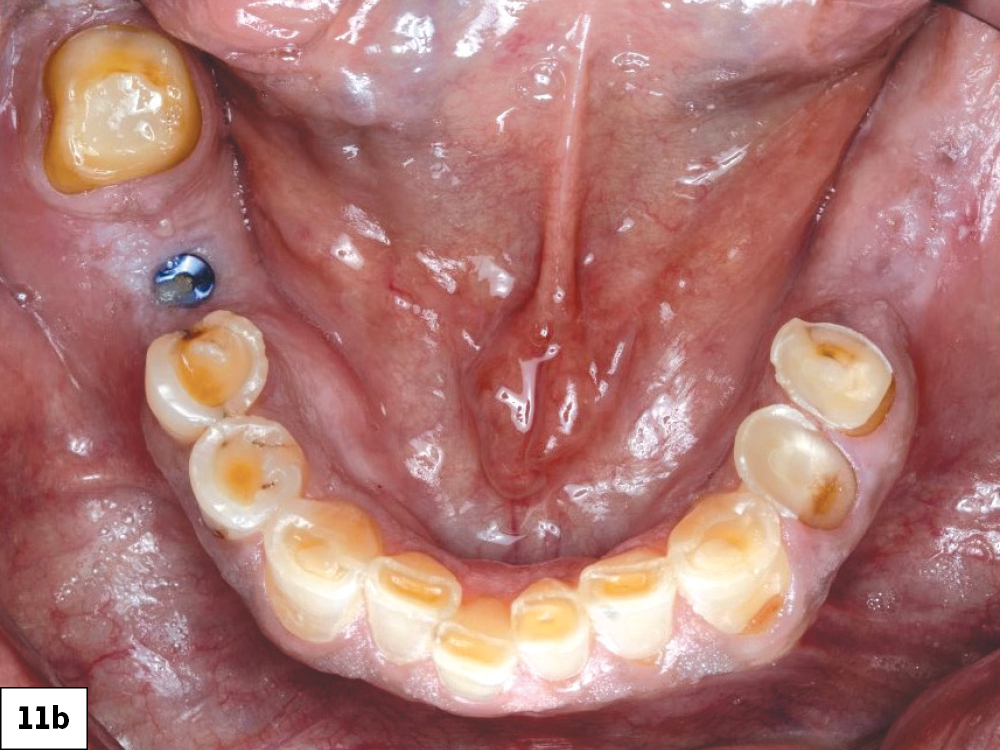
Pretreatment occlusal view of the mandibular arch showing severely worn and missing teeth with visible implant cover screw.
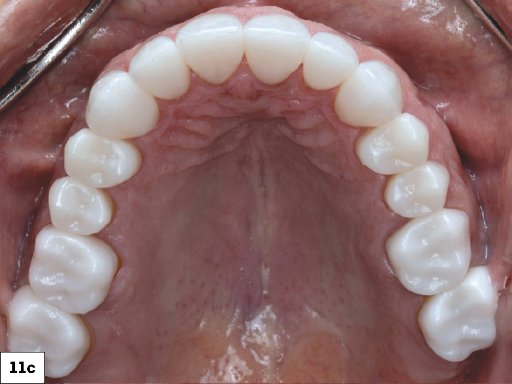
Post-treatment occlusal view of the maxillary arch with completed full-mouth BruxZir restorations.
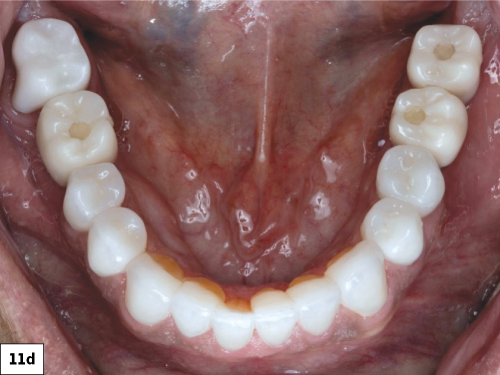
Post-treatment occlusal view of the mandibular arch with completed BruxZir crowns, veneers, and implant restorations.

Figures 11a–11d: The occlusal views help complete the story, showing how the patient’s restorations come together across both arches.

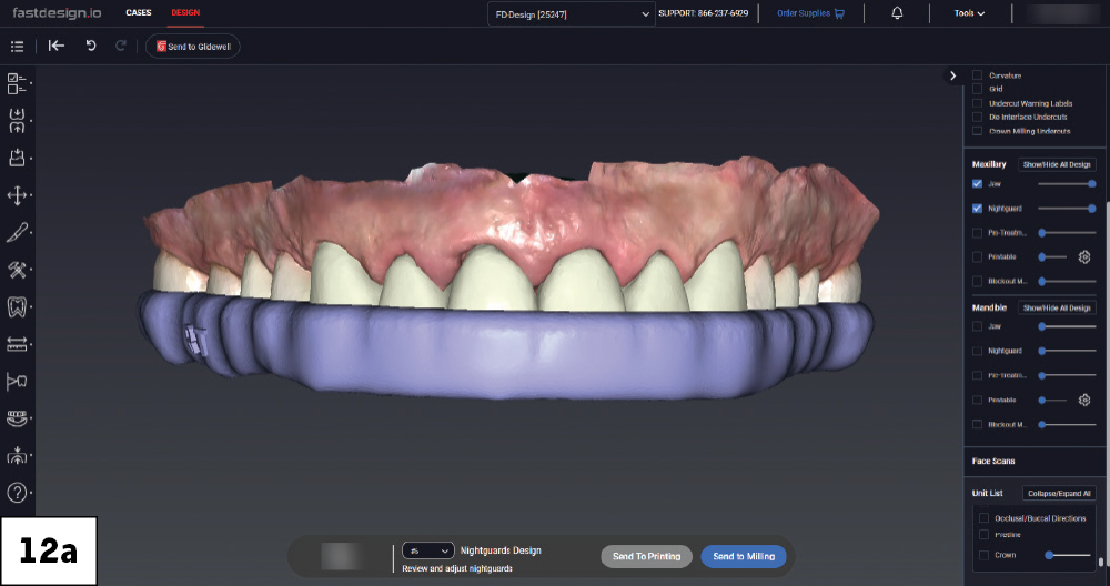
Nightguard design over the maxillary arch displayed in fastdesign.io, lateral view.
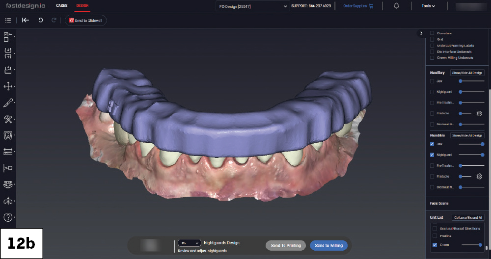
Nightguard design over the mandibular arch displayed in fastdesign.io, lateral view.
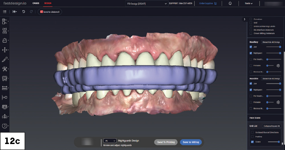
Full-mouth nightguard design shown over both arches in occlusion in fastdesign.io, facial view.

Figures 12a–12c: Upon completion of the full-mouth rehabilitation, the occlusion was carefully evaluated and equilibrated until the patient was comfortable. As with most restoration patients, I scanned the definitive maxillary and mandibular tooth positions and bite relationship to fabricate a protective nightguard chairside using the fastprint.io™ 3D Printing Solution.

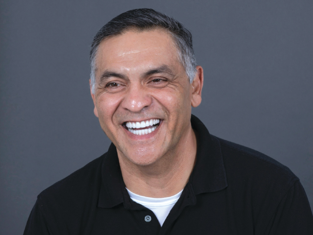
Portrait of a smiling male patient showing his fully restored smile following complete full-mouth rehabilitation.

Figure 13: Roger’s full-mouth restoration restored his bite, transformed his smile and gave him a renewed sense of confidence.

CONCLUSION

This case highlights the importance of a structured diagnostic and restorative approach when managing severe generalized tooth wear. Through digital planning, a diagnostic wax-up and a bonded mock-up, I was able to establish a predictable outcome that restored function, occlusion and esthetics. The final rehabilitation provided the patient with a functional, stable and natural-looking smile.

All third-party trademarks are property of their respective owners.