Periodontal Photo Essay: Is Closed-Flap Crown Lengthening a Biologically Sound Procedure?

January 31, 2011
 image
Daniel Melker, DDS

Objective

The objective of this article is to discuss the biological aspects of bone and the changes that occur when it is infringed upon either through disease or during the correction of certain periodontal conditions. It will discuss the correction of osseous defects and why these procedures are necessary to create a long-term stable environment. When a comparison is made between certain periodontal problems stemming from either biologic width invasion or periodontal disease with closed-flap crown lengthening, similarities suggest that closed-flap crown lengthening is an unsound biologic procedure.

Case 1

Case 1 Figure 1

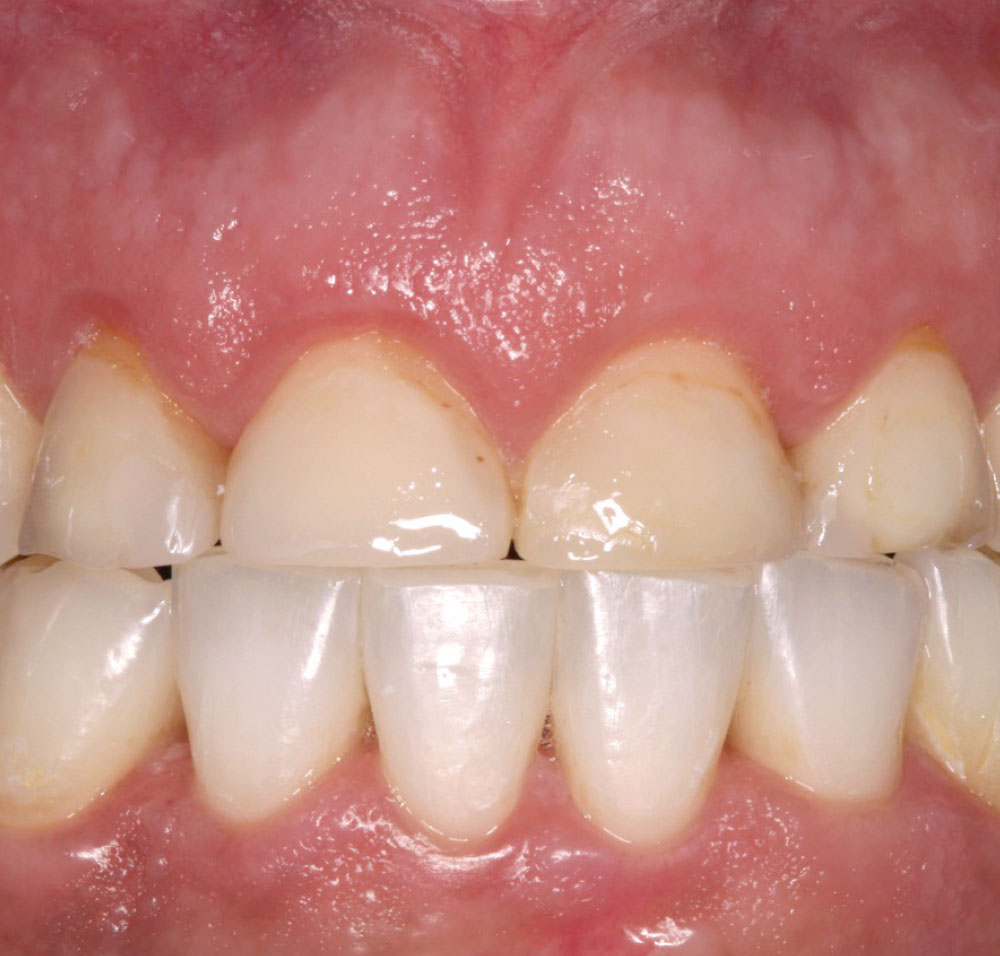
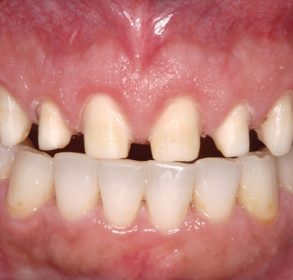
Figure 1: In order to change the length of the clinical crowns for a new restorative commitment, crown lengthening was advised. When doing closed-flap crown lengthening, marks are made on the laser tip to determine the amount of bone that needs to be removed to create space for a new crown. Soft tissue can also be removed when necessary. The major problem with such a procedure is the inability to remove the troughs created by the vertical removal of bone.

Case 1 Figure 2

Figure 2: The restorative doctor and periodontist discussed the case and determined that 1 mm of length would be needed on the incisal edges. An appropriate formula was used for the surgical procedure: biologic width, approximately 3 mm; clinical crown length, 10 mm; added porcelain, 1 mm. The total length needed from the existing incisal edge to the bone = 13 mm. Note: Six weeks postoperatively, it will be determined if touch-up surgery will be necessary to correct any biologic changes.

Case 1 Figure 3

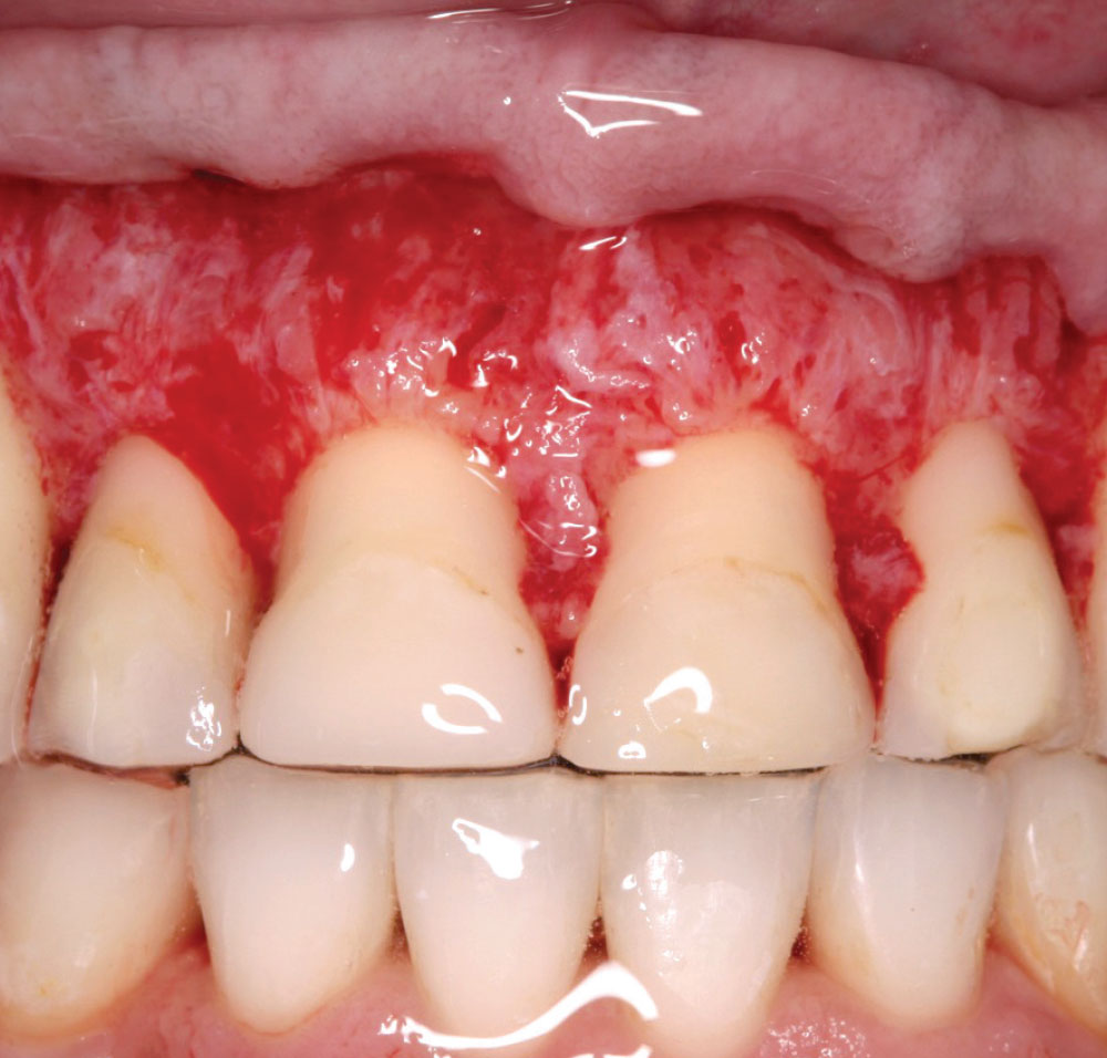
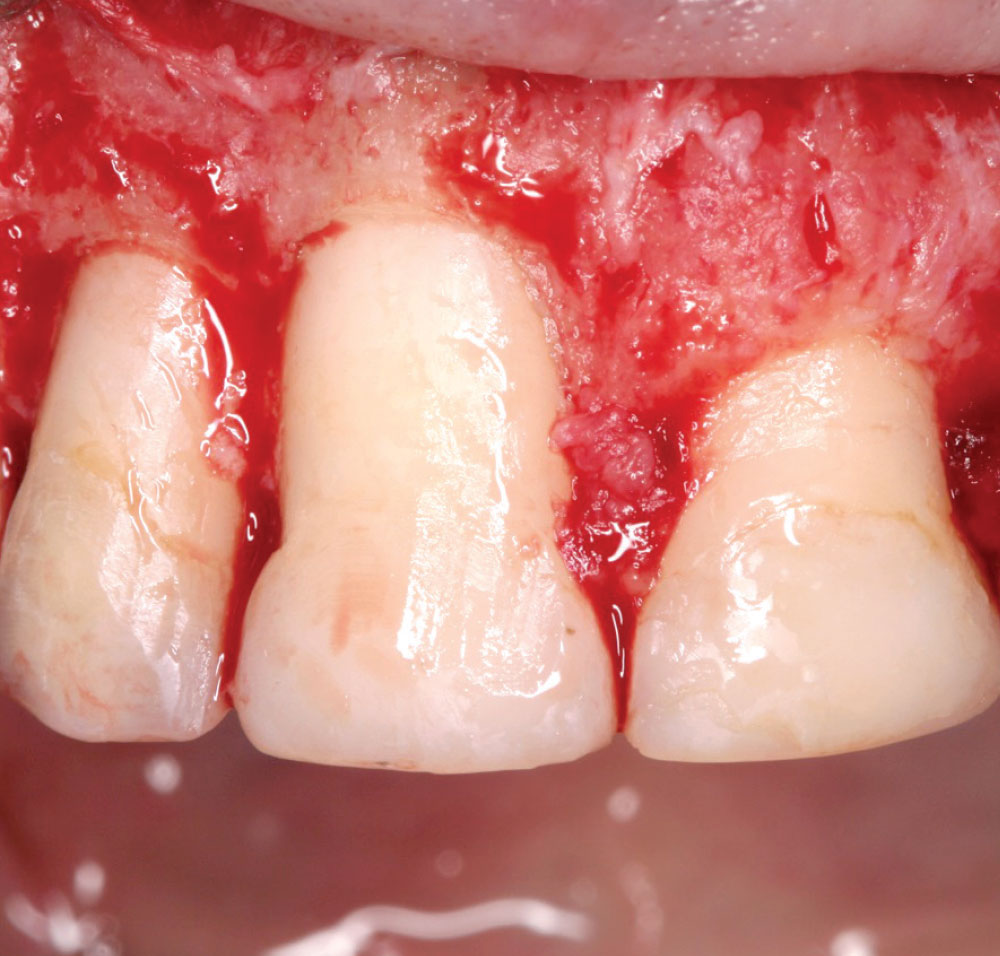
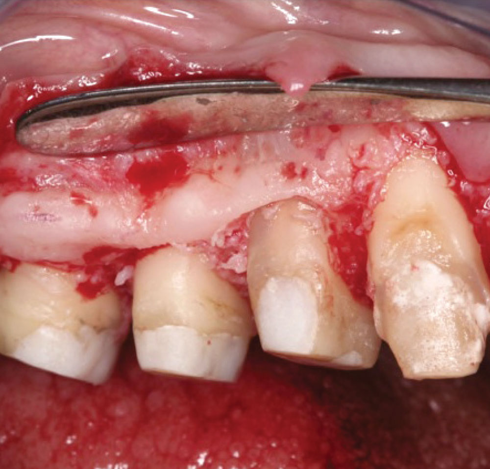
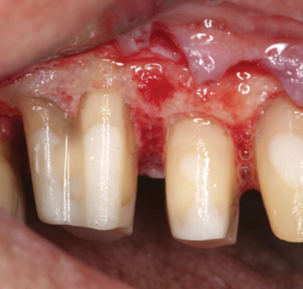
Figure 3: During crown lengthening, troughs in the bone occur that are similar to the periodontal vertical defects caused by endotoxins released by bacteria. Notice the significant defect caused between teeth #7 and #8; this is unavoidable.

Case 1 Figure 4

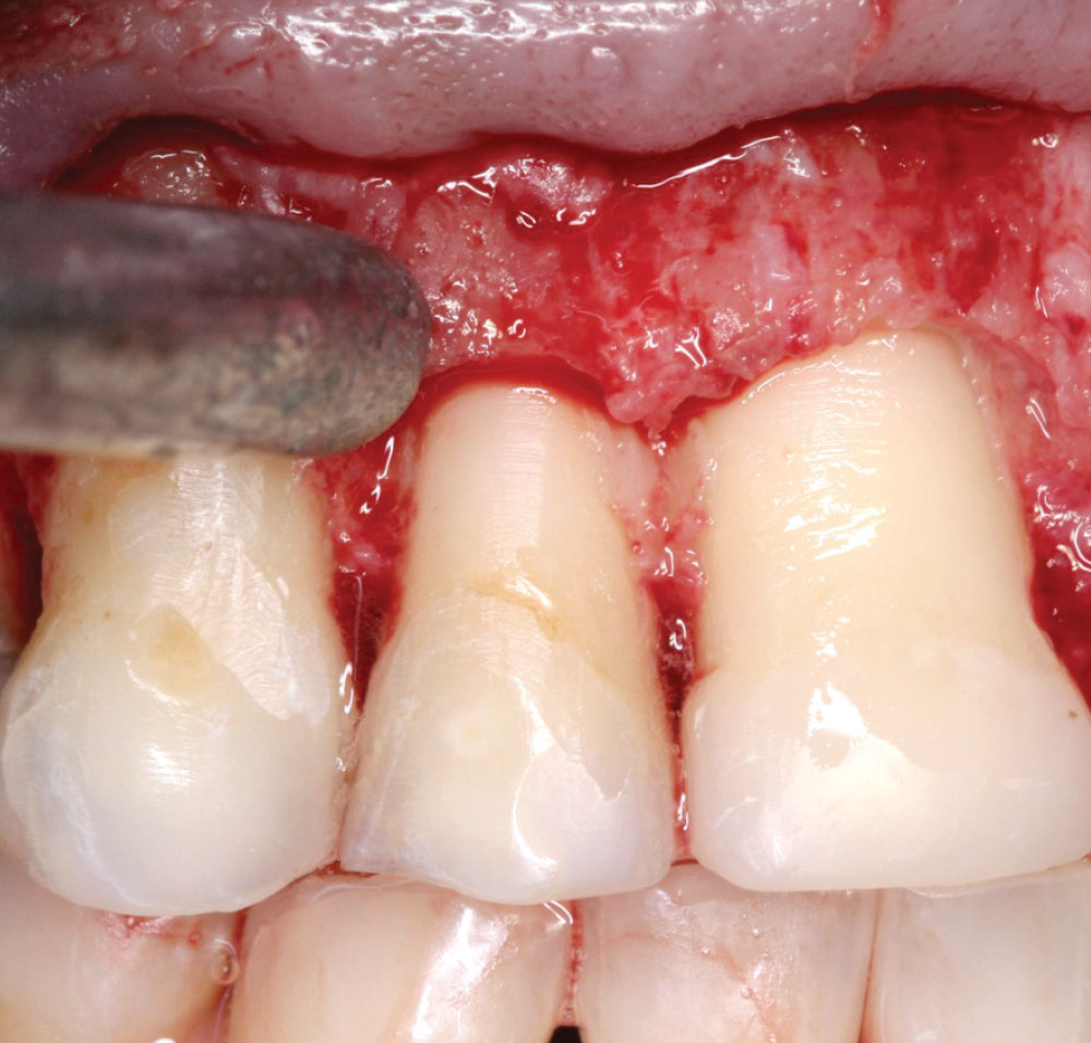
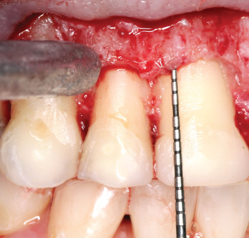
Figure 4: A probe reveals the trough created in the bone due to crown lengthening. The probe shows the vertical defect involving line angles caused by the crown lengthening. The thicker the bone, which is common in the interproximal, the greater the resulting defect. Herein lies the problem with closed-flap crown lengthening: Without the ability to remove the troughs created by lengthening the teeth, serious long-term consequences can occur due to the lack of uniformity between bone and soft tissue. It is critical for bone to mimic soft tissue when contouring. The surgeon must create a parabolic architecture. Note: Horizontal access and visibility are necessary to remove the troughing caused by crown lengthening.

Case 1 Figure 5

Figure 5: Crown lengthening is completed on teeth #7 and #8.

Case 1 Figure 6

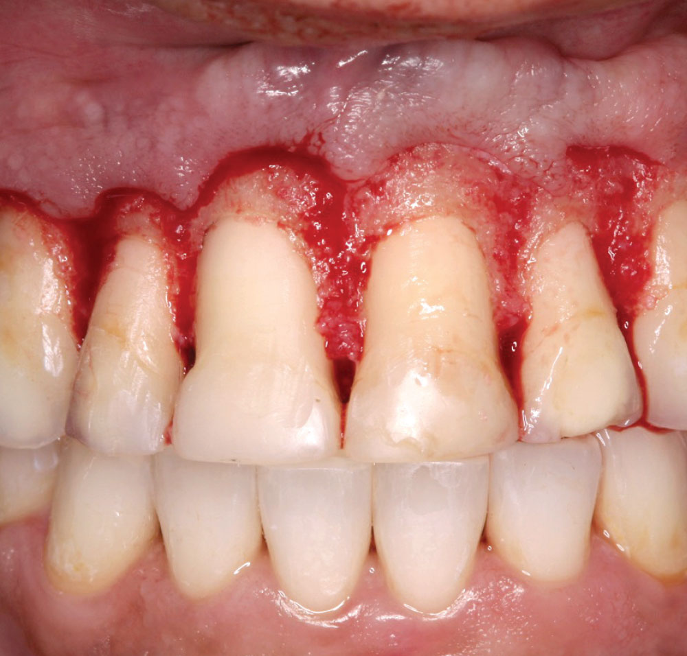
Figure 6: Notice how the bone and soft tissue mimic each other. The existing length of teeth #8 and #9 is now 13 mm from the bone to the incisal edge. This allows for 3 mm of biologic width and 10 mm for the clinical crown, with the new crown adding 1 mm to the incisal edge.

Case 1 Figure 7

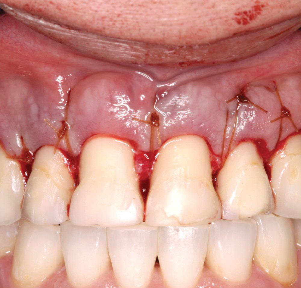
Figure 7: The tissue is sutured into place using a 5-0 chromic gut. Referring back to the original discussion on biologic width and clinical crown length, the tissue is placed where the remaining tooth structure is 10 mm. Notice there is an abundance of connective tissue remaining. Without the ability to remove the troughing created by the vertical removal of bone, the author finds closed-flap crown lengthening to be biologically unsound. Horizontal access and visibility are needed to create a sound biologic surgical procedure.

Case 1 Figure 8

Figure 8: Day of impression. Notice the tissue is slightly red. Due to her teaching schedule, the patient could not accommodate normal postoperative appointments. (The author and case surgeon would have preferred to see her several weeks before her impressions to reduce any inflammation, as this is the doctor’s responsibility. Subgingival chlorhexidine would be used to reduce minor inflammation.)

Case 1 Figure 9

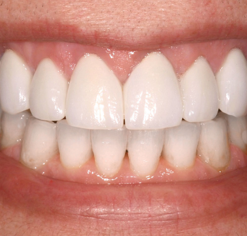
Figure 9: Final restorations with mild gingival irritation. Over time, the author expects the tissue to improve, although reducing inflammation prior to impression taking is the preferred method.

Case 2

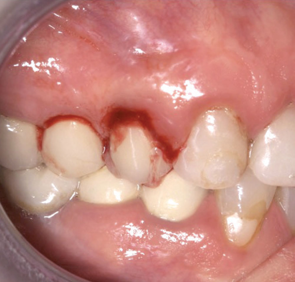
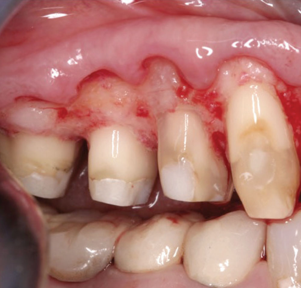
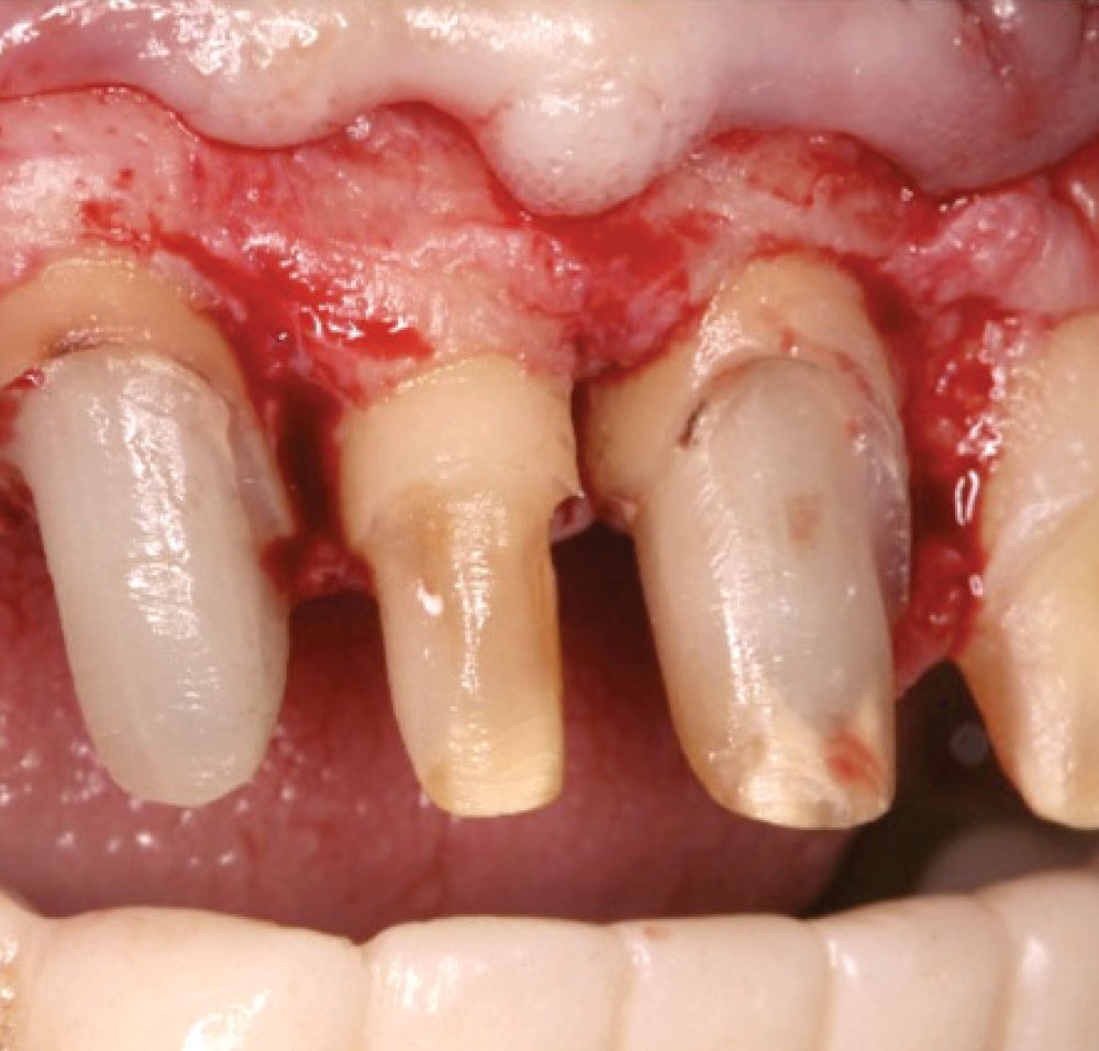
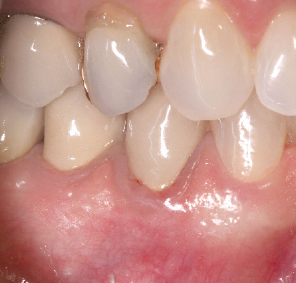
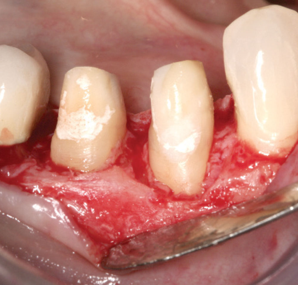
In this case, you will notice that the defect, which is caused by biologic width invasion, mimics the defect caused by closed-flap crown lengthening in the first case. Both are biologically unsound.

Case 2 Figure 1

Figure 1: Below tooth #5, the existing crown is violating the biologic width.

Case 2 Figure 2

Figure 2: Reflection of a flap exposes a created defect on the buccal of tooth #5, where biologic width invasion has occurred. To correct the defect, horizontal removal of bone is necessary, as well as the creation of bone architecture that mimics the soft tissue.

Case 2 Figure 3

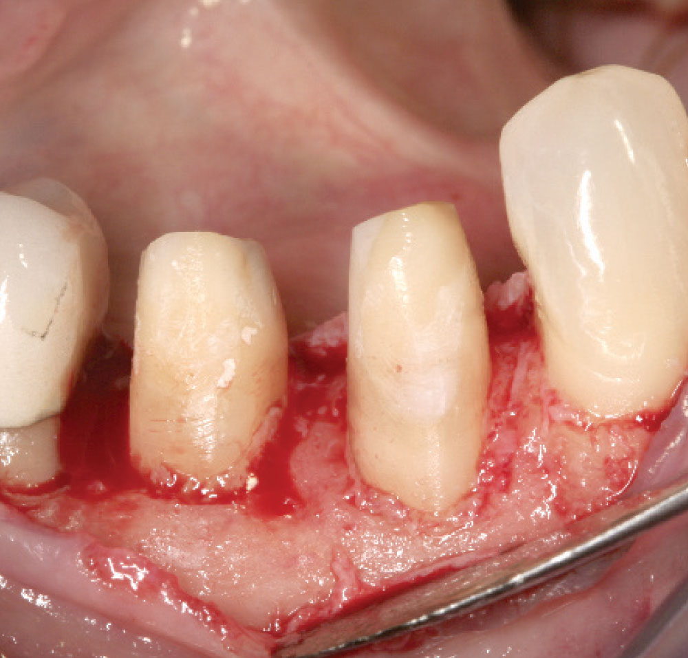
Figure 3: This shows the ideal osseous and soft tissue architecture after proper bone contouring to remove the troughs. The crown is violating the biologic width. This crown will be removed and a core and a provisional will be placed.

Case 3

Case 3 Figure 1

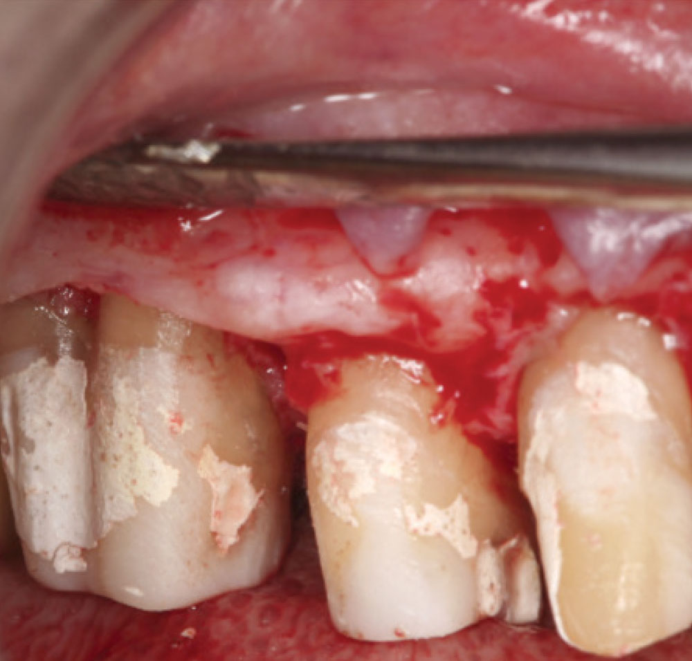
Figure 1: Crown lengthening is necessary to create a space for the biologic width. The author believes that visibility is critical for properly treating bone. A flap is required to see the underlying structures for crown lengthening.

Case 3 Figure 2

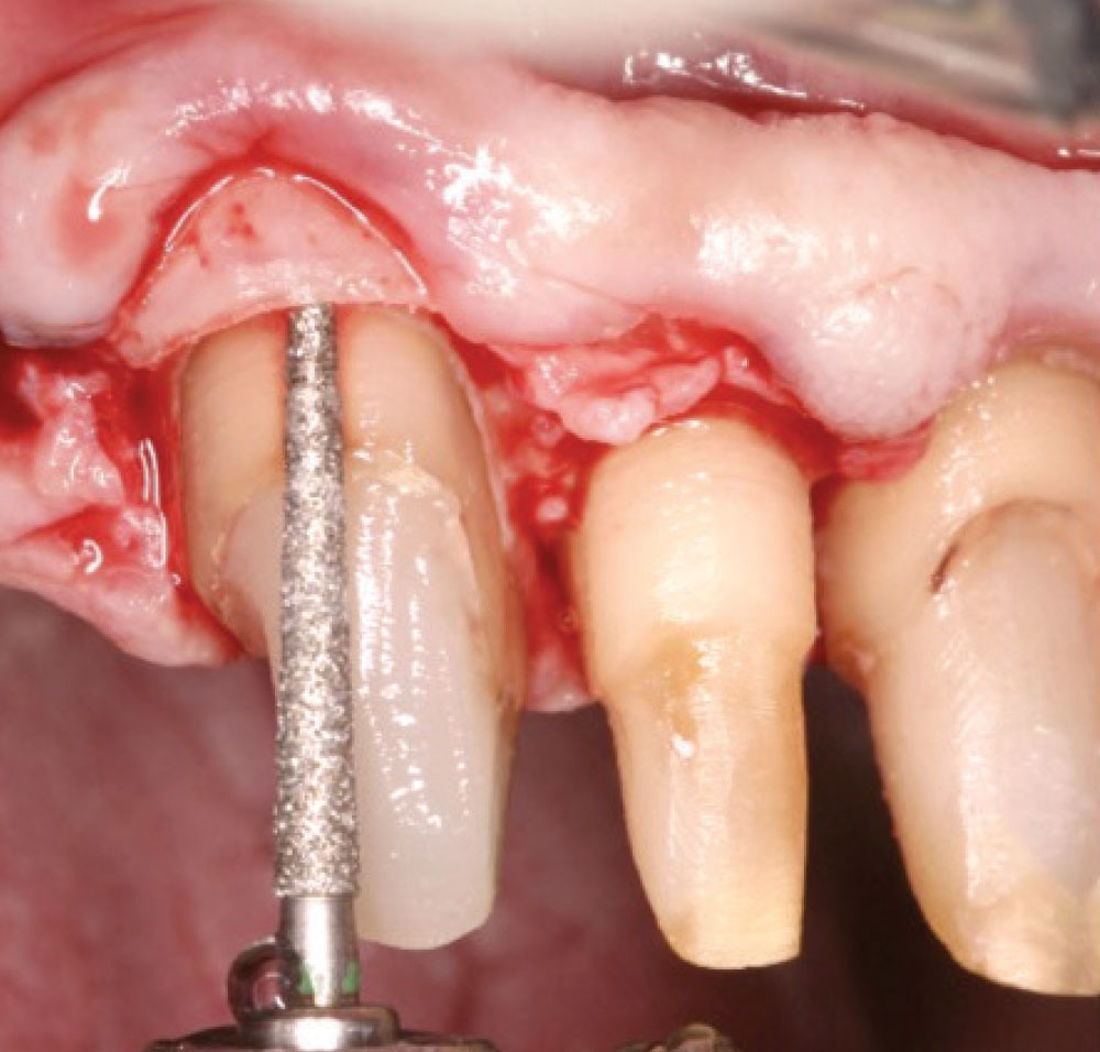
Figure 2: Regardless of the instrument — bur or laser — used when crown lengthening is performed, bone is removed. Unless the tip of the bur or laser is exactly the same dimensions as the bone to be removed, a trough will be created when there is a greater thickness of bone than tip diameter. This is a biologically unsound result. The bur is left in place to show the crater that is created as the bone is removed.

Case 3 Figure 3

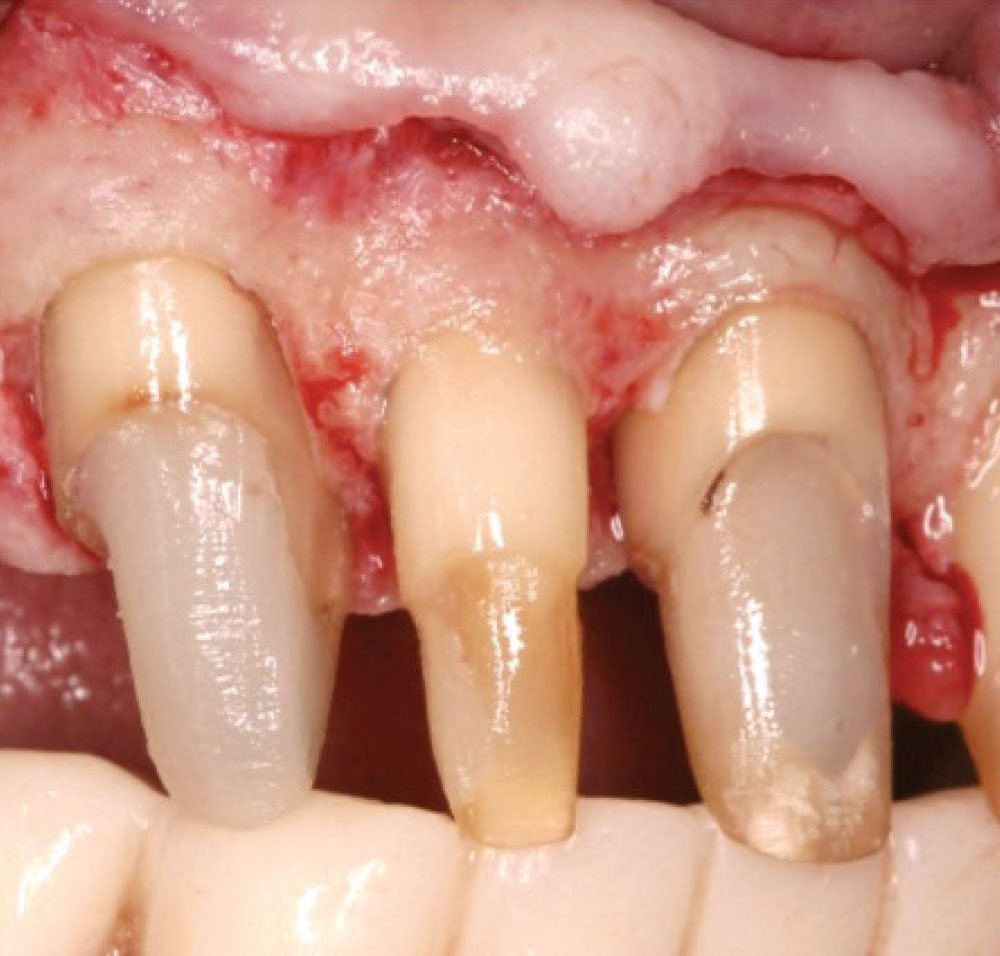
Figure 3: Using the bur or laser horizontally allows the crater to be removed and an ideal osseous architecture to be created. Notice that the bone and soft tissue mimic each other.

Case 4

Case 4 Figure 1

Figure 1: The existing crown on tooth #28 violates the biologic width. There are periodontists who say that if the biologic width is invaded, the bone will remodel to accommodate the infringement on this area. In 35 years of treating biologic width invasion, the author has consistently seen osseous defects associated with such violations. No remodeling is noted.

Case 4 Figure 2

Figure 2: Upon reflection of the tissue, a cratered defect is noted, presumably associated with the biologic width invasion. This type of defect must be removed to create an environment for the bone and soft tissue to closely adapt for minimal probing depth.

Case 4 Figure 3

Figure 3: The defect is removed and the osseous support will now conform to the parabolic architecture of the soft tissue as it heals. Thus, the bone mimics the soft tissue and minimal pocket depth will be present upon complete healing.

Case 5

Case 5 Figure 1

Figure 1: With the tissue reflected, the ravages of periodontal disease on the bone can be seen clearly. A reverse architecture is visible. This means that rather than the bone conforming to the contours of the tissue, it is irregularly shaped, thus causing a discrepancy between the soft tissue and the bone, resulting in a periodontal pocket.

Case 5 Figure 2

Figure 2: After osseous contouring to remove the pocket in the bone, the present configuration will mimic the soft tissue upon healing. A minimal probing depth will remain, allowing for better long-term maintenance.

Summary

Without the ability to remove the troughing created by the vertical removal of bone, closed-flap crown lengthening is biologically unsound. Horizontal access and visibility are needed to create a sound biologic surgical procedure.

Dr. Daniel Melker is in private practice in Clearwater, Florida, and lectures nationwide. Contact him at 727-725-0100.