The American College of Prosthodontists estimates that more than 36 million Americans do not have any teeth. These numbers are expected to grow in the next 20 years. In addition, some degree of tooth loss affects the majority of adult Americans, and the number of partially edentulous patients is expected to increase to greater than 200 million over the next 15 years.
Changes to the oral anatomy as a result of having no teeth include reduced vertical dimension of occlusion (VDO), and reduction of lower face height and mandible rotation. It is also important to note that edentulous patients are susceptible to reduced size and tone of pharyngeal musculature, and, over time, macroglossia (i.e., a large tongue), which may complicate airway issues dramatically.
It has been shown that the ratio of edentulism is six times higher in low-income communities. Studies also show that the condition is higher in women, and other factors include old age, education, access to dental care and insurance coverage.
There have been many studies that show the implications of tooth loss for patients’ overall health and quality of life. Nutritional effects include reduced consumption of fruits, vegetables and fiber, increased risk of obesity, and cardiovascular and gastrointestinal disorders. The effect of edentulism on the heart is particularly notable. One group of researchers followed 7,674 subjects for 12 years and found that the risk of cardiovascular mortality is dependent on the number of teeth a patient has.
Obstructive Sleep Apnea and Edentulism
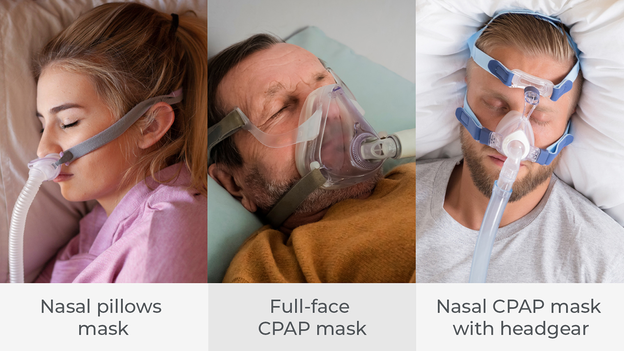
Primary treatments for obstructive sleep apnea (OSA) include continuous positive airway pressure (CPAP) and oral appliance therapy (OAT). CPAP therapy, which is often referred to as the gold standard of care, consists of a bedside blower attached via tubing to some form of patient interface — a mask or nasal pillows — that delivers a column of air into the patient’s airway. This column of air forms a pneumatic splint supporting the tissues of the airway, so that the patient’s airway doesn’t collapse during sleep.
CPAP therapy is integrated into the sleep diagnostic protocol and is the first therapy that most patients experience. Successful treatment with CPAP is often more difficult for edentulous patients because of the challenges of finding patient interfaces that fit well and can be worn every night, all night. Patients are considered compliant with CPAP therapy when they use their CPAP four hours or more per night, five nights per week. Most CPAP devices include software that monitors patient compliance to give the respiratory therapist the objective information needed to manage patient care.
CPAP is a part of a standard sleep diagnostic protocol and would generally be fitted and calibrated in the sleep diagnostic lab during a split-night study. A split-night study is an overnight polysomnogram (PSG) performed in a sleep lab while being continuously monitored by a credentialed technologist. It includes an initial two-hour period of sleep study to establish clinically significant sleep apnea, followed by a CPAP titration study where a therapeutic pressure is established based on the patient’s sleep quality under treatment.
The key element to a successful titration study is that the patient is required to be comfortable enough with the CPAP interface to fall asleep and stay asleep while the air pressure is being adjusted. This process is expected to take three hours. PSG is usually performed at night, when most people sleep, though some labs can do the test at other times of the day in order to accommodate shift workers and people with circadian rhythm sleep disorders.
The PSG monitors many body functions, including the following:
- Brain activity — An electroencephalogram (EEG) establishes sleep stages and monitors seizure activity.
- Eye movements — Electrooculography (EOG) measures eye movements. Eye movements and brain activity establish REM sleep.
- Muscle activity or skeletal muscle activation — Electromyography (EMG) is a good indicator of limb movement disorders and jaw parafunction during sleep.
- Heart rhythm — An electrocardiogram (ECG or EKG) is used to monitor issues with the heart during sleep.
- Respiratory airflow — Airflow is the key diagnostic feature of sleep apnea, which is characterized as a cessation of breathing for 10 seconds or longer.
- Respiratory effort — Respiratory effort will inform the clinical team if the patient is trying to breathe by measuring the excursions of the chest and stomach. If there is effort but no airflow, apnea is considered obstructive. If there is no airflow and no effort, the apnea is considered central in nature.
- Pulse oximetry — Pulse oximetry is the measurement of the amount of oxygen in the blood. In a sleep study, this parameter is used to monitor the changes in oxygen saturation over time that occur with sleep apnea or respiratory conditions, such as chronic obstructive pulmonary disease (COPD) or sleep apnea.
- Snoring — Snoring can be measured or noted by the sleep tech.
Continuous Positive Airway Pressure (CPAP) Therapy
Andre Puleo, vice president at Sommetrics and a trained sleep technologist, had the following observations when discussing the challenges faced by denture patients who are treated with CPAP:
“Key to a successful CPAP experience is getting a comfortable mask that forms a good seal and doesn’t leak. If the patient has no teeth, the lower edge of the nasal mask can slip and cause a leak. These leaks often cause patients to overtighten their masks, leading to fit and function issues. Full-face masks are a solution; however, the patient typically will have problems getting a seal around the chin. It is also very common to have edentulous patients’ mouths drop open during sleep, in which case the column of air flows out of the mouth. In the sleep lab and at home, we would use a chin strap to hold the mouth closed to achieve a lip seal and a therapeutic pressure.”
It is clear that patient compliance becomes more challenging as the patient interface becomes more invasive.