Biologic Shaping from a Restorative Perspective
May 1, 2013
 image
Daniel Melker, DDS
Biologic Shaping from a Restorative Perspective

In today’s world of advanced dental procedures and technology, traditional or classic dental principles can easily be lost. This may especially be true with the decision-making process of saving teeth. Implants are wonderful options when appropriate, but they should not be selected when a tooth can be saved using a predictable perio or restorative protocol that yields excellent long-term prognoses. Too often today, good teeth are being removed in favor of implant placement that is occurring in a clinical environment of inadequate bone and soft tissue, as well as biomechanical compromise. Biologic shaping and soft tissue grafting offer a classic, proven methodology for treating teeth with absolute predictability.

Often our restorative treatment plans lead us to subgingival margins, furcation involvement, root flutes and concavities, in addition to a multitude of complex issues. Many of the issues we face are in the subgingival environment and require periodontal corrective procedures to return the foundation to a healthy state. Traditionally, crown lengthening was indicated for deep subgingival margins, not only to facilitate impression making but also to correct biologic width infringements. Biologic shaping is a periodontal corrective procedure reported in the literature1 that may complement traditional crown lengthening, yet it differs from traditional crown lengthening in the following ways:

  1. Traditional crown lengthening moves the bone away from the margin. Biologic shaping moves the margin away from the bone.
  2. Traditional crown lengthening requires osseous surgery to re-establish the biologic width. Biologic shaping may require minor osseous surgery, but it generally avoids major osseous surgery and still re-establishes biologic width because you have the choice to locate your restorative margin coronal to the old restorative margin (0.5 mm apical to the core is the coronal extent).
  3. Traditional crown lengthening may open furcations and render a poor prognosis. Biologic shaping preserves the integrity of the furcation because aggressive osseous surgery was not needed.
  4. Traditional crown lengthening does not eliminate flutes, concavities or root clefts, leaving the postoperative lengthened crown at risk for disease recurrence due to increased susceptibility for plaque, calculus and caries formation. Biologic shaping leaves the subgingival area as smooth as glass; there are no areas for plaque, calculus or caries to hide.
  5. Traditional crown lengthening worsens crown-to-root ratio. Biologic shaping maintains crown-to-root ratio.
  6. Traditional perio is about pockets and probing. Biologic shaping is about preserving bone, smoothing out the rough spots, and making restorative dentistry predictable and a joy to perform.

The concept of biologic shaping is presented in the case that follows. The procedure stresses a 360-degree removal of tooth surface irregularities as well as all cementoenamel junctions (CEJs) and existing margins. An important aspect of the procedure is to remove any concavities or furcation involvements. Once the root surfaces are perfectly smooth, the flap is placed just coronal to the osseous surface and sutured in place. After 12 to 14 weeks of healing, the restorative dentist simply places a new margin just coronal to the gingival collar, which allows for a perfect impression to be taken. This case also features the specific correction of a mesial concavity on an upper first bicuspid.

Case Presentation

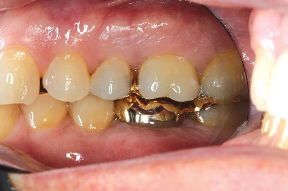
Figure 1: This patient will undergo a maxillary full-arch restoration to correct occlusal issues and mild periodontal disease. When performing definitive restorative procedures, it is critical to have an ideal periodontal foundation to restore. There was an initial discussion on whether to restore the bicuspids. After review of occlusal issues, it was decided to include the bicuspids in the provisional phase of treatment.

Figure 1: This patient will undergo a maxillary full-arch restoration to correct occlusal issues and mild periodontal disease. When performing definitive restorative procedures, it is critical to have an ideal periodontal foundation to restore. There was an initial discussion on whether to restore the bicuspids. After review of occlusal issues, it was decided to include the bicuspids in the provisional phase of treatment.

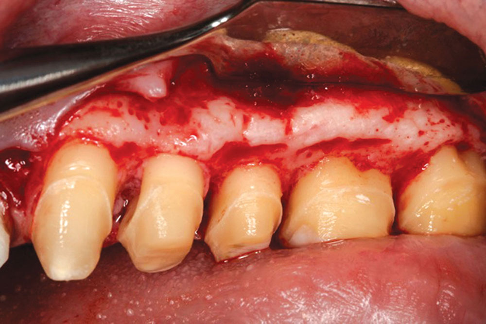
Figure 2: Upon reflection of the tissue with a full-thickness flap due to the existing thick bone, the tooth surfaces exhibited calculus located in concavities.

Figure 2: Upon reflection of the tissue with a full-thickness flap due to the existing thick bone, the tooth surfaces exhibited calculus located in concavities.

Figure 3, 7

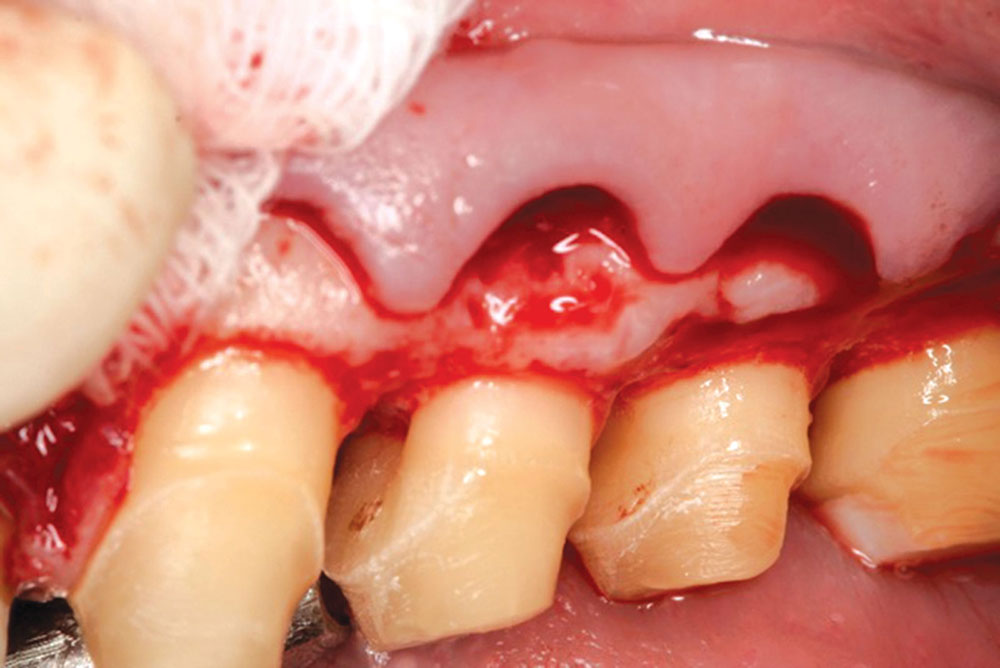
Figure 3: From a slightly different angle, the irregular contours of the bone can be seen. Osseous contouring will be necessary to create contours that will be compatible with the soft tissue when it is replaced. Once the flap is reflected, a split-thickness dissection is used to preserve the periosteum for suturing of the flap and for stability.

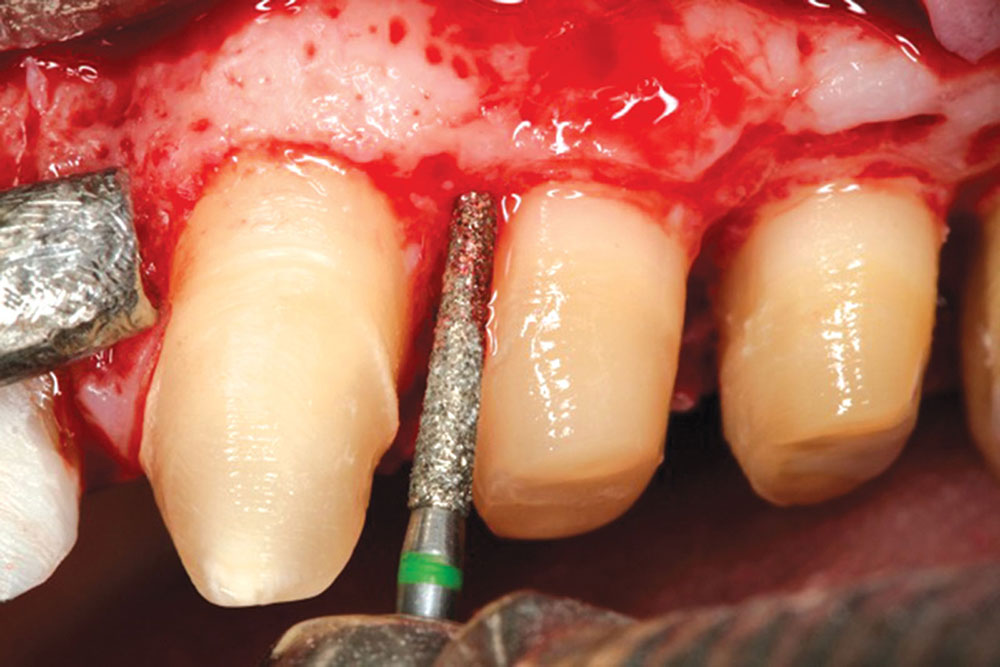
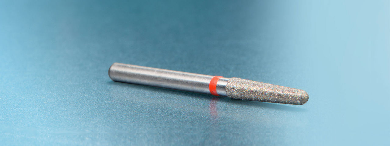
Figure 4: Using a C847-016 diamond bur (Axis Dental; Coppell, Texas), the tooth surface is gently smoothed to remove any irregularities of the root surface, as well as all CEJs. The concavity on the upper first bicuspid is also removed by gently blending the line angles approximating the concavity. Removal of the middle tooth surface of the bicuspid was avoided so as not to deepen the concavity.

Figure 4: Using a C847-016 diamond bur (Axis Dental; Coppell, Texas), the tooth surface is gently smoothed to remove any irregularities of the root surface, as well as all CEJs. The concavity on the upper first bicuspid is also removed by gently blending the line angles approximating the concavity. Removal of the middle tooth surface of the bicuspid was avoided so as not to deepen the concavity.

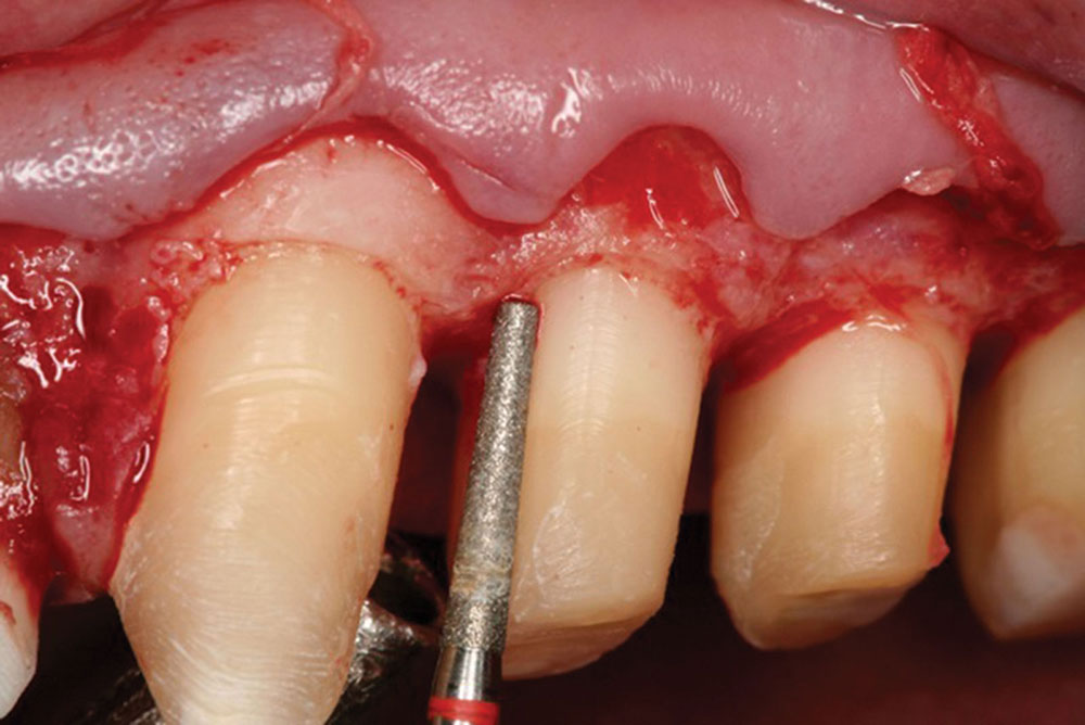
Figure 5: Once the gross removal of tooth structure is completed, an F847-016 diamond bur (Axis Dental) is used to smooth the root surface.

Figure 5: Once the gross removal of tooth structure is completed, an F847-016 diamond bur (Axis Dental) is used to smooth the root surface.

Biologic shaping and soft tissue grafting offer a classic, proven methodology for treating teeth with absolute predictability.

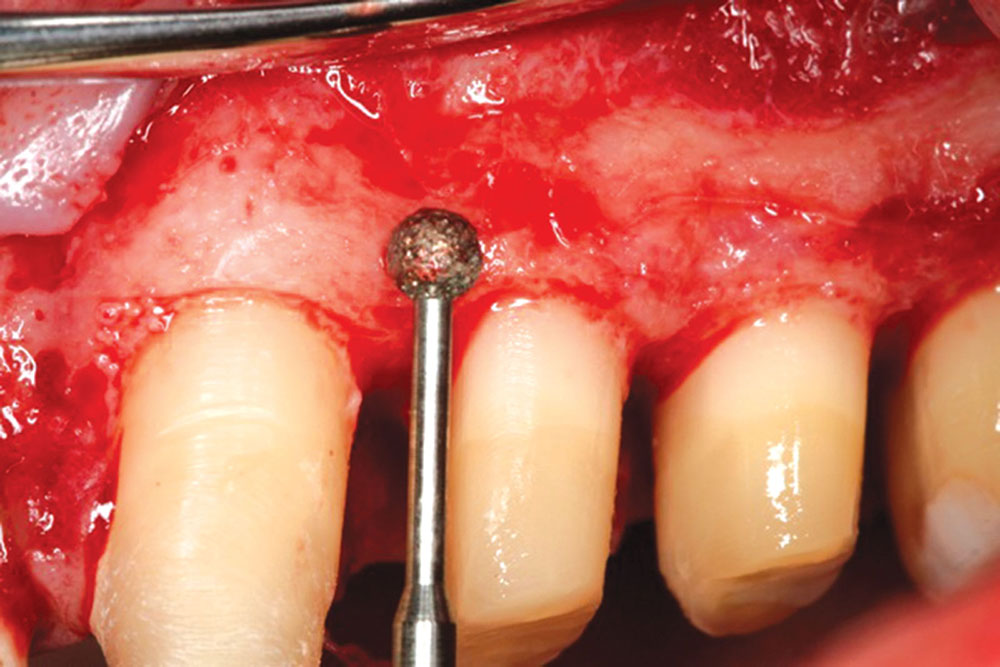
Figure 6: A C801L-023 diamond round bur (Axis Dental) is then used to properly contour the bone to mimic the soft tissue. The term for this procedure is “creating a parabolic architecture,” and it is the key to forming an ideal interface between bone, tooth and tissue. This phase of the surgery helps to avoid the formation of pockets between the bone and soft tissue when the tissue is replaced.

Figure 6: A C801L-023 diamond round bur (Axis Dental) is then used to properly contour the bone to mimic the soft tissue. The term for this procedure is “creating a parabolic architecture,” and it is the key to forming an ideal interface between bone, tooth and tissue. This phase of the surgery helps to avoid the formation of pockets between the bone and soft tissue when the tissue is replaced.

Figure 3, 7

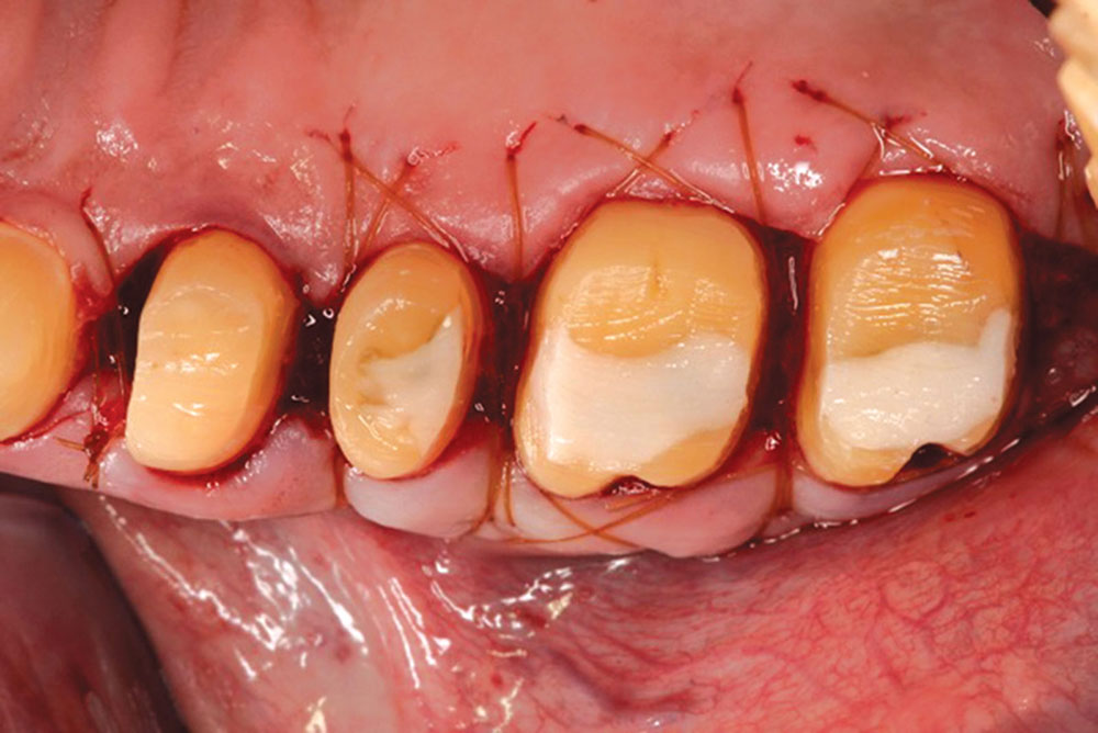
Figure 7: Upon completion of the biologic shaping and osseous contouring, an ideal foundation is created over which the soft tissue can be sutured in place.

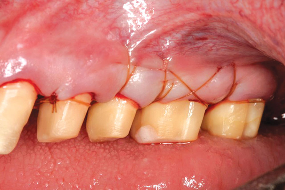
Figure 8: 5-0 chromic gut suture material is used to replace the flap just coronal to the osseous underlying foundation. An important aspect of suturing the flap is to involve the periosteum as an attachment apparatus for the suture. The suture grabs the periosteum apically to allow for perfect placement of the flap so that no movement or displacement of the flap can occur. There is no need for any dressing to be placed.

Figure 8: 5-0 chromic gut suture material is used to replace the flap just coronal to the osseous underlying foundation. An important aspect of suturing the flap is to involve the periosteum as an attachment apparatus for the suture. The suture grabs the periosteum apically to allow for perfect placement of the flap so that no movement or displacement of the flap can occur. There is no need for any dressing to be placed.

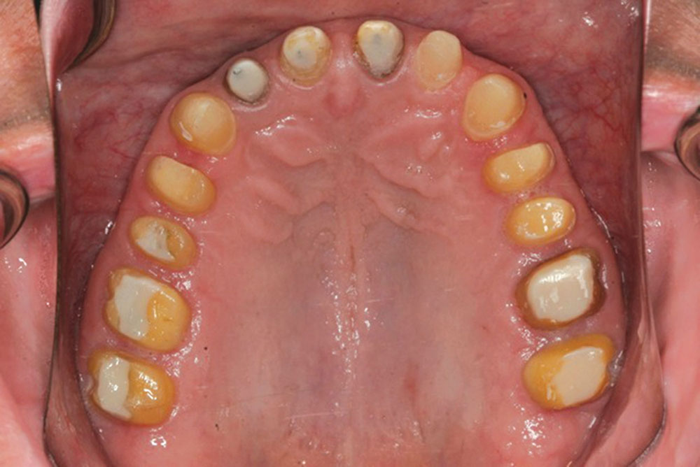
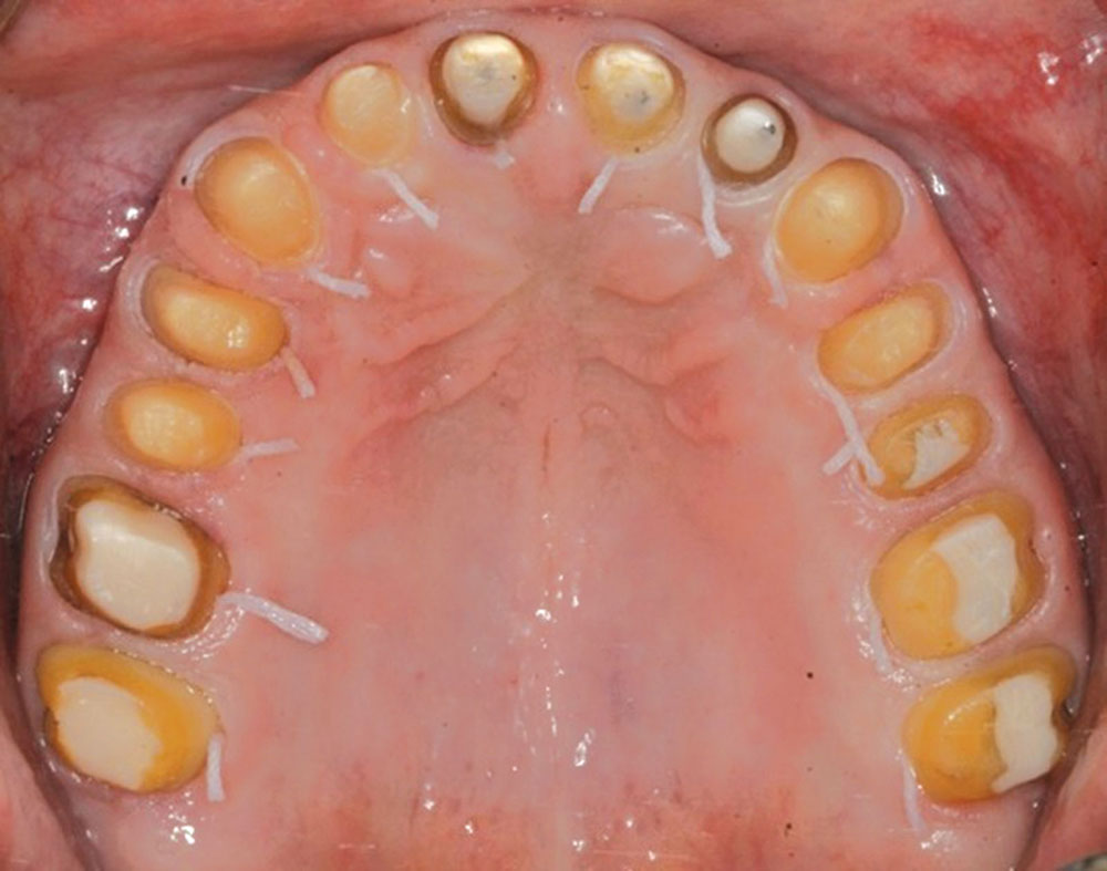
Figure 9: An occlusal view showing as much primary closure of the flaps as possible. This allows for decreased discomfort in the healing phase. Also note that no CEJs are present on any of the teeth. A recent article by Rapley and Cobb, et al.2 demonstrated with electron microscopy that the CEJs tend to hold biofilm and that these areas can be a source of periodontal breakdown. It is the belief of the author that by removing the CEJs, we are treating a cause of future breakdown, thus changing the environment for long-term maintenance.

Figure 9: An occlusal view showing as much primary closure of the flaps as possible. This allows for decreased discomfort in the healing phase. Also note that no CEJs are present on any of the teeth. A recent article by Rapley and Cobb, et al.2 demonstrated with electron microscopy that the CEJs tend to hold biofilm and that these areas can be a source of periodontal breakdown. It is the belief of the author that by removing the CEJs, we are treating a cause of future breakdown, thus changing the environment for long-term maintenance.

Biologic shaping is about preserving bone, smoothing out the rough spots, and making restorative dentistry predictable and a joy to perform.

Figure 10: The day of the reline appointment after four weeks of healing. The provisionals will be closed to fit the teeth, leaving 1 mm of space between the provisional and the tooth surface to allow for future biologic width growth in a coronal direction. No prepping of the tooth surface is done at this appointment.

Figure 10: The day of the reline appointment after four weeks of healing. The provisionals will be closed to fit the teeth, leaving 1 mm of space between the provisional and the tooth surface to allow for future biologic width growth in a coronal direction. No prepping of the tooth surface is done at this appointment.

Figure 11: Impressions day, 12 weeks post-op. All margins are placed just coronal to the gingival collars. A size 7/00 SilTrax® cord (Pascal International; Bellevue, Wash.) is placed in the sulcus to allow for the lab technicians to trim the dies.

Figure 11: Impressions day, 12 weeks post-op. All margins are placed just coronal to the gingival collars. A size 7/00 SilTrax® cord (Pascal International; Bellevue, Wash.) is placed in the sulcus to allow for the lab technicians to trim the dies.

 The procedure stresses a 360-degree removal of tooth surface irregularities … all CEJs and existing margins.
Figure 12
Figure 13
Figure 14

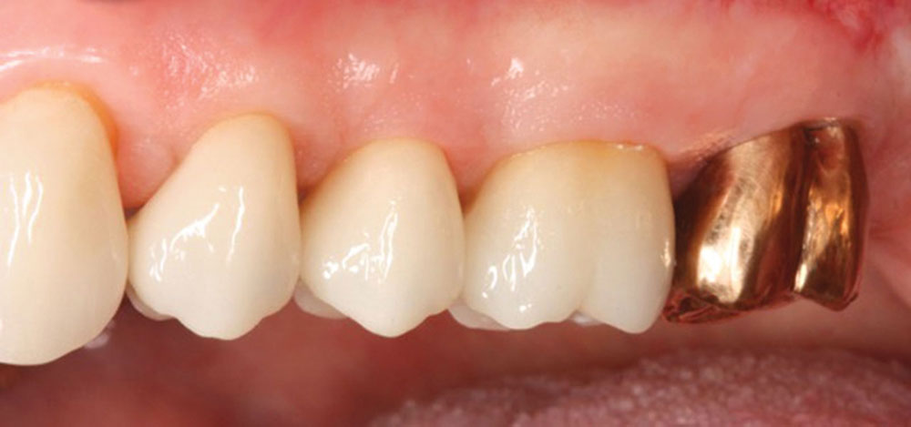
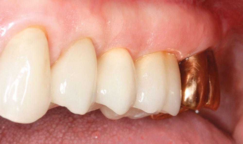
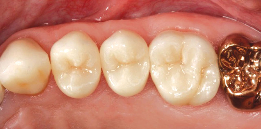
Figures 12–14: Final restorations placed. All are IPS e.max® crowns (Ivoclar Vivadent; Amherst, N.Y.) with the exception of full-coverage gold on the second molars. All margins are supragingival. Ideal health exists between the crowns and the soft tissue with no inflammation present. (Restorations courtesy of Dr. Howard Chasolen of Sarasota, Florida.)

Dr. Daniel Melker is in private practice in Clearwater, Florida, and lectures nationwide on periodontics and prosthodontics. Contact him at 727-725-0100.

References

  1. ^Melker DJ, Richardson CR. Root reshaping: an integral component of periodontal surgery. Int J Periodontics Restorative Dent. 2001 Jun;21(3):296-304.
  2. ^Satheesh K, MacNeill SR, Rapley JW, Cobb CM. The CEJ: a biofilm and calculus trap. Compend Contin Educ Dent. 2011 Mar;32(2):30, 32-7.