2201 Dupont Dr., Irvine, CA 92612
© 2020-2026 Glidewell. All rights reserved.
800-854-7256 USA
Learn clinically proven tips for full-arch data acquisition methods like CBCT scans, intraoral scans and more


Restoring a full arch is one of the most complex and rewarding procedures in implant dentistry — but the success of surgery and the final prosthesis hinges on what happens before the first drill touches bone. Data acquisition for the full-arch is the foundation of prosthetically driven, patient-specific treatment planning. Without accurate, complete records, even the best surgical skills and lab work can lead to compromised results.
This guide distills key concepts from advanced full-arch training into a practical reference for clinicians. Whether you’re placing FP1, FP2, FP3 restorations or removable options, these steps will help you plan with the end in mind, avoid costly errors, and deliver predictable function, esthetics and phonetics for your patients.
What You’ll Learn in this Article
Four Essential Records for Every Full-Arch Case
In full-arch dentistry, there is no one-size-fits-all protocol. Every patient presents with unique anatomy, esthetic demands, functional needs and financial considerations. Skipping a step or working with incomplete data introduces risks: malpositioned implants, violated transition zones, ill-fitting prostheses and unhappy patients.
A restorative-based, “crown-down” philosophy ensures surgical planning supports the desired final prosthesis. Just as you wouldn’t build a house without architectural drawings, you shouldn’t place implants without a restorative blueprint.
Regardless of restorative type (FP1–FP3 or RP4–RP5), four core records are non-negotiable:
Together, these records form the blueprint for both surgical and restorative success. They ensure that every clinical decision — from implant placement to final esthetics — is grounded in complete, accurate information rather than assumptions. Let’s take a deeper look at each of these four essential records for full-arch cases.
Field of view (FOV) is the first key consideration when taking CBCT scans. For full-arch planning, aim for a CBCT unit that can accommodate up to 18×16 or larger FOV, capturing at least from the inferior orbital rim to the chin.
Regarding resolution there are a few points to keep in mind. Smaller voxels (e.g. 75 μm) provide crisp detail. Larger voxels (>300 μm) can obscure anatomy but are less radiation. A balance in the middle of 150 μm is generally good for the full arch as it provides enough detail while minimizing radiation.
Here are some common issues you need avoid when taking these scans:
Here are some special considerations:

Preoperative CBCT image with a balanced voxel taken with patient out of occlusion.
For both dentate and edentulous patients, scans must capture all relevant landmarks:
Keep these key intraoral scanning tips in mind:

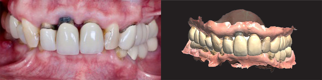
Preoperative intraoral scans showing upper and lower arches.
A precise bite helps evaluate & determine the vertical dimension and occlusal relationships, impacting restorative space and function. Here are the key points to remember:
As a general tip, if you are testing a new vertical dimension of occlusion (VDO) with significant changes, have the patient wear a try-in appliance at the proposed new vertical for at least two weeks.
Example of a physical photo and scan for verification of bite.
There are five crucial photos necessary for the full arch:
These five photos are extremely important. Treatment planning considers lip mobility, smile line and transition zone. High smile line measurement informs implant positioning and the amount of bone reduction needed.
3D facial scans are the emerging digital standard, and allow 360° visualization and creation of a dental avatar. For example:
Dental avatar created through facial photos and scans to determine and evaluate facial planes, midline, and the thirds of the face.
Once records are validated these are the steps we recommend you take:
Doing this ensures surgical outcomes align with esthetic and functional goals.
In full-arch rehabilitation, data acquisition isn’t just the first step — it defines every decision throughout the entire process. High-quality CBCT, precise intraoral scans, accurate bite registration and comprehensive facial records give you control over the process.
Just remember that with a complete dataset, you can:
In short: Plan with the end in mind — and start with the right data!
Related Dental Articles
Send blog-related questions and suggestions to hello@glidewell.com.
2201 Dupont Dr., Irvine, CA 92612
© 2020-2026 Glidewell. All rights reserved.