Clinical Case Report: Full-mouth Reconstruction with BruxZir and BruxZir Anterior
Introduction:
BruxZir® Anterior is a highly esthetic zirconia ceramic designed to satisfy the esthetic and functional requirements of the anterior region of the mouth. BruxZir Anterior has an average flexural strength of 650 MPa with translucency and color similar to natural dentition. BruxZir Anterior requires less reduction than monolithic glass ceramic restorations and is kind to natural opposing dentition during occlusal and parafunctional activity. Indications include single-unit crowns and three-unit bridges with one pontic. Prep requirements are conservative – only 0.8 mm of reduction is required, although 1.25 mm is ideal. BruxZir Solid Zirconia with flexural strength of 1200 Mpa was used to restore posterior molars.
Case Presentation:
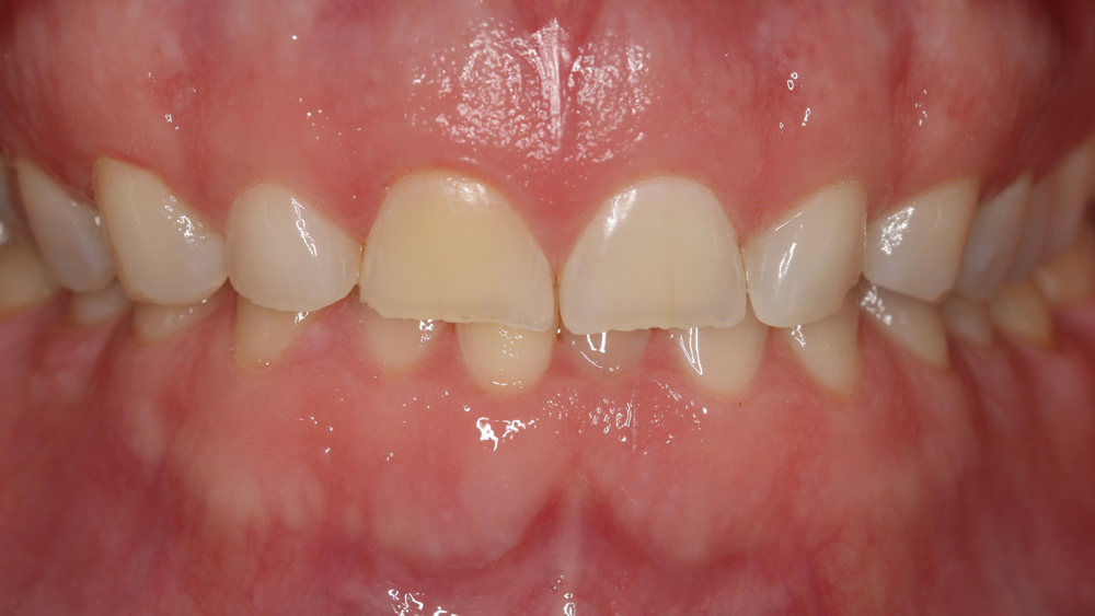
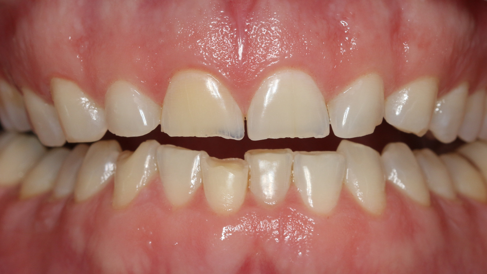
A forty-four year old male patient presented with worn upper and lower dentition due to years of bruxism, resulting in about 2 mm loss of vertical affecting his smile and other facial features (Figure 1a,b). The patient was concerned about the compromised esthetics and the long-term functionality of his teeth. Preliminary PVS impressions, x-rays and photographs were taken of his teeth to help formulate a treatment plan.
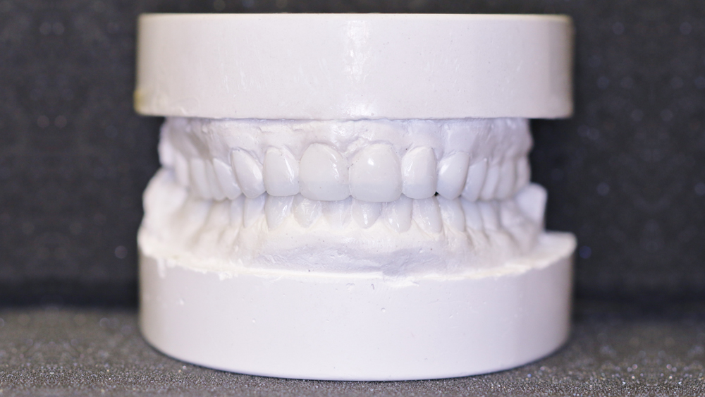
The models were poured and mounted on an articulator and the bite was opened by 2 mm to achieve an ideal tooth length. The patient did not want his teeth to look too long – he said, “I want my teeth to look natural and still fit my personality.” Once the stone models were mounted, a wax-up was done to simulate the final size, form and shape of the teeth (Figure 2).
Clinical Procedure:
Second Appointment
The finished working models were shown to the patient (Figure 2). He liked the size and esthetics and gave his approval to proceed. The shade was discussed and agreed upon with the patient settling on the B1 VITA shade.
To temporarily open the bite by 2 mm, 12 posterior teeth (including 1st molars, 2nd molars and 2nd pre-molars) were slightly roughened with a medium-grit football shaped diamond bur. The posterior teeth had three amalgam fillings, six composites and one PFM crown. The amalgams were replaced with composites. All were etched with 37% phosphoric acid, rinsed, and bonded with Scotchbond Universal (3M) followed by the application of a composite (Camouflage, shade A2, Glidewell Laboratories). Enough composite was added to the teeth to achieve a 2 mm vertical opening. The composite was light cured, the occlusion was checked in centric and the composite was adjusted until a 2 mm vertical opening was achieved.
Third Appointment
Two weeks after establishing the new vertical, the patient reported no discomfort with his bite and he was scheduled to start with the treatment. The plan was to prepare a total of 16 teeth, including two upper and lower molars as well as two upper and lower bicuspids in the right and left quadrants.
The patient was scheduled for a five-hour appointment. At that appointment the upper and lower right quadrants were anesthetized and the molars were prepared for restorations made from BruxZir Full Zirconia Crowns and Bridges (Glidewell Laboratories), while the bicuspids were prepared for BruxZir Anterior (Glidewell Laboratories) restorations. Upon completion of tooth preparations, temporaries were made at the newly established vertical using Luxatemp shade B1 (DMG) temporary material.
The tissues were packed with a #1 cord where indicated and full-arch impressions were taken of both the upper and lower arches using Aquasil putty (DENTSPLY/Caulk) for the tray material and Aquasil XLV light-body material. A bite registration (Futar Fast, Kettenbach) was also taken of the prepared right quadrants at the pre-selected vertical opening. While the temporaries were being readied, the upper and lower left quadrants were anesthetized.
All eight teeth were prepared for BruxZir anterior and posterior crowns as described earlier for the right side. Full-arch upper and lower impressions were taken. The impressions were examined and found to be suitable to send to the laboratory. The right bite registration was used to obtain a left bite registration at the same vertical opening. The full-arch impressions taken at first for the right side were used as back up impressions in case any tooth preparation was not clearly defined. Several photographs were taken and study models were sent as well as specific directions to assist the lab in completing the preparations of the crowns to achieve a satisfactory outcome.
Fourth appointment:
The patient did not experience any problems during the three-week temporization period and was very pleased with the fit, comfort and esthetics of the temporaries. The molar crowns were fabricated by Glidewell Laboratories using BruxZir Full-Strength Crowns and Bridges, while the bicuspids were fabricated with BruxZir Anterior. The esthetics of all restorations was excellent; one could not detect a difference between the molar and bicuspid crowns. The crowns were tried in for fit, shade and occlusion and received the approval of the patient for their cementation. The teeth and the internal surface of the crowns were treated with Scotchbond Universal (3M) bonding agent prior to applying RelyX Ultimate Adhesive Resin Cement (3M) in each crown.
The four crowns were seated one quadrant at a time, the margins flash cured for not more than 1-2 seconds, the excess cement removed from around each crown, and the teeth carefully flossed before final curing each crown. This procedure was repeated for all four quadrants. The occlusion was checked and selectively adjusted when needed. The need for adjustment was minimal. Once everything looked satisfactory, the patient was scheduled for the next appointment at which time the upper and lower anterior teeth would be prepared.
Fifth appointment:
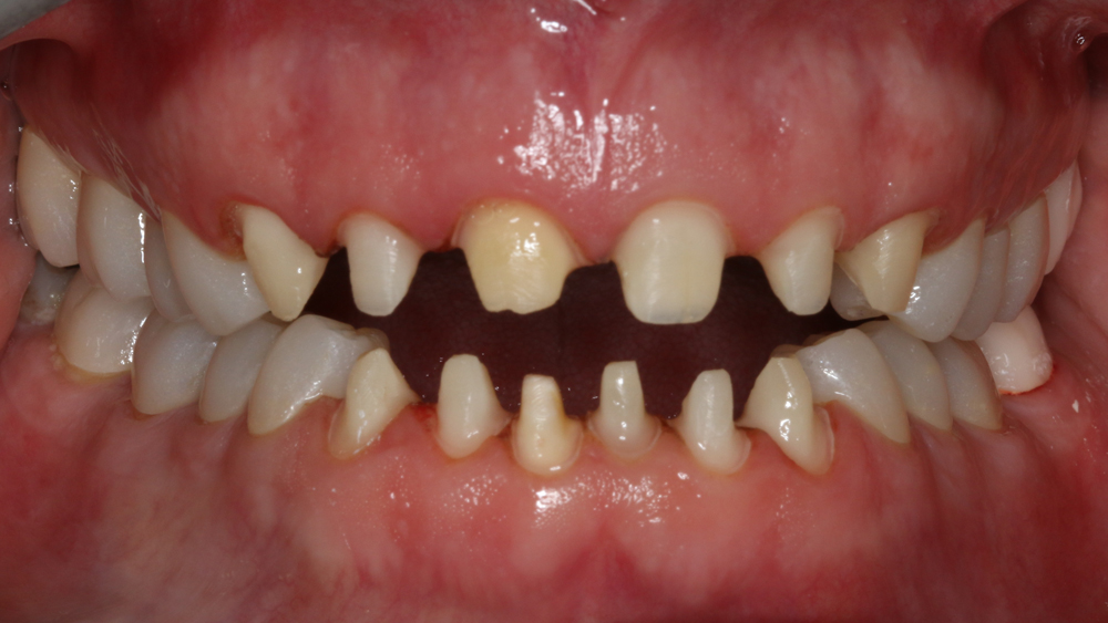
The upper anterior (#6-11) and lower anterior (#22-27) teeth were anesthetized and prepared for BruxZir Anterior crowns. Since the bite was open, almost no reduction was needed at the incisal edges. Circumferential tooth reduction was kept to a minimum because the BruxZir Anterior material has excellent strength and good translucency (Figure 3).
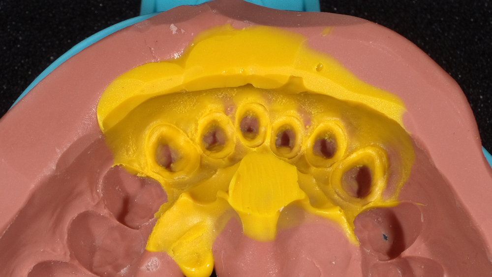
Once the preparations were done, full-arch impressions were taken using Aquasil putty for tray material and Aquasil XLV light-body material (Figure 4). A bite registration was also taken to help in properly mounting the case. The impressions, bite registration, models and photographs of the prepared teeth were all sent to Glidewell Laboratories to assist in completing the case accurately. The front teeth were temporized using Luxatemp shade B1 (Figure 5).
Sixth Appointment:
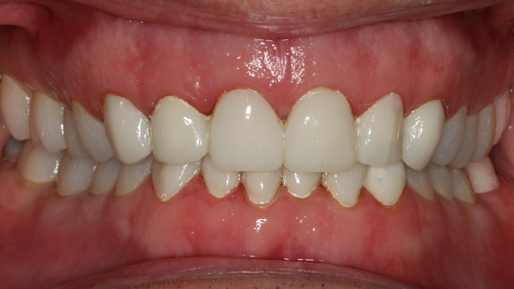
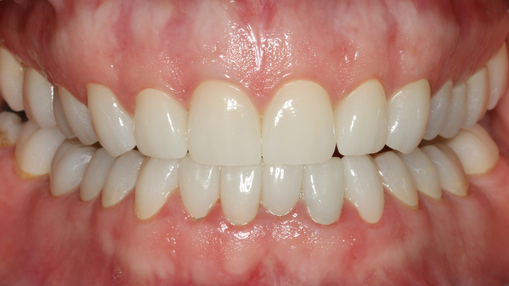
The patient was anesthetized, the temporary crowns removed and the BruxZir Anterior crowns fitted using Stabiliner (Crown Delta Corp) try-in silicone liner. The patient was pleased with the esthetics and requested cementation of the crowns. The crowns were cemented with Scotchbond Universal and RelyX Ultimate Adhesive Resin Cement (3M Oral Care). The cement was flash-cured, the excess removed, teeth flossed, and everything was post-cured. The occlusion was checked and adjusted where needed. Final photographs were taken as shown in Figure 6.
Summary:
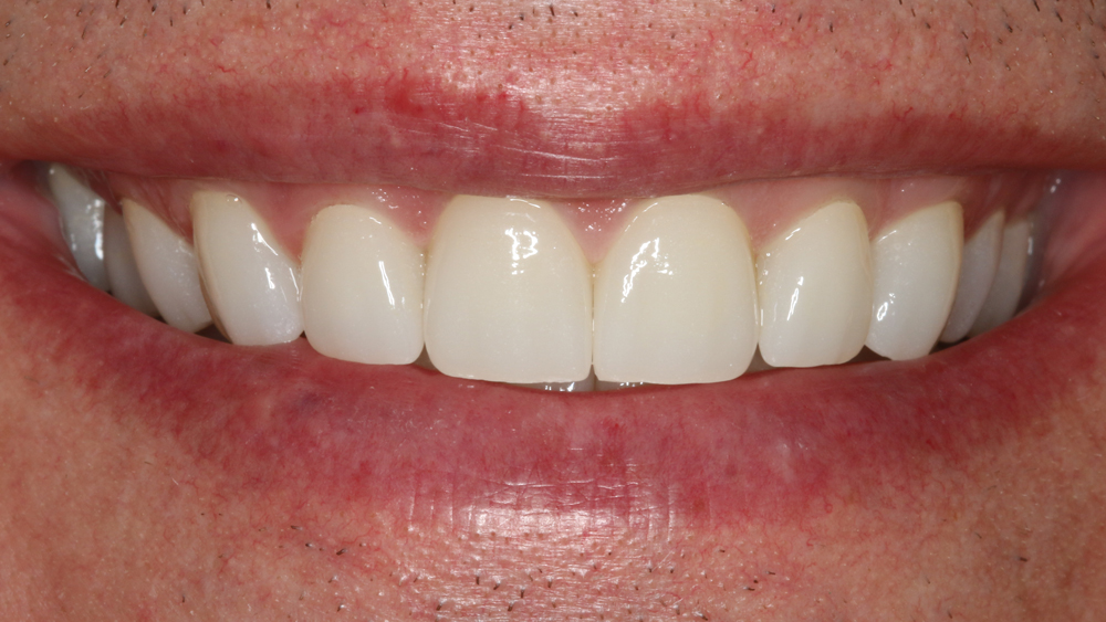
The patient was pleased with the final outcome and the resulting esthetics. Three months after completing the case, the patient is doing very well, the tissues are healthy and he is very pleased with his winning smile (Figure 7).
THE DENTAL ADVISOR 3110 West Liberty, Ann Arbor, Michigan 48103 (800) 347-1330 connect@dentaladvisor.com, www.dentaladvisor.com
©2016 Dental Consultants, Inc.

