Restoring Long-Lost Smiles
For the first time in many years, Maria is all smiles. It is a dramatic difference from where she was just four months ago. When she arrived in my office, she was so self-conscious I could barely get her to open her mouth. Simple daily tasks like talking and eating had become problematic due to a fractured tooth that was barely attached by an endodontic post. It was so mobile that during our initial visit, Maria was afraid it would fall out while she spoke.
After losing both parents at a young age, Maria lived with her adopted parents before moving out to live on her own when she was 17. Even though her root canal had failed years prior to our visit, she had been unable to have the tooth evaluated. As she took the first steps toward addressing the years of neglect, I looked forward to providing this deserving patient with a functional and esthetic solution to her problem.
BRIDGING THE GAP WITH BRUXZIR® ESTHETIC
After conducting a thorough diagnostic examination, a vertical root fracture was noted, leaving the #9 central incisor malaligned and unstable. Due to the presence of significant vertical bone loss, extensive treatment for guided bone regeneration and a connective tissue graft would have been required prior to implant placement. After the patient was consulted on these factors, she opted against implant treatment. Once we discussed the remaining options, she decided on a bridge from #8–11 and a veneer on #7. Because she was congenitally missing tooth #10, placing a bridge to restore the edentulous area required preparing the adjacent canine and reshaping it to look like the missing lateral. I selected BruxZir® Esthetic Solid Zirconia as the best material to restore her beautiful smile. Not only does BruxZir Esthetic have superior strength compared to similar all-ceramic materials such as IPS e.max®, but it also has a translucent, natural-looking appearance. Sometimes clinicians think it is risky to do an all-ceramic bridge, but with a strong material like BruxZir Esthetic that has an average flexural strength of 870 MPa, doctors can confidently seat an anterior bridge that will produce long-lasting results. BruxZir has become such a popular material for dentists that it has been utilized to successfully fabricate more than 1.2 million bridges.
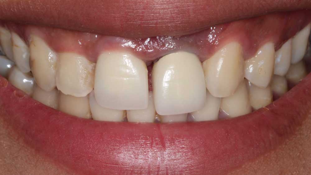
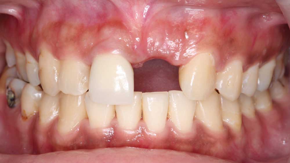
Figure 1: During Maria’s initial examination, I noticed her central incisor #9 was out of alignment, unstable and extruded.
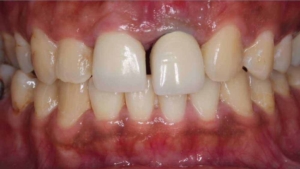
Figure 2: Upon further evaluation, I determined that additional restorations were failing, including a composite on lateral incisor #7.
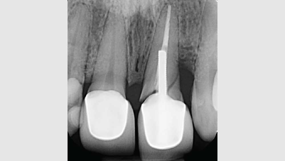
Figure 3: A previous root canal and post left tooth #9 compromised, resulting in a sizeable fracture. Because this fractured tooth went untreated for an extended time, vertical and horizontal bone loss were evident.
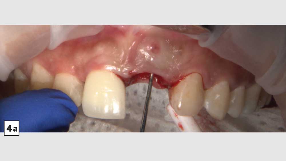
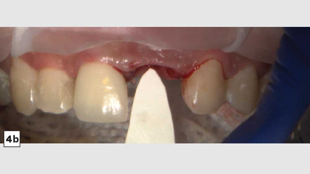
Figures 4a, 4b: Due to the hopeless state of tooth #9, I extracted it and removed the source of the infection. Upon removal, significant bone loss was observed, as depicted by the periodontal probe. I used the “ice cream cone” guided bone regeneration technique, including the use of a Newport Biologics™ Resorbable Collagen Membrane (Glidewell Direct; Irvine, Calif.) precut in a cone shape. I then filled the socket with Newport Biologics Mineralized Cortico/Cancellous Allograft Blend bone grafting material (Glidewell Direct) to regenerate the buccal plate of the fresh extraction socket. This guided bone regeneration technique required approximately three months for the site to heal.
Figures 4a, 4b: Due to the hopeless state of tooth #9, I extracted it and removed the source of the infection. Upon removal, significant bone loss was observed, as depicted by the periodontal probe. I used the “ice cream cone” guided bone regeneration technique, including the use of a Newport Biologics Resorbable Collagen Membrane (Glidewell Direct; Irvine, Calif.) precut in a cone shape. I then filled the socket with Newport Biologics Mineralized Cortico/Cancellous Allograft Blend bone grafting material (Glidewell Direct) to regenerate the buccal plate of the fresh extraction socket. This guided bone regeneration technique required approximately three months for the site to heal.
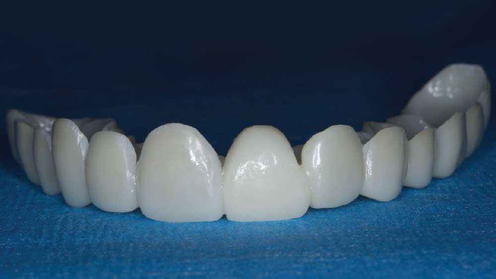
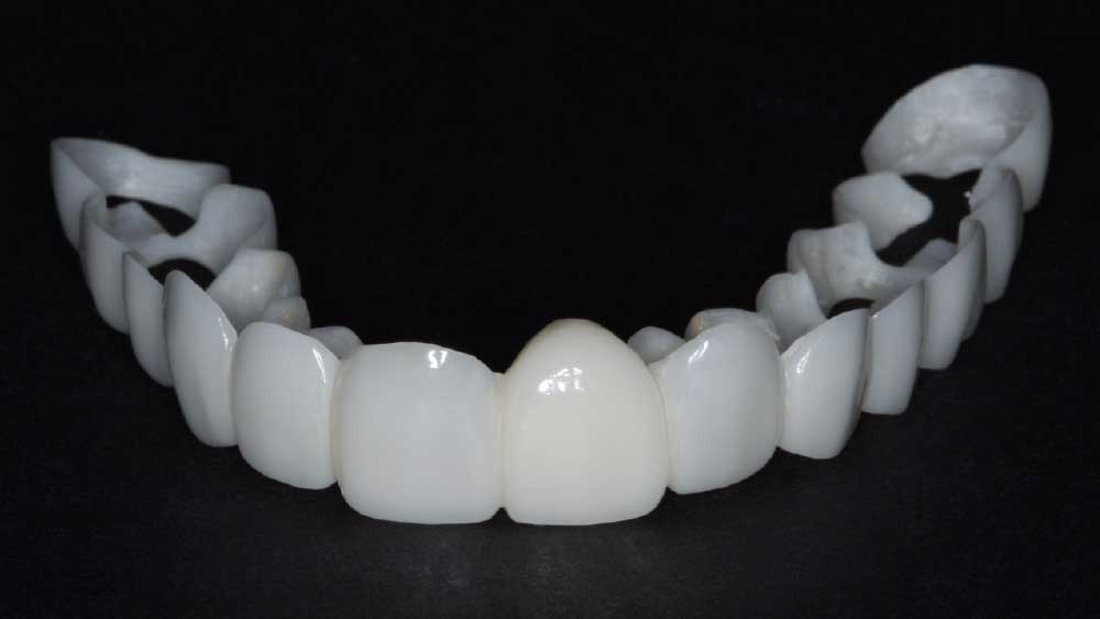
Figure 5: The extraction site left an unusually large space because the patient was also congenitally missing her upper left lateral and her canine #11 was now shifted into that position. Therefore, I scanned the patient with the iTero Element® 2 Intraoral Scanner and asked the lab to fabricate a Smile Transitions™ full-arch provisional so the patient and I could get a visual indication of the design of the final restoration. The pontic in the #9 position could then guide the healing of the tissue.
Figure 6: For the first 72 hours, I kept the #9 pontic of the Smile Transitions appliance 2 mm away from the tissue so it would not interfere with the healing process of the surgical site.
Figure 7: I instructed the patient to return to my office after 72 hours so that I could increase the height of the pontic and construct an ovate design that would begin contouring the tissue to create the appearance that the final restoration is naturally emerging from the gingiva.
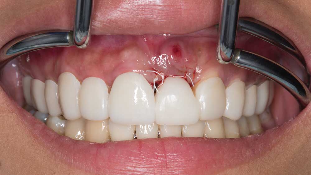
Figure 8: After three months of wearing the Smile Transitions appliance, the pontic had nicely contoured the tissue and the patient was ready to move forward with the final restorative treatment.
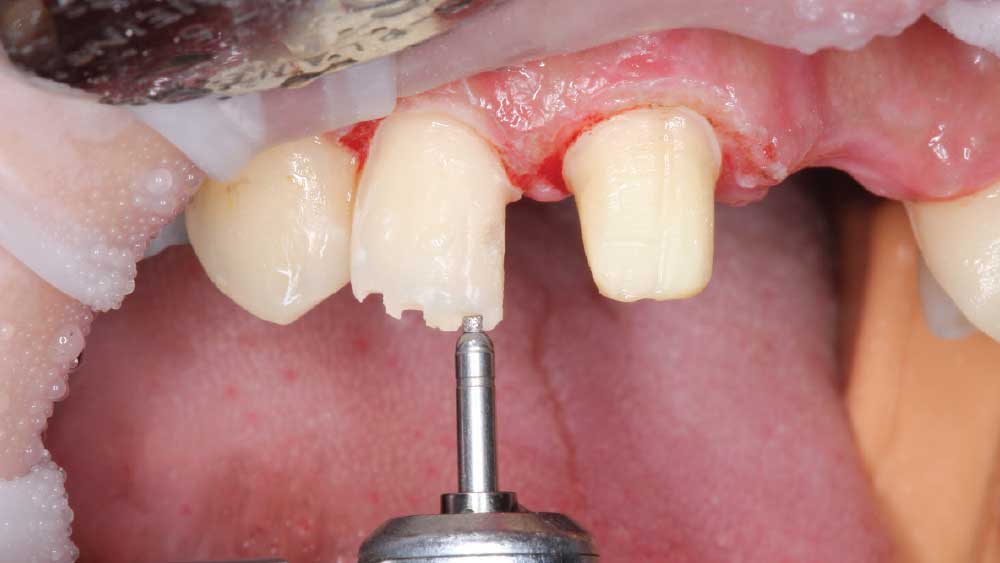
Figure 9: Due to a large, failing composite restoration, I prepared tooth #7 and placed a BruxZir Esthetic veneer. I removed the existing crown on tooth #8 and used that tooth as an abutment to support the BruxZir Esthetic bridge for teeth #8–11. Because tooth #10 was missing and tooth #11 was in its position, I prepared tooth #11 and shaped it to support a crown that resembled a lateral.
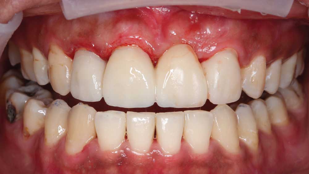
Figure 10: After preparation, I performed a gingivectomy in the apical region of teeth #7 & #8 to even out the gingival height. I then relined and cemented BioTemps® Provisionals (Glidewell Laboratories; Newport Beach, Calif.), which were fabricated by the lab and helped manage her tissue during the healing process. I also continued performing gingivoplasty to the pontic area as needed.
Figure 11: The patient came in for a series of post-procedural appointments until I was satisfied that the tissue had healed nicely and created a harmonious gingival display. Once this process was complete, she was ready to have an impression taken for her final restoration.
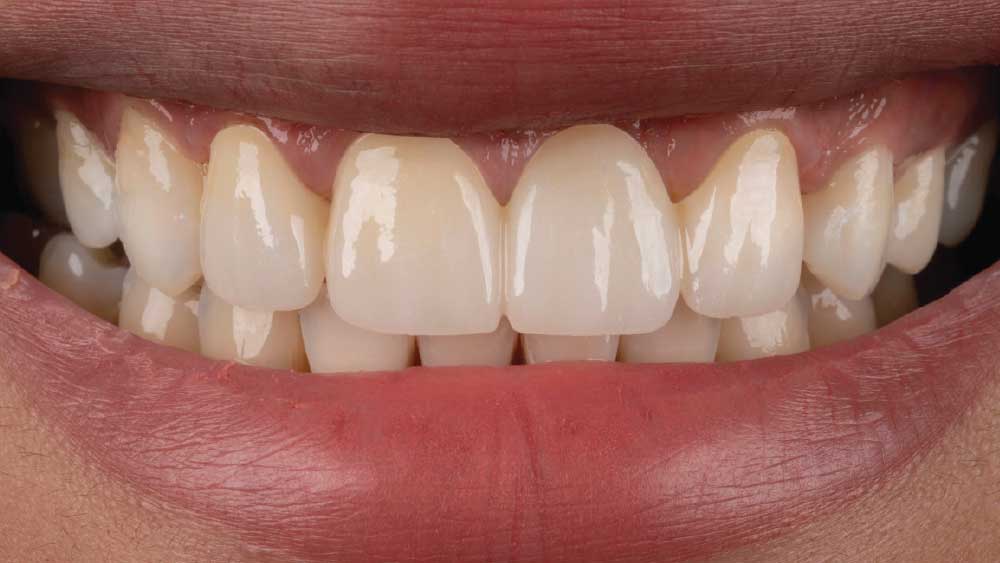
Figure 12: The patient received a BruxZir Esthetic bridge to replace extracted tooth #9 because she did not want an implant. The canine #11 was then shaped to appear as if it were #10 in the bridge span and the bicuspid #12 was cosmetically shaped to resemble the canine as it occupied that position. To harmonize her smile, I redistributed the space by making the centrals narrower. I finished her smile by adding composite to #12 to proportion the incisal embrasure to better resemble a canine.
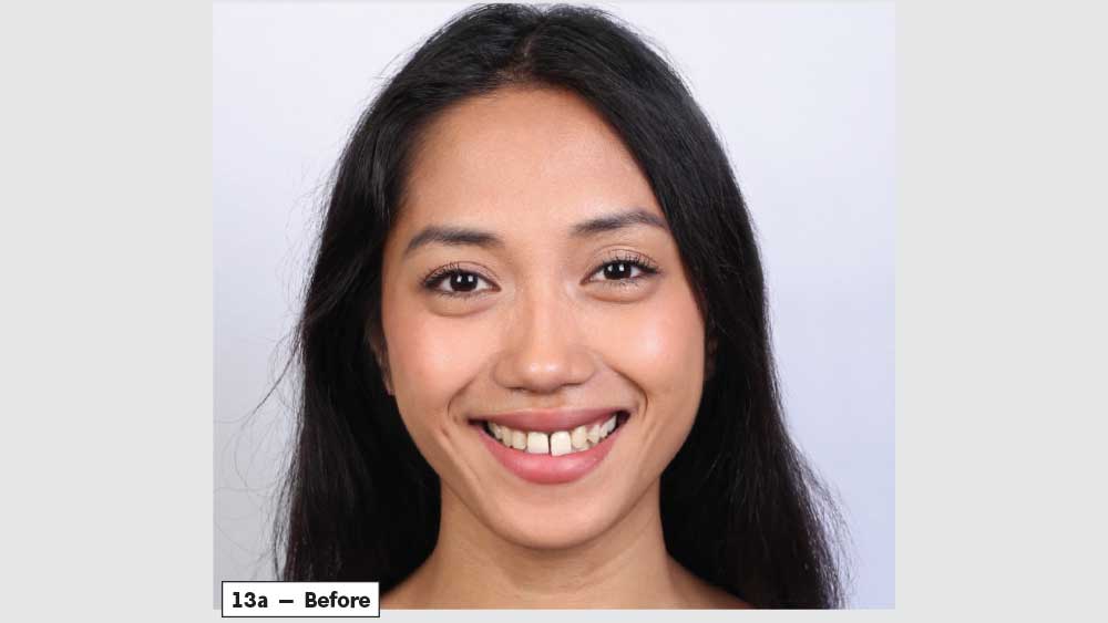
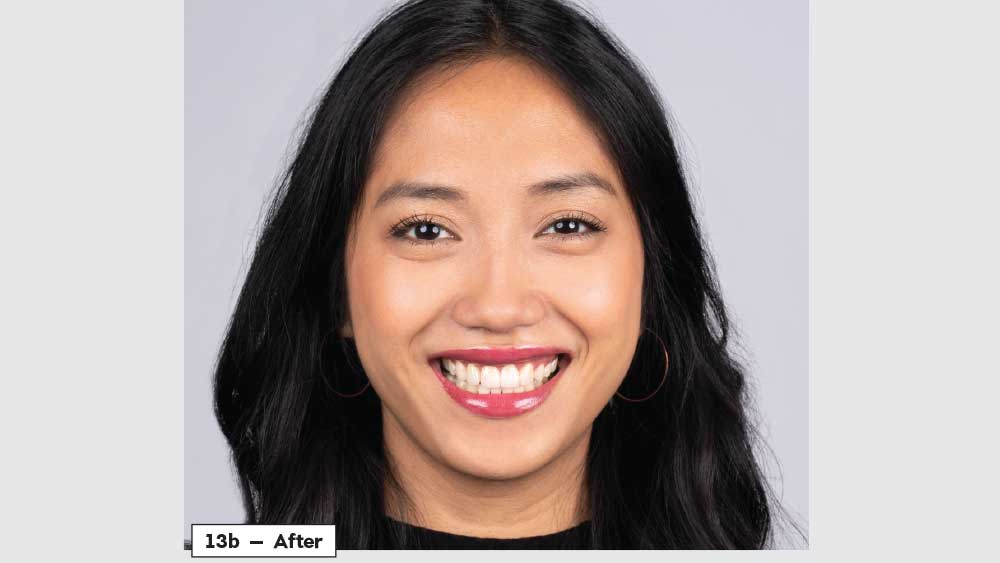
Figures 13a, 13b: Before-and-after images illustrate the dramatic transformation. The goal was to create a natural-looking smile. I finished her treatment by whitening her teeth to create a vibrant appearance.
- 1
- 2
- 3
- 4
- 5
- 6
- 7
- 8
- 9
- 10
- 11
- 12
- 13
- 14
- 15
CONCLUSION
It was a four-month restoration process but a worthwhile journey. The outcome radically transformed Maria’s life. She no longer struggles to eat certain foods, nor does she lack confidence in her appearance. Instead, she feels great about herself and is able to talk confidently in front of people. Her BruxZir Esthetic veneer and bridge have given her a new lease on life. Now she is no longer afraid to share her smile with the world.
IPS e.max is a registered trademark of Ivocalar Vivadent. iTero Element is a registered trademark of Align Technology, Inc.