‘The Mother of All Black Triangles’ Case
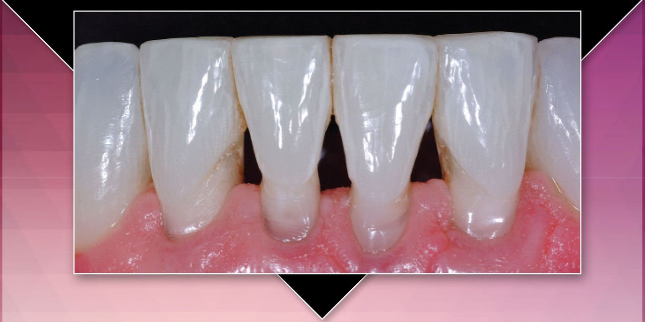
Sometimes a particular case comes along that appears, at first, to be overwhelming. The case described in this article fits that description. However, when this patient emailed my office and inquired about the possibility of flying across the country to have me treat him, I had fortunately done many cases involving hundreds of teeth using the matrix system that I developed to treat dentition afflicted with black triangles, albeit none of this magnitude. I felt absolutely confident that we could achieve a good outcome. The trick was to disassemble the case into bite-sized pieces.
This case presents many excellent questions and the additional challenge of severe facial abrasions. I will first review the background of black triangles and of lower incisor complications, and then proceed with the presentation of the clinical procedures used to treat this particular patient.
BLACK TRIANGLES: PREVALENCE AND PATIENT ATTITUDES
One third of adults have unesthetic black triangles, which are more appropriately referred to as "open gingival embrasures."1 Besides being unsightly and prematurely aging the smile, black triangles are prone to accumulate food debris and excessive plaque.2 A recent study of patient attitudes found patient dissatisfaction with black triangles to rank quite high among esthetic defects, ranking third following carious lesions and dark crown margins.3 If you go online and search "dental black triangles," you will be able to view hundreds of black triangle questions from patients, as well as patient complaints and lawsuits resulting from adult orthodontic cases and post-periodontal therapy papilla loss. This clinical and esthetic dilemma demands more attention from our profession. The caveat is that, until now, there has been no disciplined minimally invasive approach for treatment. Today, instead of improvising and struggling, I have developed a specific, predictable protocol to treat this problem.
LOWER INCISOR ESTHETICS
The esthetics of the lower teeth are often overlooked or simply ignored by many dentists. To this point, a fellow passenger seated next to me on a recent flight was intrigued by the photos that were on my laptop. He asked: "Why do dentists only seem to treat the upper teeth when the lower teeth look all jacked up? Do they think no one notices? It looks ridiculous to have perfect top teeth and ugly bottom teeth!" In addition, as we age, the lower incisors become more visible as the facial muscles lose their tension on the lower lip.
LOWER INCISOR CHALLENGES AND ETHICS
Lower incisors present their own unique restorative challenges. The incisal edge is broad and thin mesiodistally. The root, in contrast, is very broad buccolingually. Imagine a butter knife that has been permanently twisted at 90° in the middle of the blade. This anatomic curiosity creates demanding draw/path-of-insertion issues for a porcelain laminate or full-crown preparation. A lower incisor with significant recession leads to a mutilatory tooth preparation for porcelain. When I had an opportunity to show this case to the top ceramists in Toronto, Ontario, and Seattle, Washington, their answer was refreshingly candid: "Dr. Clark, to treat this case properly with porcelain laminates would require you to mutilate these teeth."
WHY DO SO MANY DENTISTS MISTRUST COMPOSITE TO TREAT BLACK TRIANGLES?
Like many clinicians, Michael’s (the patient in question) dentist in North Carolina hadn’t heard of Bioclear® Matrix (Bioclear Matrix Systems; Tacoma, Wash.), and was unfamiliar with injection molding of composites. Therefore, he was leery of treating Michael with "bonding." At that point, Michael decided to cross the country for a different solution because porcelain veneers and periodontal surgeries did not appeal to him as ideal treatments. After he saw my "Black Triangle" and "Restoratively Driven Papilla Regeneration" articles on the internet and videos on YouTube, he opted to fly to the West Coast for treatment.
After spending many hours working with manufacturers and tens of thousands of dentists, I compiled a list of the top five composite and porcelain fallacies that have steered dentists away from minimally invasive composite treatments for black triangles, or have doomed their previous attempts, leaving them gun-shy to try it again. Here’s the list:
- "Acid-etching cleans the tooth." False. Phosphoric acid barely touches plaque. Biofilm is so tenacious, and we forget that phosphoric acid removes the mineral, not the organic component of tooth surfaces. Biofilm is organic, not a mineral. This residual biofilm at the margins is likely the No. 1 reason why Class V and interproximal composites turn brown at the margins. No bonding agent can bond to biofilm, and most dentists are leaving biofilm on their hard-to-access margins.
- "A stronger dentin bonding agent is the answer." False. They (the manufacturers) keep selling us new and improved dentin bonding agents with higher and higher dentin bond strengths. The problem is twofold; first of all, in a case like this, most dentists are bonding to plaque, calculus and contaminated dentin, and no current resin bonds to biofilm. Secondly, with an approach using the Bioclear Matrix, uncut, blasted and rinse-etched (with phosphoric acid) enamel is leveraged to provide the bulk of the retention, and reliance on the dentin is lessened. We can trust enamel bonding. The key is in the design of the Bioclear Matrix and the ability to "wrap" the tooth with a seamless composite jacket.
- "A full crown is better." False. If you were the patient with otherwise healthy teeth, would you choose full crowns? Consider that a full crown destroys 70% of coronal tooth volume, with a 10% to 20% chance of eventual resultant pulpal death.
- "A porcelain veneer is better than bonding." In a case like this, false. First, porcelain veneers cannot reach far enough to the lingual — the space is blocked from view, but becomes a plaque trap on the lingual. Secondly, bonding a veneer to this much cervical dentin should make you nervous. Very nervous.
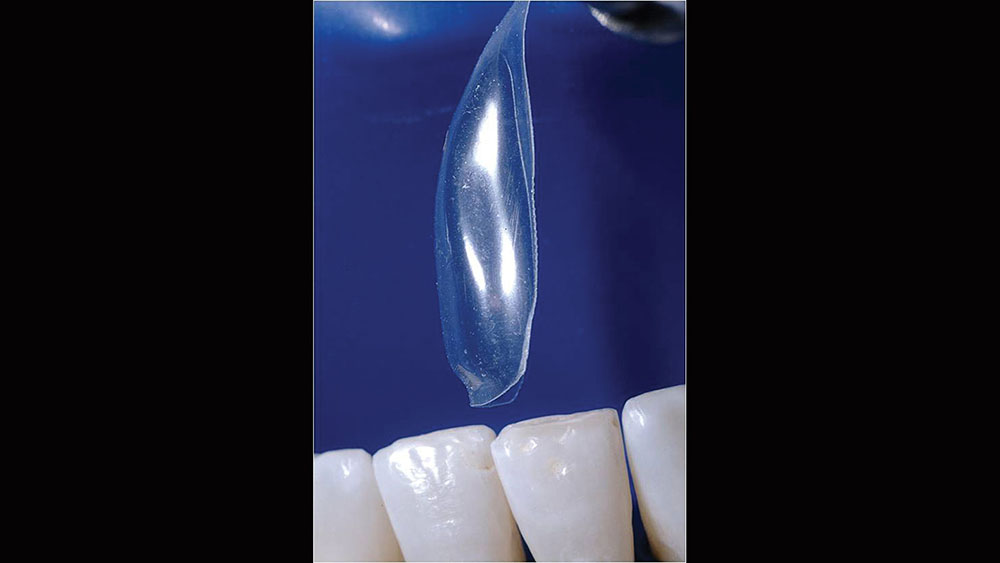
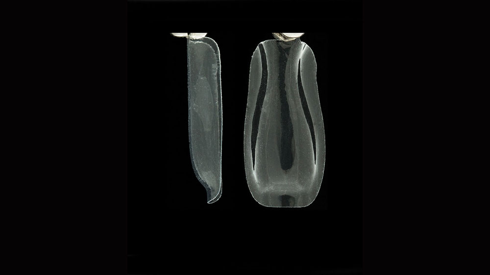
- "Direct bonding is too difficult." In the past this may have been true. But today, false. In the modern resin era, we utilize anatomic Bioclear matrices coupled with an injection molding filling technique with, for example, a universal nanocomposite, thus creating an ideal flowable/paste interlace.
CASE WORKUP
First, I consulted two renowned microscope-equipped periodontists. Normally, I would have immediately excluded the surgical option based on this patient’s situation; but in this case, because of the severity of the attrition of the embrasures, I felt that second and third opinions were warranted. In addition, if a follow-up surgical approach were needed, the periodontist would already be on board.
Noted periodontist Dr. Peter Nordland summarized this patient: "Dear David, the papillae height across the lower anterior teeth is located at the same level as all of the other adjacent papillae. This means that the individual papillae are not deficient, but instead, the patient has suffered incisal edge wear and extrusion of the incisors. Although root coverage could be very predictable, I would recommend a restorative solution as you have so beautifully shown in the Bioclear video. My experience is that surgical papilla reconstruction is most predictable in situations where the papilla has been surgically abused previously."
CASE PRESENTATION
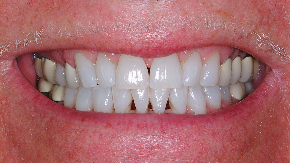
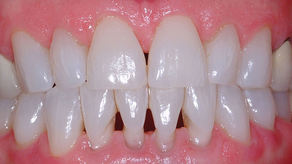
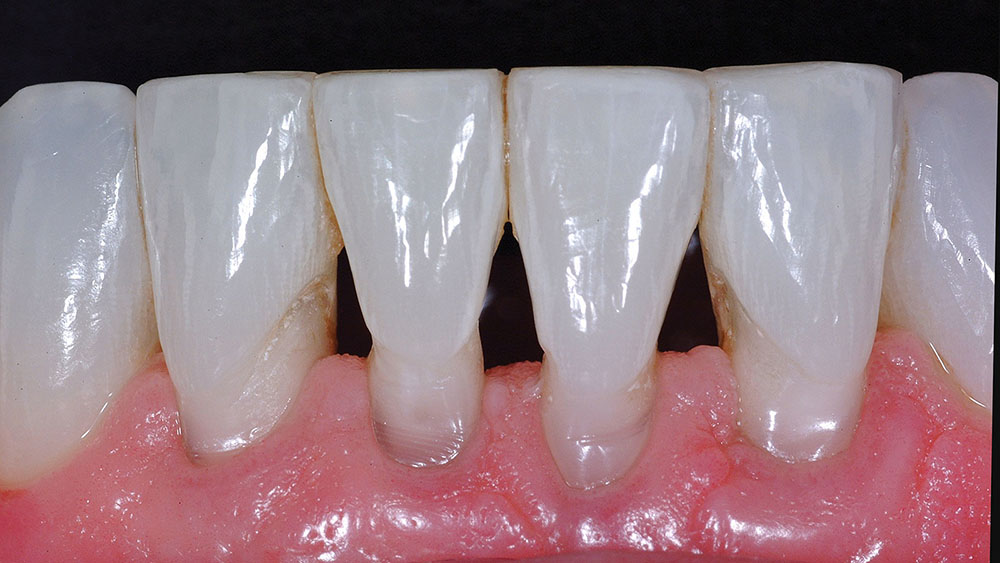
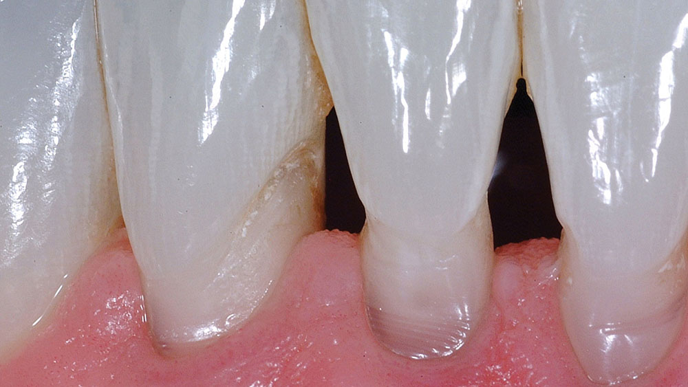
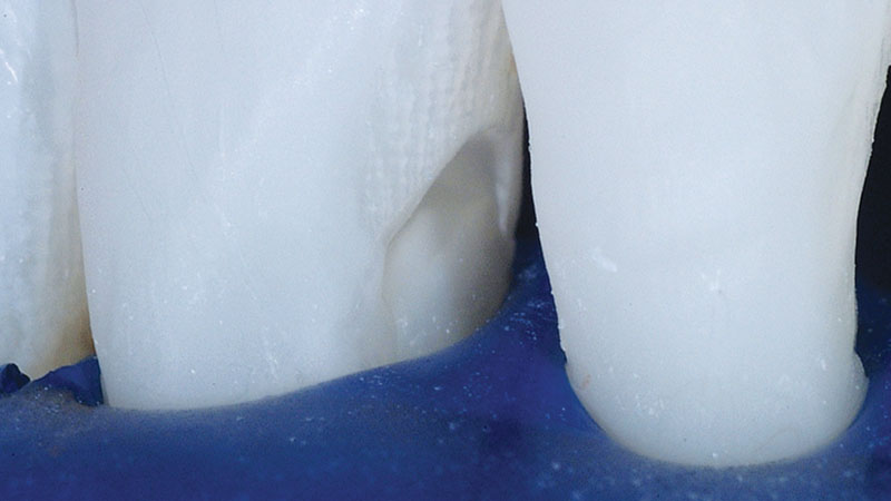
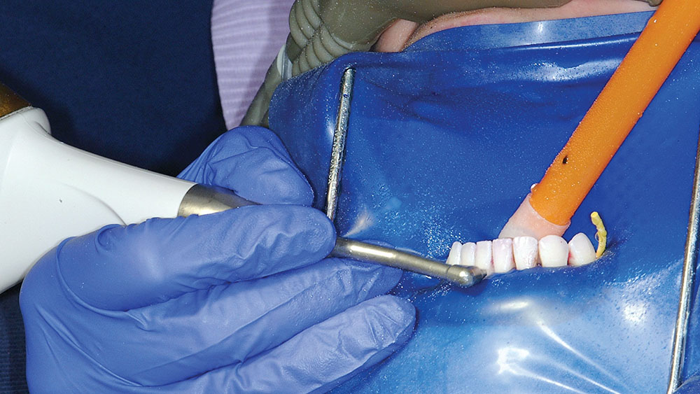
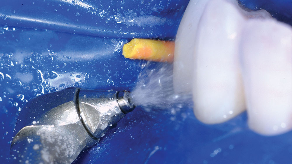
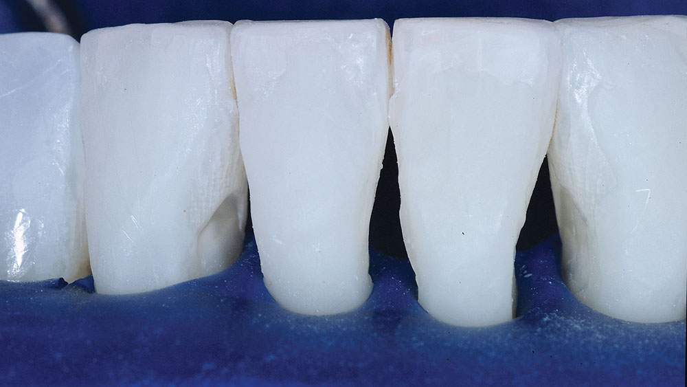
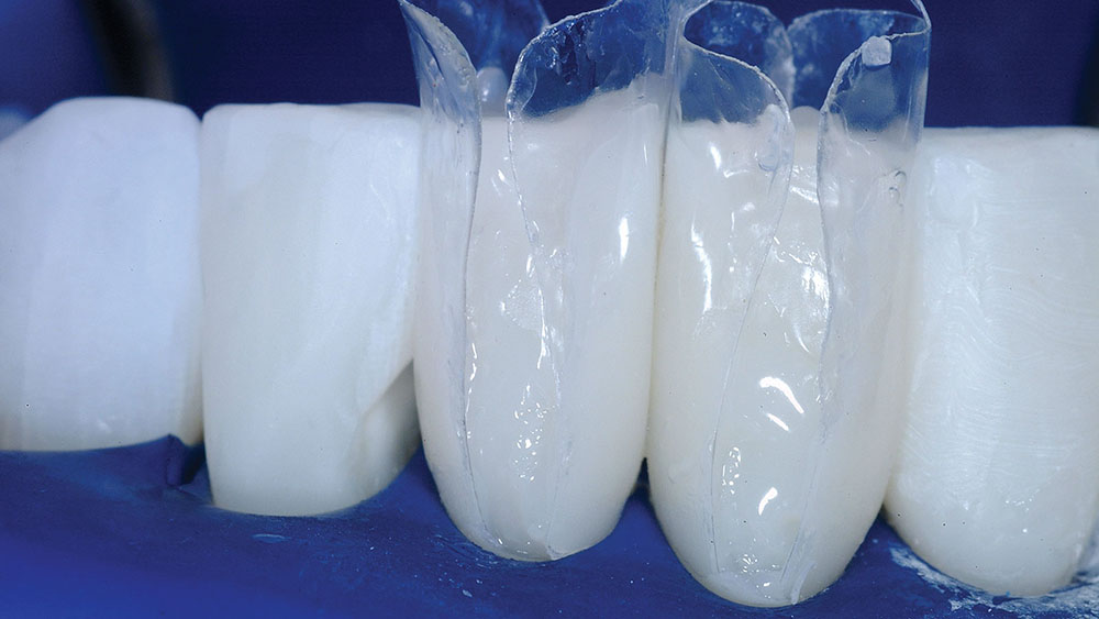
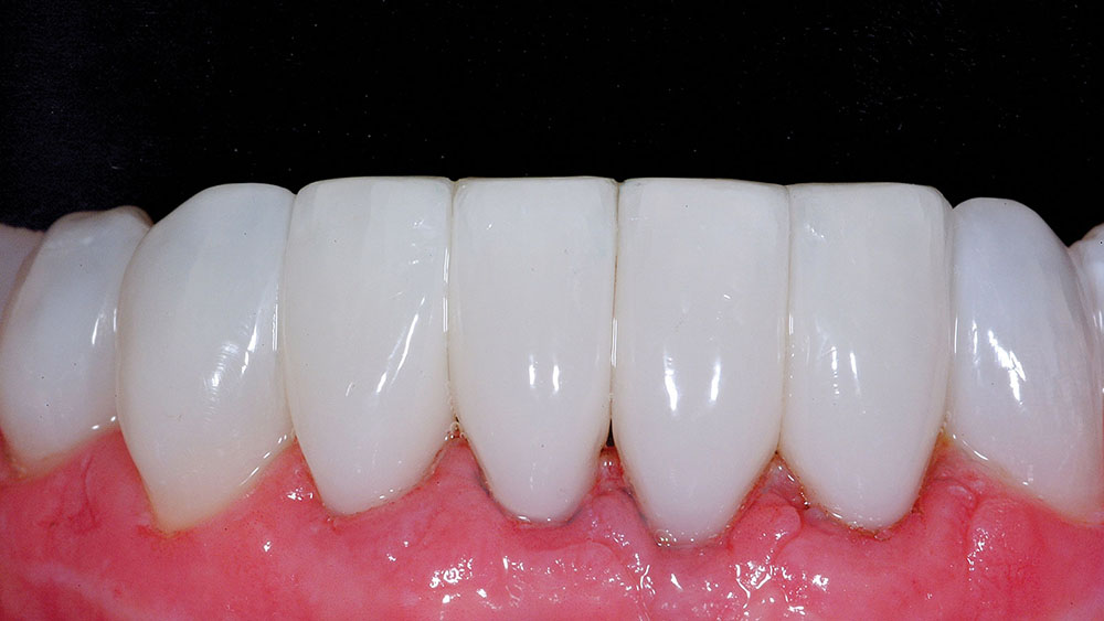
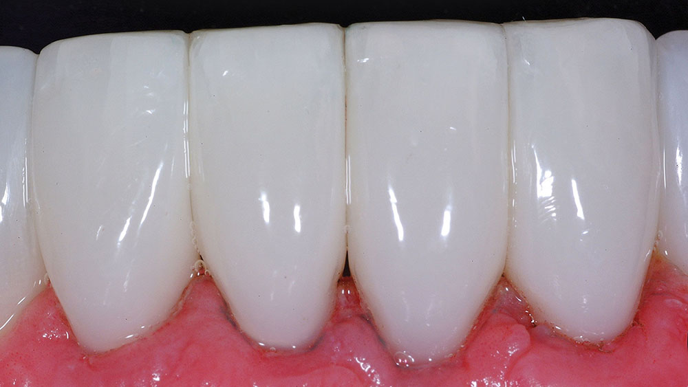
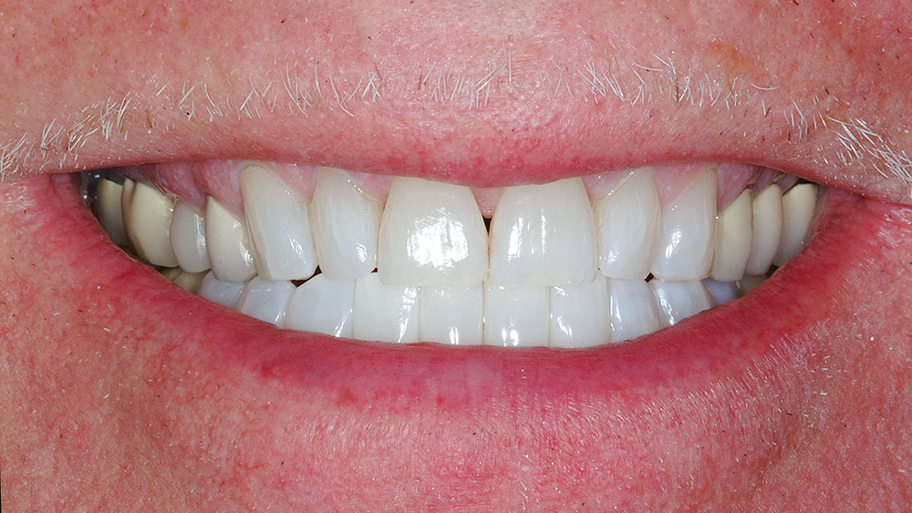
Figure 1 shows the functional and esthetic dilemma. The retracted view (Fig. 2) shows the magnitude of the black triangles on the lower. The patient’s first priority was treating the lowers, and he would return to the West Coast in a few months to treat the upper black triangles. Facial abrasions and recession tripled the complexity of treatment (Fig. 3). Blasting, which is application of a mild abrasive with an air-water mix, is an absolute necessity for this treatment (Figs. 4–7). Once the facial abrasions are restored up to the line-angle areas, a rubber dam is placed.
Blasting, which is application of a mild abrasive with an air-water mix, is an absolute necessity for this treatment.
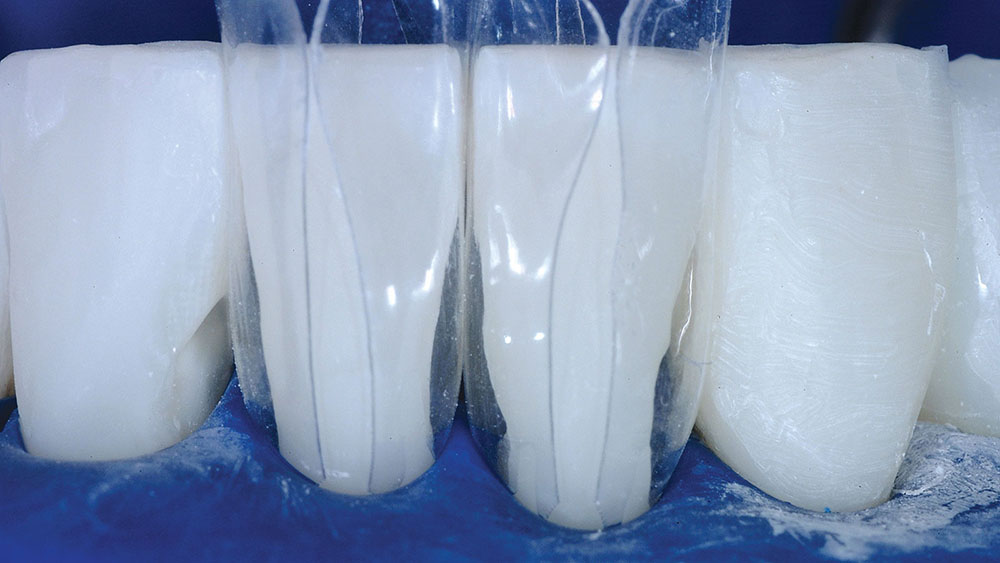
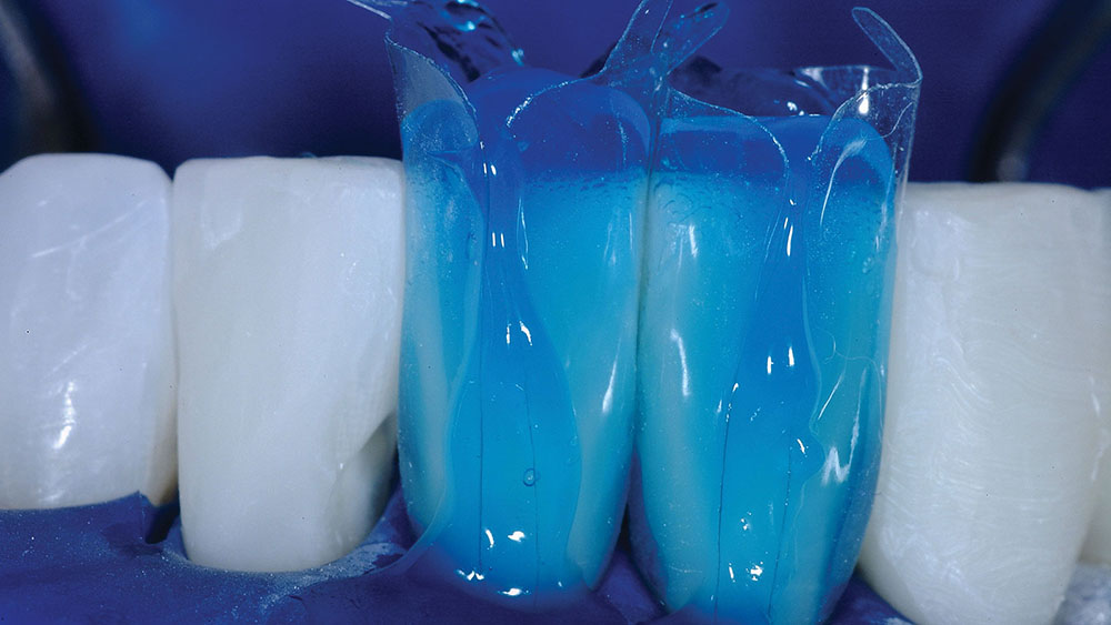
The interproximal areas are nicely managed with the rubber dam and the DC-203 Bioclear matrices together (Figs. 8–15). It is not recommended to try to treat the facial abrasions at the same time that the matrices are in position. The Bioclear method is almost the inverse of the old flat-matrix technique. The facial surfaces are left with some excess because this is the loading area. The interproximals, when molded, will require little or no finishing. Immediate postoperative views demonstrate the dramatic emergence profiles, mirror finish and regenerated papillae (Figs. 16–18). Dentists and periodontists often ask these patients: "Are these veneers? Are these crowns?" No. This is done with an injection molding technique performed with high-level magnification, injecting a universal nanocomposite (in this case, Filtek™ Supreme Ultra [3M™ ESPE™; St. Paul, Minn.]) (flowable and paste) into the Bioclear matrix, and polishing all with Jazz Polishers (SS White® Burs; Lakewood, N.J.).
Having a mirror-smooth composite finish makes everyone happy: the patient, the soft tissue and especially you, the clinician.
THE MIRROR FINISH: TAKING THE CASE FROM GOOD TO GREAT
Having a mirror-smooth composite finish makes everyone happy: the patient, the soft tissue and especially you, the clinician. The matte or grainy finishes of the past collect lipstick, biofilm, stain and feel like cheap dentistry to the patient’s tongue. In our traditional mindset, only porcelain stayed smooth. Those days need to end now; composite has come of age. The first step is to use a microfill that holds its shine. I am nearly always disappointed at how miserable the composite finishing systems are that I am asked to evaluate, and how disappointing many of the composite finishes that are presented in dental journals and magazines can be. The folks at Kerr, 3M ESPE and SS White have commented that they have never seen polishes like the ones I show in my lecture. That’s probably because most doctors adopt a manufacturer’s "system," and frankly, those systems are mediocre at best and grossly overcomplicated. To learn about my unique mirror polish, visit the dentistrytoday.com video library to view Dr. David Clark’s 3-Step Perfect Composite Polish Technique.
TABLE
CASE WORKUP:
- Appropriate treatment plan with appropriate fees
- Treat and price facial abrasions independently
- Preoperative whitening
- Pro-Banthine administered at beginning of appointment
CLINICAL PROCEDURE:
- Anesthetize, then pack Ultrapak® #00 cord (Ultradent Products Inc.; South Jordan, Utah) cord soaked in hemodent on facial and interproximal areas of teeth with facial abrasions (#23–26)
- Blast with Bioclear Prophy Plus, scale away stubborn calculus, then reblast with aluminum trihydroxide powder
- Apply disclosing solution
- Continue blasting until all biofilm is gone and surface dentin has been stripped away
- Acid-etch the entire tooth with 37% phosphoric acid etchant
- Restore facial surfaces with flowable and paste with the "Clark Class V profile … big, fat and full," then stop at the line angles
- Place rubber dam, quickly grind back gross excess areas
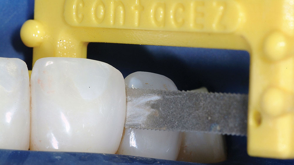
- Lighten and clean contact areas with red or yellow ContacEZ to allow the somewhat delicate Bioclear Matrix to slide between the teeth
- Reblast
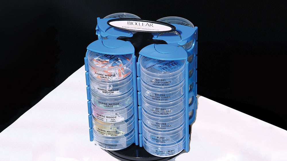
- Place Bioclear Matrices (DC-203 for larger spaces near incisors, A-103 for smaller spaces near incisors, and A-102 for canines and bicuspids near smaller spaces), re-acid-etch entire tooth. Seal large areas of dentin with bonding agent, then light-cure.
- Injection mold with bonding resin, then Filtek Supreme Ultra Flowable chased with Filtek Paste all in sequence without light-curing until the end
- Gross finish with carbide burs, flame diamonds, and a coarse Soflex Disc (3M ESPE)
- The Clark 30-second, 3-step polish: (1) Marginate with brownie, (2) Matte finish with coarse pumice and cup, (3) High shine with Jazz Polisher (SS White Burs)
SUMMARY
Before the emergence of Bioclear Matrix and a disciplined approach to composite treatment of black triangles, many treatments ended with significant compromise in periodontal health. Many cases debonded soon after placement; others suffered problems with stain. Nonetheless, our patients are hopeful for a better solution. The interdental papilla serves as both a functional and esthetic asset. Anatomically ideal interproximal composite shapes that are mirror smooth can serve as a predictable scaffold to regain this valuable gingival architecture. Clean enamel surfaces can be leveraged to permanently retain the restorations. However, the reader is cautioned that if one is to attempt this elective procedure using no magnification, without a strict adherence to dentin detoxification with a blasting appliance, and using a flat matrix, then nontreatment or referral is recommended. Our profession can change its thought processes, retrain its hands and expand its armamentarium to perform techniques that were previously impossible.
Dr. Clark founded the Academy of Microscope Enhanced Dentistry, and he is a course director at the Newport Coast Oral Facial Institute in Newport Beach, California. He is co-director of Precision Aesthetics Northwest in Tacoma, Washington, and an associate member of the American Association of Endodontists. He is a 1986 graduate of the University of Washington School of Dentistry. Dr. Clark is proud to lecture and give hands-on seminars internationally on a variety of topics, as well as serve on the board of CR (formerly CRA). He also developed the Bioclear Matrix System, which allows for biomimetic restoration of teeth using single-phase injection molding and minimally invasive preparation styles. He can be reached at drclark@microscopedentistry.com and bioclearmatrix.com.
Disclosure: Dr. Clark has a financial interest in the Bioclear Matrix System.
Reprinted by permission of Dentistry Today, ©2012 Dentistry Today.
References
- ^Kurth JR, Kokich VG. Open gingival embrasures after orthodontic treatment in adults: prevalence and etiology. Am J Orthod Dentofacial Orthop. 2001 Aug;120(2):116-23.
- ^Ko-Kimura N, Kimura-Hayashi M, Yamaguchi M, et al. Some factors associated with open gingival embrasures following orthodontic treatment. Aust Orthod J. 2003 Apr;19(1):19-24.
- ^Cunliffe J, Pretty I. Patients’ ranking of interdental "black triangles" against other common aesthetic problems. Eur J Prosthodont Restor Dent. 2009 Dec;17(4):177-81.

