Single-Unit Implant Restoration Milled with BruxZir® NOW SRC
October 6, 2025
 image
Justin Chi, DDS, CDT
Single-Unit Implant Restoration Milled with BruxZir NOW SRC

In-office milling is a quickly evolving option for the modern dental practice. With the introduction of a same-day screw-retained option, BruxZir® NOW SRC milling blocks, there’s even more opportunity for optimizing single-unit restoration workflows. In this case study, I’ll demonstrate how this product can be used as part of an efficient workflow for tooth replacement in the posterior. When combined with guided surgery and in-office crown design, this procedure offers reliable results while saving money and significantly cutting down on chair time for dentists.

Case Report

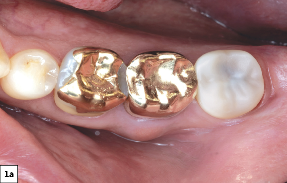
Pain in tooth #18 (Top View)
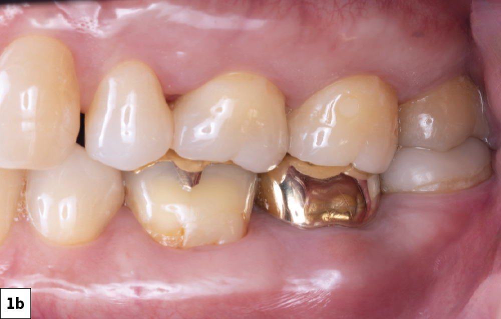
Pain in tooth #18 (Side View)

Figures 1a, 1b: The patient presented with complaints of pain in tooth #18. He previously had endodontic treatment done and presented with swelling of the facial gingiva, leading me to believe infection was present.

Radiograph of tooth

Figure 2: A radiograph of the tooth confirmed the failing root canal. Because of the significant radiolucency around the furcation and mesial root, we decided the best option was extraction and grafting to establish a good foundation for future implant treatment.

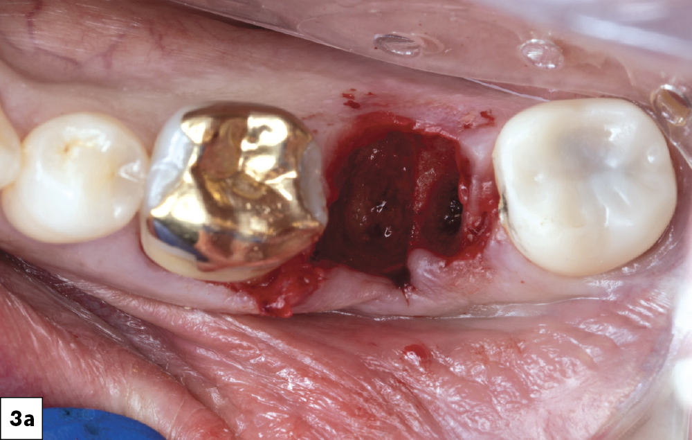
Tooth #18 extracted
Cleaning #18 socket
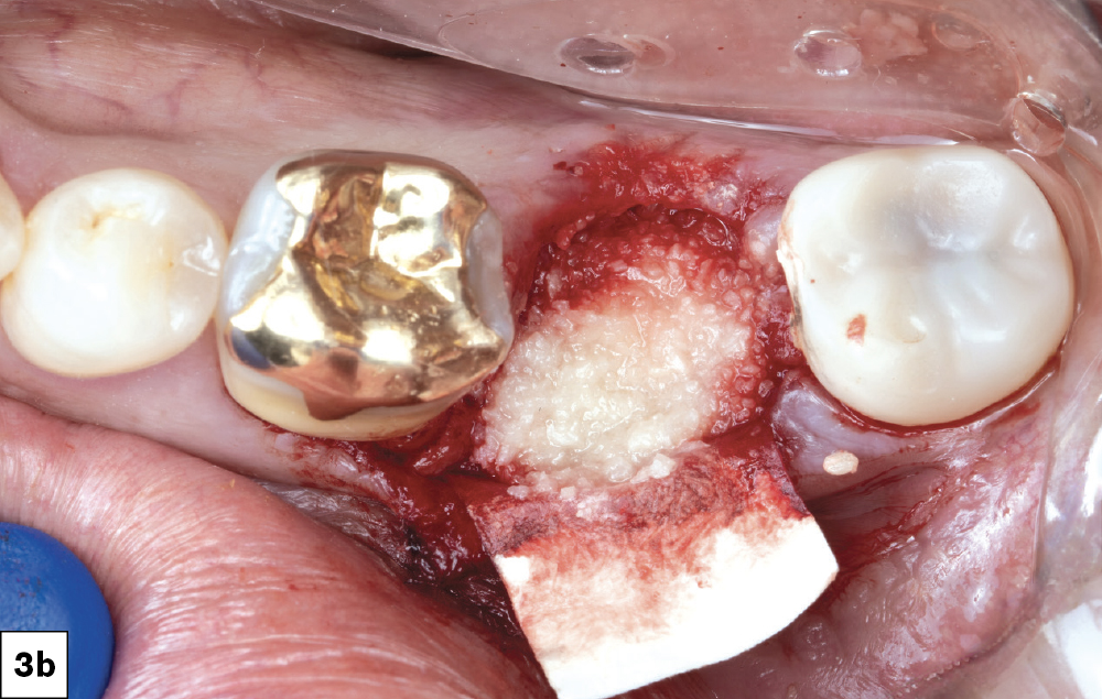
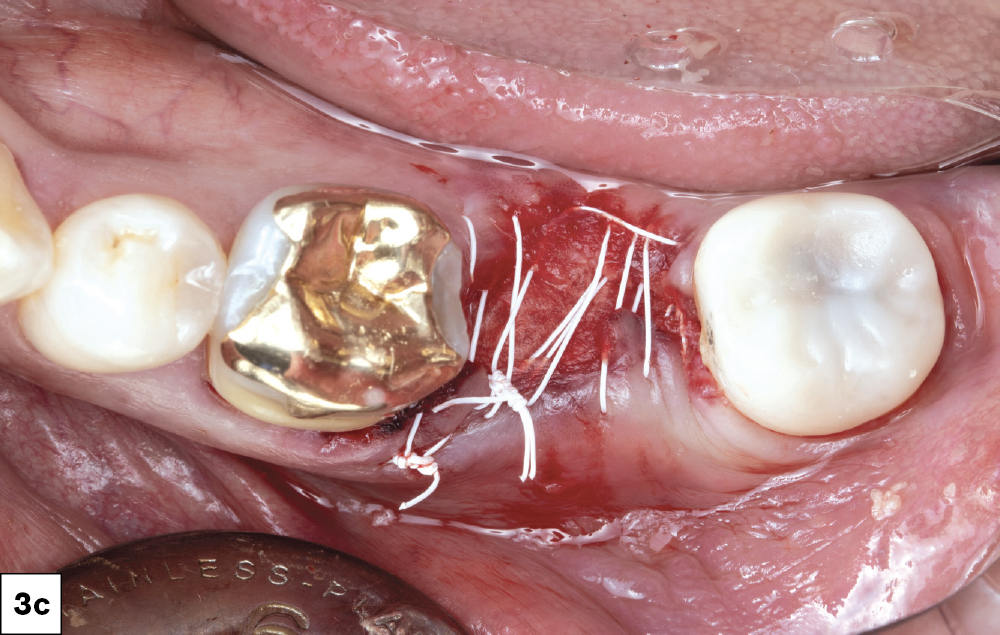
Stitched #18 socket

Figures 3a–3c: Tooth #18 was atraumatically extracted, and all infected tissue was removed using a curette to thoroughly scrape and clean the socket. Newport Surgical™ Mineralized Cortico/Cancellous Allograft Blend was applied to the site with a Newport Surgical Resorbable Collagen Membrane 4-6 added to prevent soft tissue ingrowth (Glidewell Direct; Irvine, Calif.). Biotex™ PTFE Sutures (Glidewell Direct) were used to close the site for a four-month healing process.

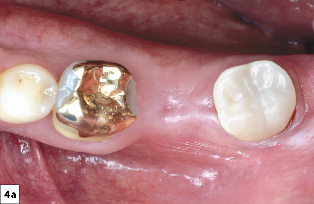
4 months after tooth extraction (top view)
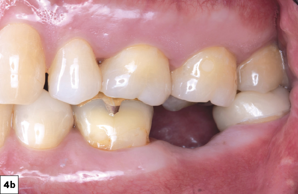
4 months after tooth extraction (side view)

Figures 4a, 4b: The site was evaluated after four months. At least 2 mm of attached gingiva was present on the facial aspect of the future implant site, meeting the minimum requirement for implant placement.

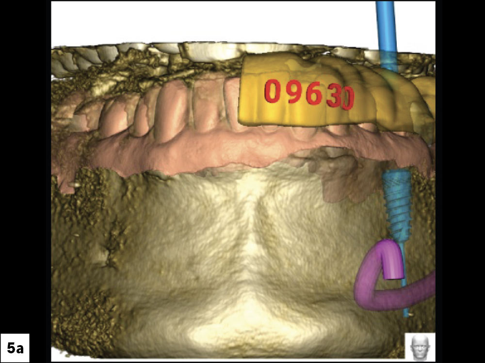
CBCT Scan (Front View)
CBCT Scan (Top View)
CBCT Scan (Left View)
CBCT Scan (Right View)

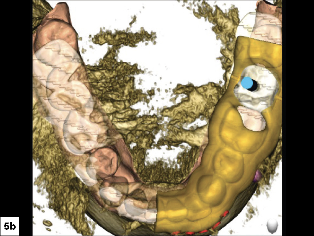
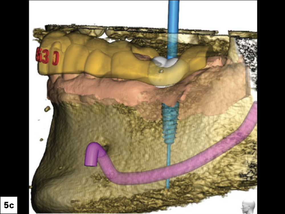
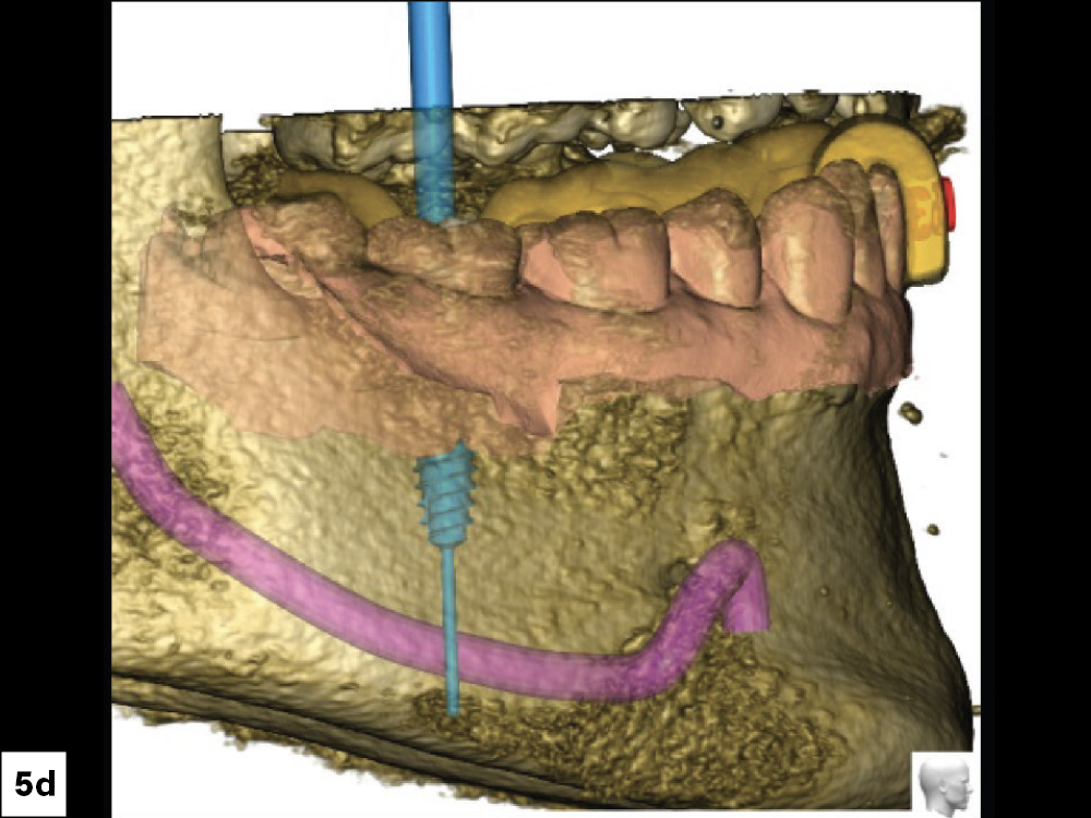
Figures 5a–5d: Treatment planning was done by Glidewell’s Digital Treatment Planning (DTP) team, which used CBCT scans to determine the ideal position of the implant.

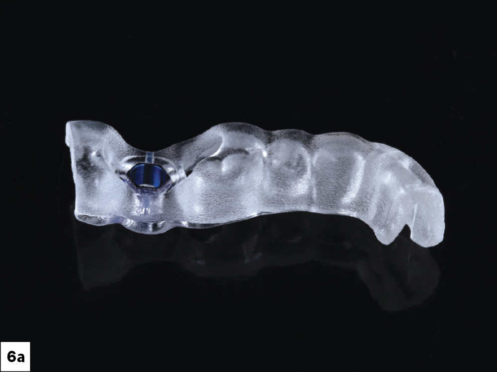
Tooth-supported surgical guide (Side View)
Tooth-supported surgical guide (Top View)

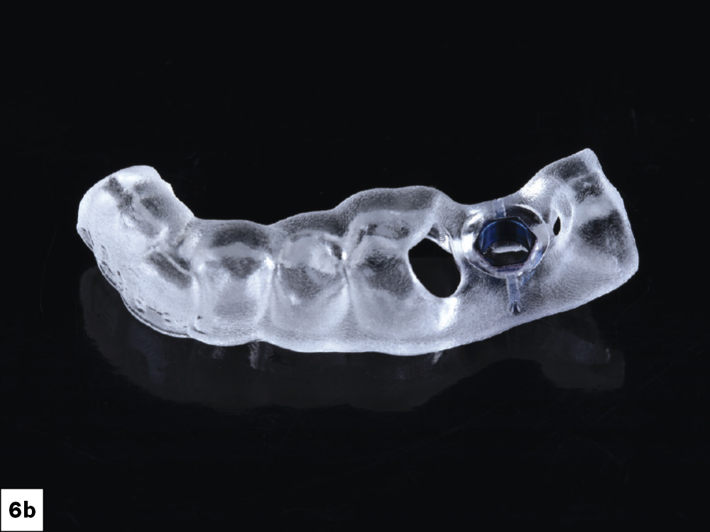
Figures 6a, 6b: A tooth-supported surgical guide was fabricated to transfer the 3D surgical plan to the surgical site.

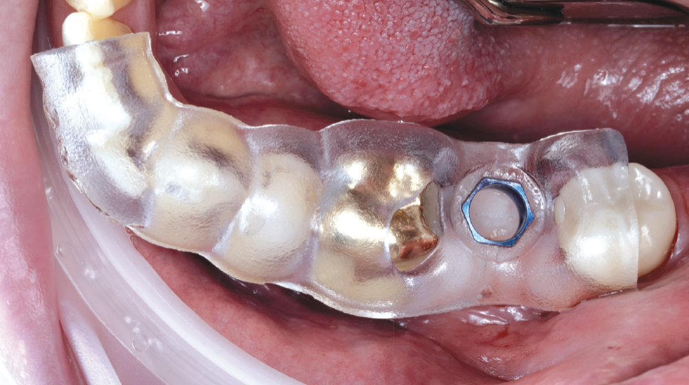
Tooth-supported surgical guide in mouth

Figure 7: Prior to surgery, the tooth-supported guide was positioned to evaluate for proper fit and contact with the supporting teeth.

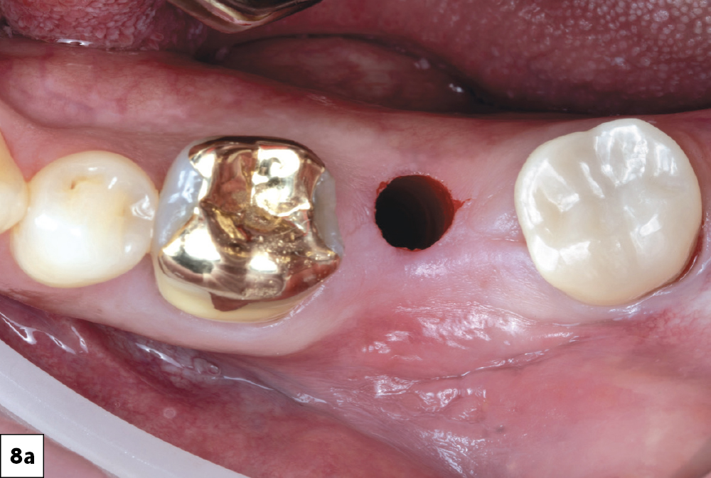
Tissue punch in socket #18
Guided drilling protocol
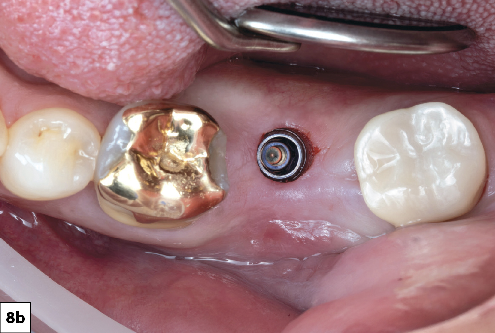
Glidewell HT Implant torqued into place

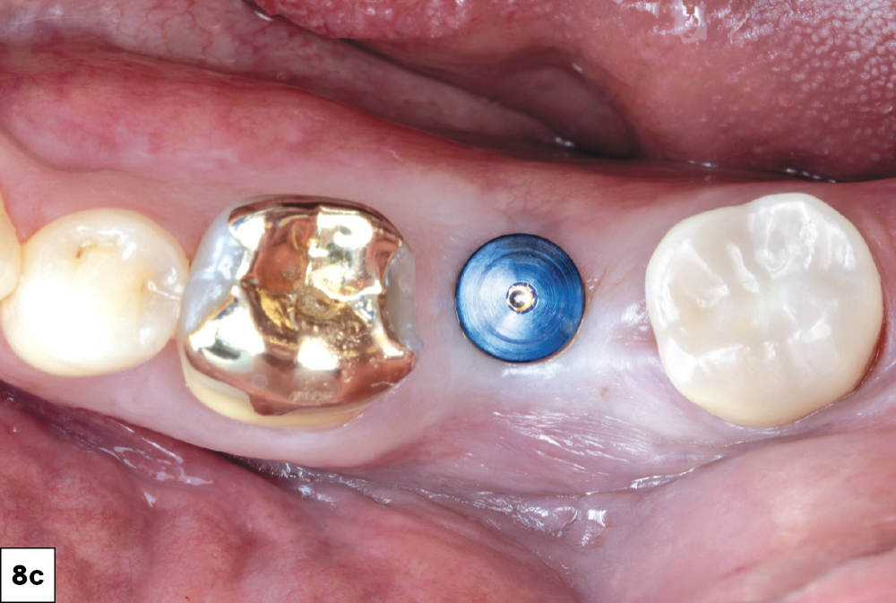
Figures 8a–8c: Due to the sufficient amount of attached gingiva and the precise positioning of the guide, a minimally invasive tissue punch was made to expose the site for the guided drilling protocol. A 5 mm Glidewell HT™ Implant was torqued into place at 45 Ncm and a Glidewell HT Implant Healing Abutment was placed for four months of healing (Glidewell Direct).

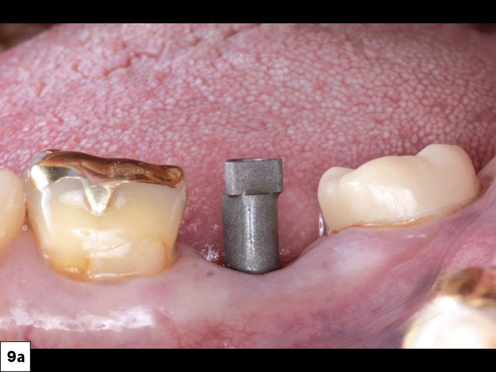
Inclusive Titanium Scan Body
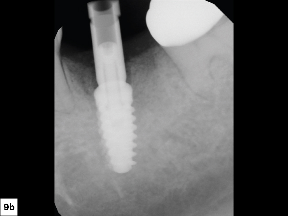
Inclusive® Titanium Scan Body in radiograph

Figures 9a, 9b: An Inclusive® Titanium Scan Body (Glidewell Direct) was placed, and proper seating was confirmed with a radiograph.

fastscan.io implant scan (top view)
fastscan.io implant scan (side view)

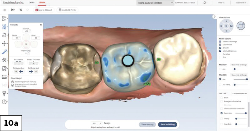
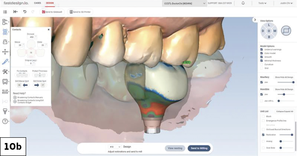
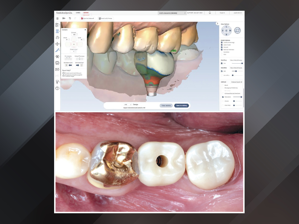
Figures 10a, 10b: The fastdesign.io™ Software and Design Station uses sophisticated AI to simplify and streamline same-visit restorations within glidewell.io™ In-Office Solution workflows. This software was used to generate proper contacts and contours of the proposed final restoration design, taking into account how the soft tissue would adapt from the implant to the coronal portion of the screw-retained crown.

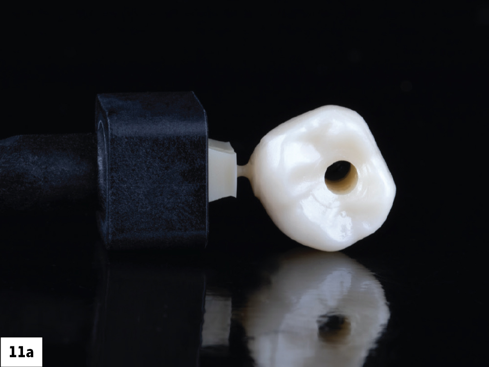
BruxZir screw-retained crown (top view)
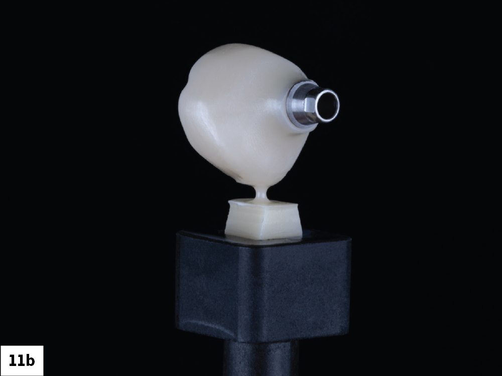
BruxZir screw-retained crown (side view)

Figures 11a, 11b: The final BruxZir screw-retained crown was milled from a block of fully sintered BruxZir NOW SRC material via the fastmill.io™ In-Office Mill. Milling time was only 35 minutes with no need for additional oven time or staining. A BruxZir™ Adjustment & Polishing Kit (Glidewell Direct) was used to achieve a high shine on the restoration.

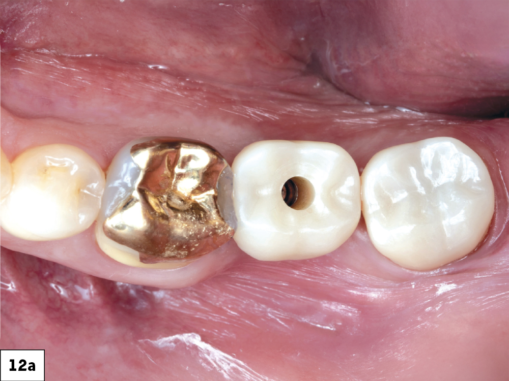
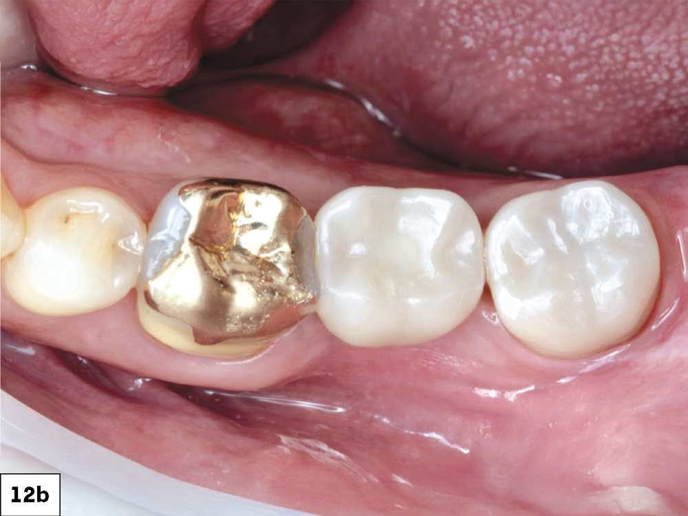
BruxZir NOW SRC in mouth with open access channel
BruxZir NOW SRC in mouth with closed access channel
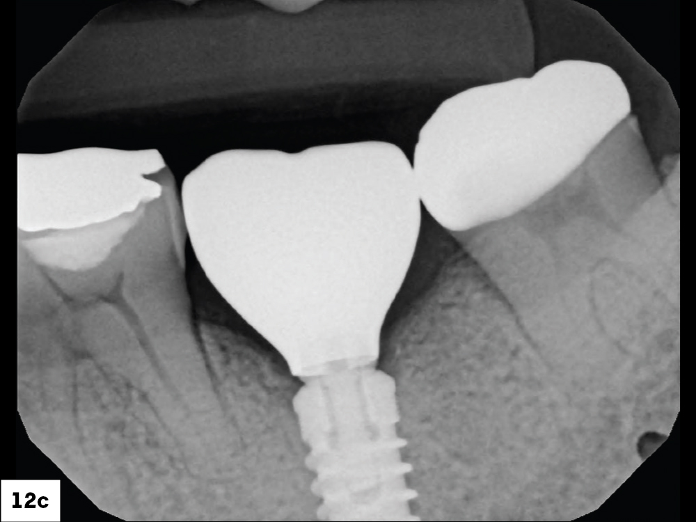
BruxZir NOW SRC in mouth radiograph

Figures 12a–12c: The screw-retained crown was seated and the access channel sealed with Teflon tape and composite. The final restoration showed ideal contacts, occlusion and emergence profile.

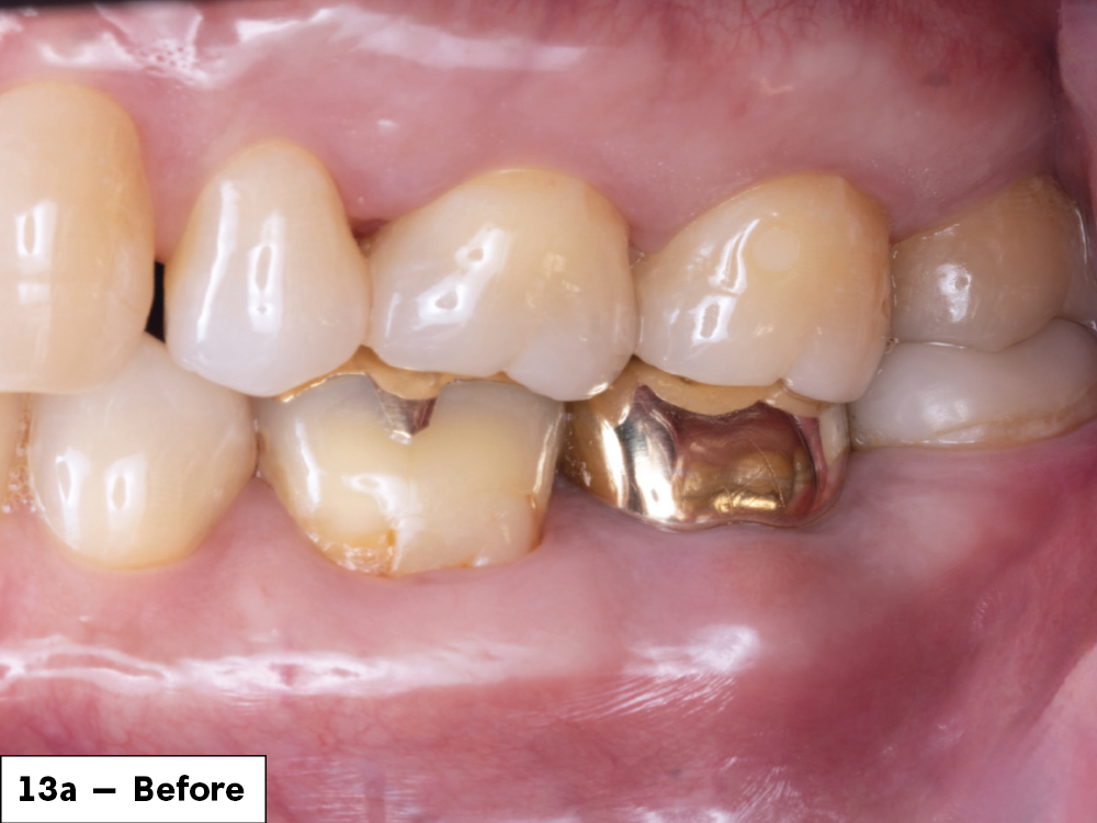
Before tooth surgery
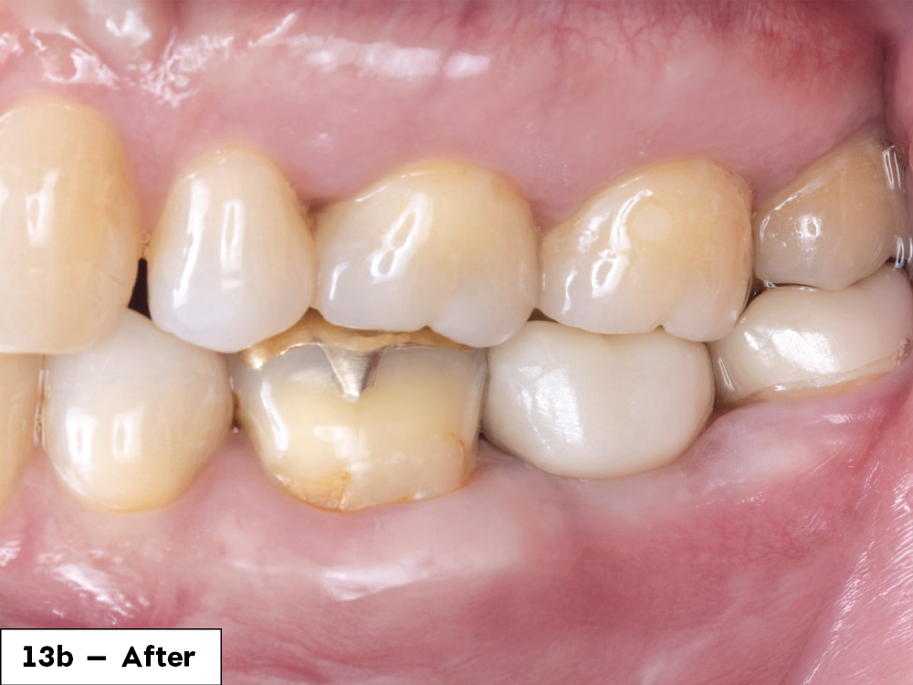
1 year after tooth surgery

Figures 13a, 13b: One year after removing the tooth and infection, the final screw-retained crown demonstrated great stability and esthetics. The patient was happy with the restored functionality of his tooth.

Conclusion

Increased sophistication of in-office crown design software gives dentists more control over restorations they mill in their own practice. More streamlined workflows in surgical and restorative phases means more predictability, repeatability, and long-term success for these essential procedures. By reducing the likelihood of surgical errors with guided implant surgery and cutting down on in-lab working times with in-office milling, clinicians can provide great services while continuing to increase revenue.