Clinical Benefits of Digitally Guided Implant Surgery
Note: The Hahn Tapered Implant System is now known as the Glidewell HT Implant System
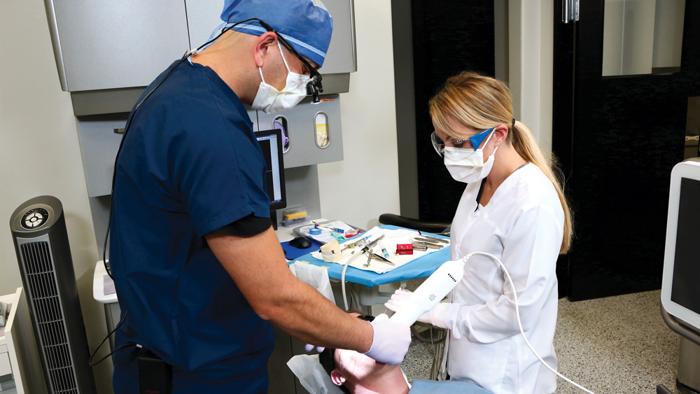
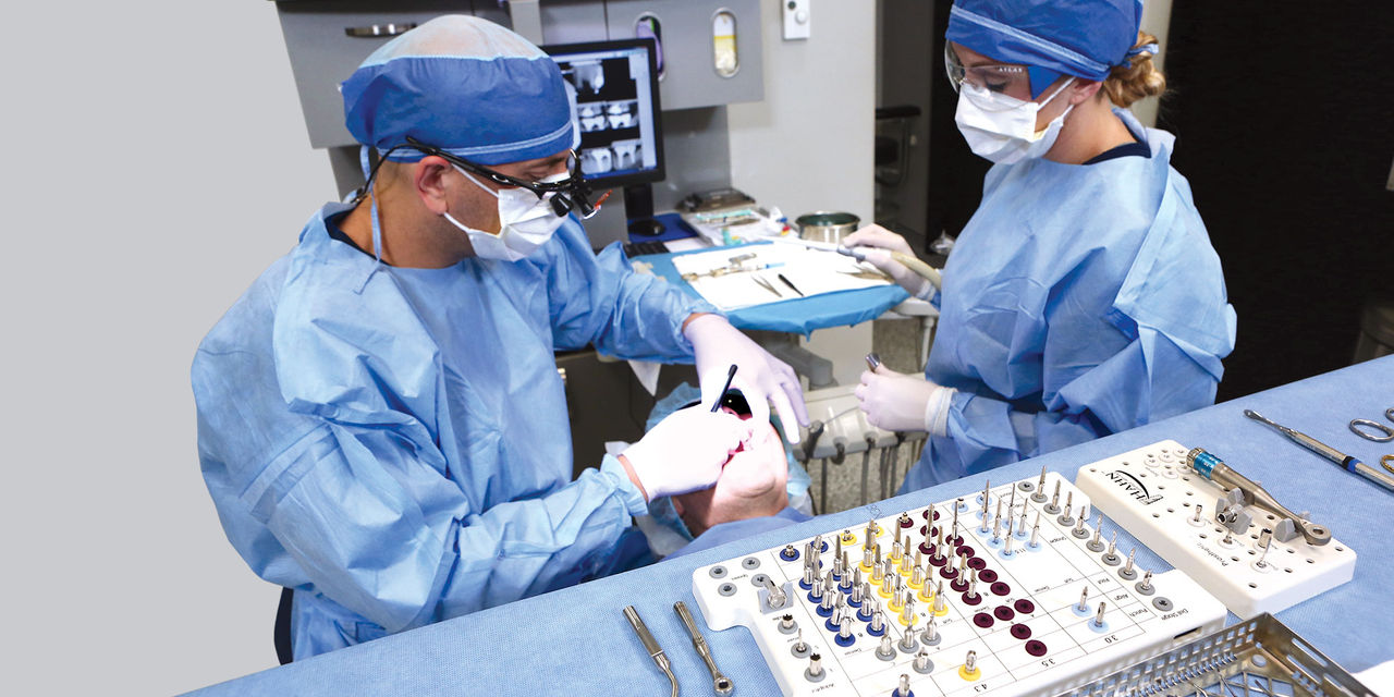
The benefits of CBCT scanning, digital treatment planning and digitally guided implant surgery are well-established. After determining the ideal implant position based on a 3D scan and the restorative goals, a surgical guide is fabricated to precisely transfer the digital treatment plan to the patient’s mouth. The surgical procedure is completed with great efficiency, reducing the chair time required to place an implant. Due to the clinical advantages of this approach, digitally guided surgery is utilized both by experienced practitioners seeking to optimize their results and by newcomers to implant placement, who benefit from increased safety and confidence.
Advantages of Guided Surgery
- Faster implant surgery reduces morbidity and fress up chair time
- Less invasive surgical procedure
- Faster healing time
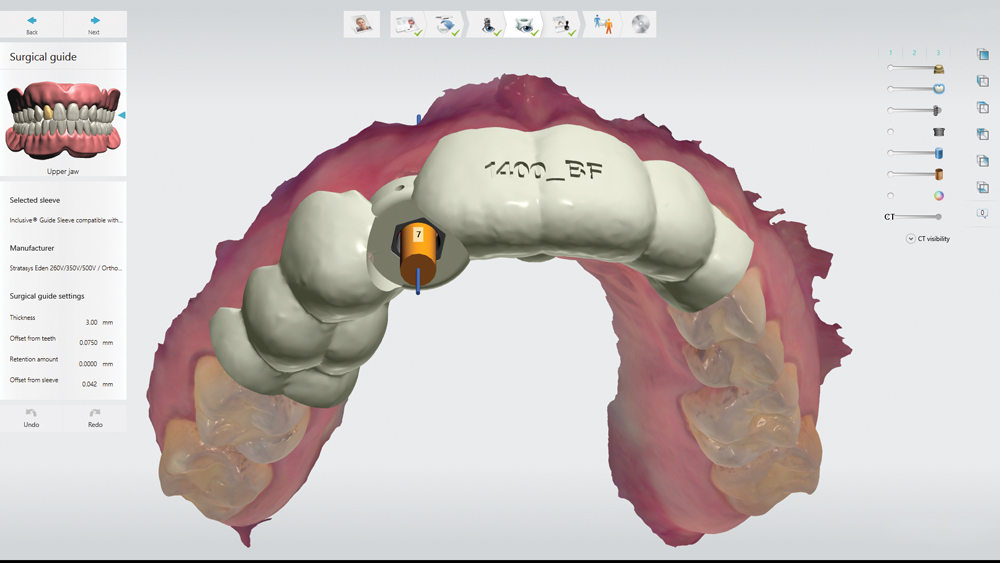
- Improved precision and predictability
- Can help avoid nearby patient anatomy and accomodate limited bone volume
- Option of immediate loading with prefabricated provisional
THE HAHN™ TAPERED IMPLANT GUIDED SURGERY SYSTEM
With the recent release of the Hahn™ Guided Surgery System (Glidewell Direct; Irvine, Calif.), clinicians can now place Hahn Tapered Implants at a digitally predetermined position, affording a high degree of precision and confidence. The Hahn Guided Surgery System includes a streamlined surgical protocol that enables clinicians at all experience levels to efficiently deliver Hahn Tapered Implants, which excel at achieving high primary stability in a wide variety of clinical situations.
Clinicians can now place Hahn Tapered Implants at a digitally predetermined position, affording a high degree of precision and confidence.
Advantages of Hahn Guided Surgery System
- Streamlined surgical protocol
- Drills are diameter-specific and include depth stops
- All-in-one surgical kit suitable for virtually any case
- Entire procedure is performed through a surgical guide
- Easy-to-follow drilling sequence
- Affordable treatment costs
- Minimal instrumentation required
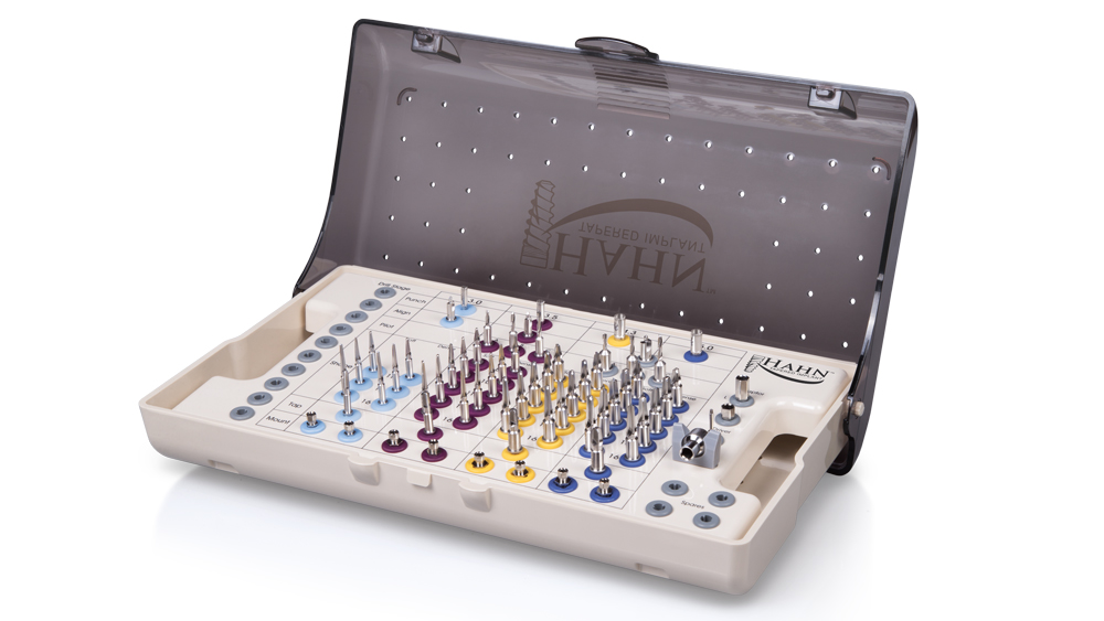
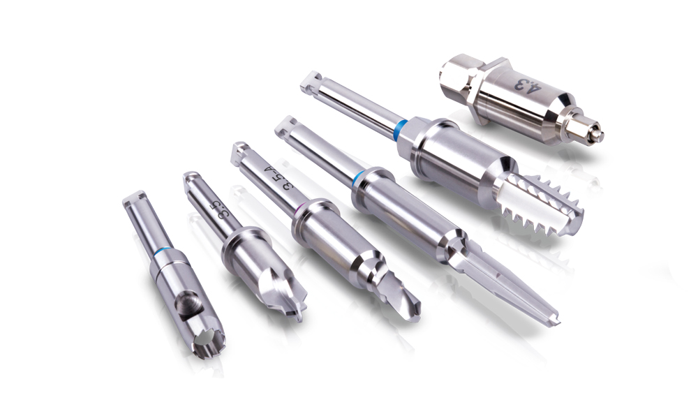
The Hahn Guided Surgical Kit (Glidewell Direct) has been designed to eliminate the need for drill keys, inserts or “spoons” commonly found in other guided surgery systems. The surgical kit’s efficient, straightforward sequence of drills creates an osteotomy in the exact shape, diameter and length of the prescribed implant, ensuring the implant ends up in the exact position determined by the digital treatment plan. Further, the entire surgical procedure, from the initial osteotomy to implant placement, is performed through the surgical guide. Whether using the Digital Treatment Planning service of Glidewell Laboratories or another source for surgical guides, specific guide sleeves are provided to enable practitioners to perform the entire surgical procedure with an efficient and fully guided protocol.
CASE REPORT
The following case illustrates the efficient, straightforward clinical workflow for placing Hahn Tapered Implants via guided surgery. A digital treatment plan is developed in which a 3.5 mm implant is positioned to support the ideal prosthetic outcome. An immediate provisional crown is designed in concert with the surgical guide and delivered at the time of surgery, helping to produce a predictable, highly esthetic restoration for a demanding case in the smile zone.
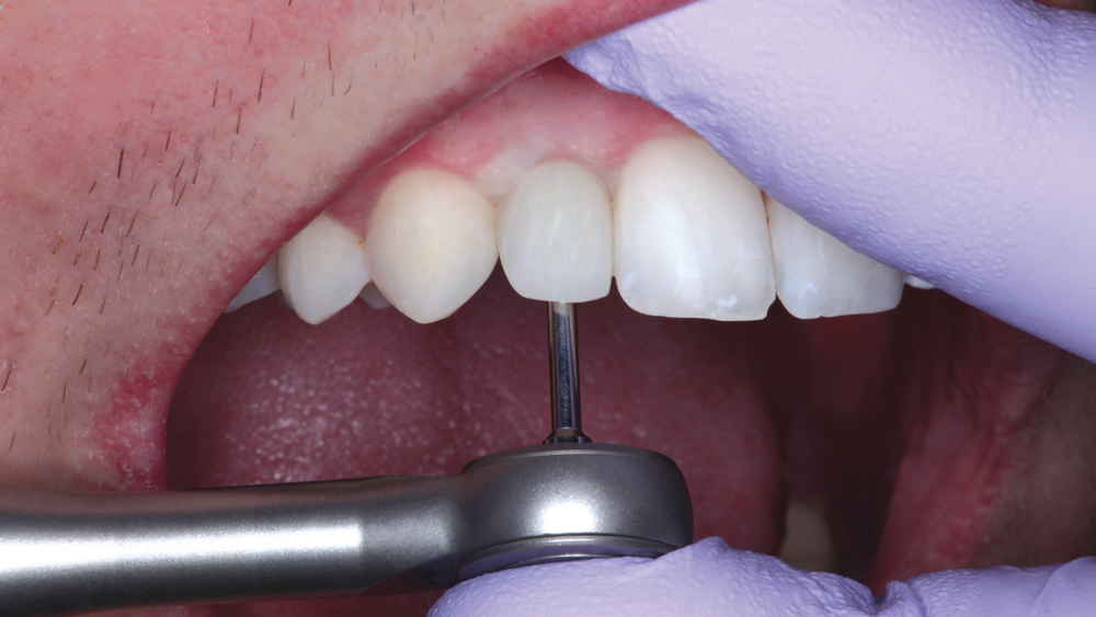
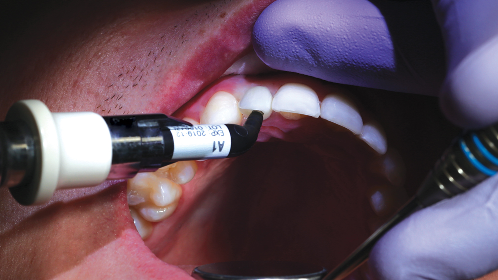
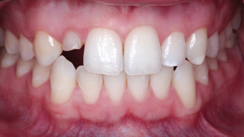
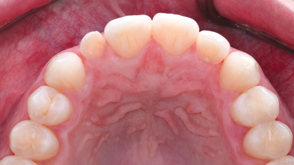
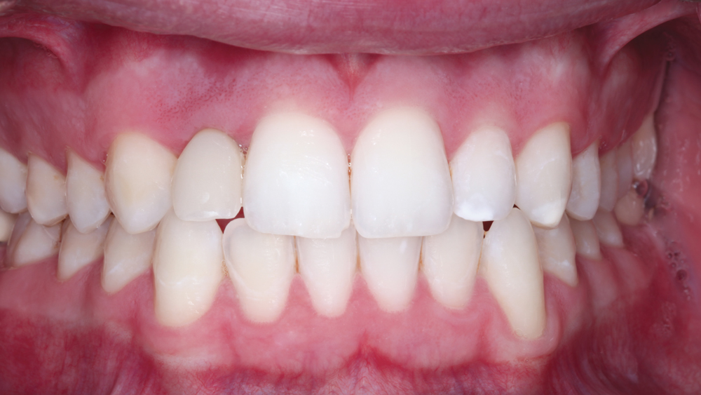
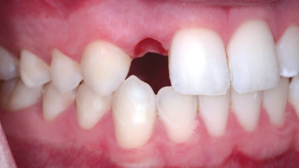
Figure 1: The patient sought a long-term solution for a retained primary tooth #D that was causing him significant esthetic concerns. The root of his upper primary lateral incisor had resorbed, and he desired a permanent replacement for the congenitally missing secondary tooth.
Figure 2: With good overall dental health, no periodontal involvement, and pink, stippled soft tissue, implant treatment was an excellent option for replacing the maxillary lateral incisor.
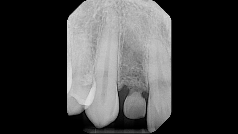
Figure 3: Radiographic view of the retained primary tooth. Although most of the root structure had vanished over time, favorable bone volume was present apical to the tooth. Tooth extraction followed by immediate placement of a dental implant was proposed to the patient, who was young and wanted to avoid preparation of the healthy adjacent teeth to support a fixed bridge. The patient accepted the treatment plan and agreed to return two weeks later for the surgical appointment.
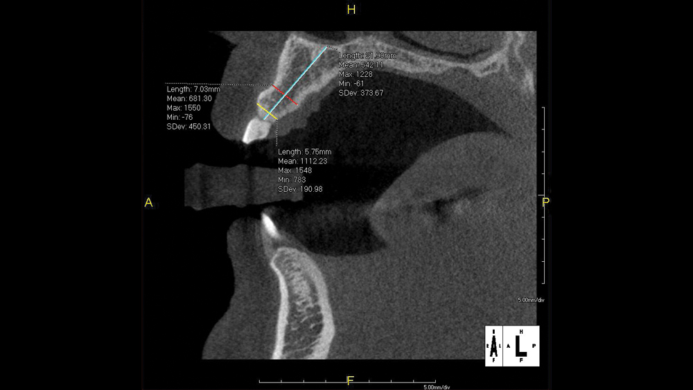
Figure 4: CBCT analysis confirmed adequate vertical and horizontal bone volume for a dental implant immediately following extraction, without the need for bone grafting. Guided surgery was selected to minimize the invasiveness of the procedure. Further, the patient’s adequate attached tissue was ideal for flapless surgery, and the tooth’s location in the esthetic zone called for a higher degree of precision and predictability to ensure the best possible esthetic outcome.
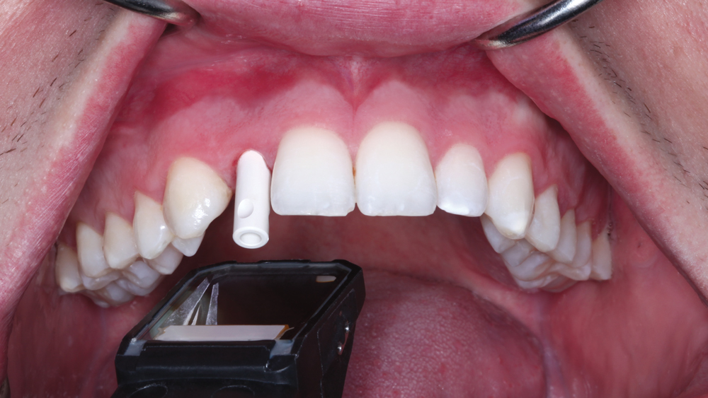
Figure 5: Full-arch scans were taken with a TRIOS® intraoral scanner (3Shape North America; Warren, N.J.). To initiate a case for the Hahn Guided Surgery System, either a highly accurate vinyl polysiloxane (VPS) impression or a full-arch digital impression is acceptable. The impression is submitted along with the CBCT scan and instructions for the digital treatment plan to the surgical guide manufacturer. In this case, a digital wax-up of the tooth was requested, as well as implant positioning that would facilitate a screw-retained crown. Additionally, an immediate provisional crown was prescribed for delivery at the time of surgery.
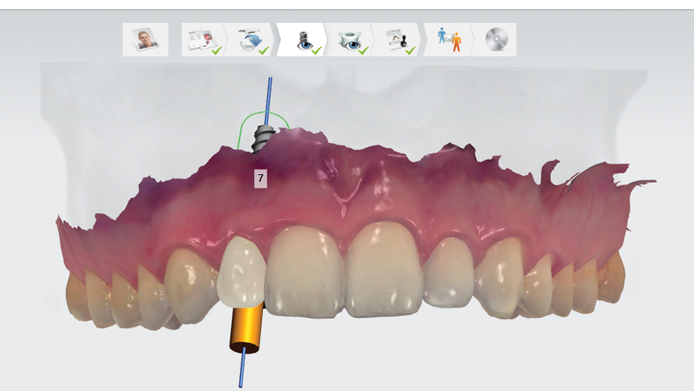
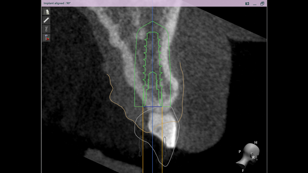
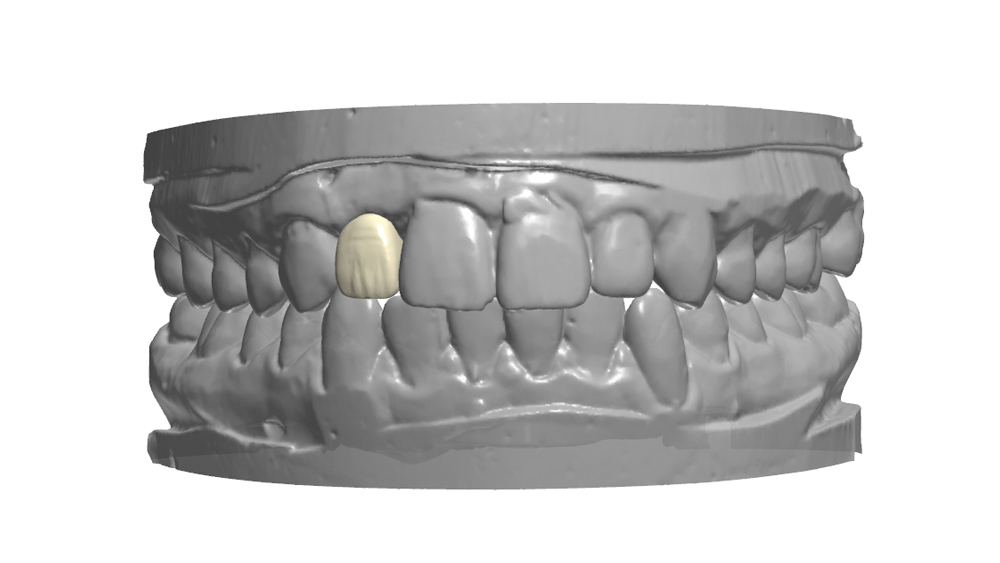
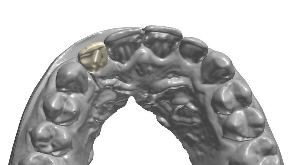
Figures 6a, 6b: After receiving the intraoral and CBCT scans, the Digital Treatment Planning (DTP) department at Glidewell Laboratories merged the data to create a virtual 3D model of the patient’s mouth, dentition, hard- and soft-tissue contours, and underlying anatomy. The DTP team proposed a digital treatment plan, including proper implant positioning for the desired method of screw-retention and an optimal emergence profile in the esthetic zone. Note that Hahn Tapered Implants can be placed using surgical guides from several guided surgery service providers.
Figure 7: The DTP team created the surgical guide design to precisely control the implant position at a distance of 2 mm from the neighboring teeth and 2.5 mm below the adjacent CEJ. The digital treatment plan and surgical guide design were sent for doctor review and were approved without the need for any modifications.
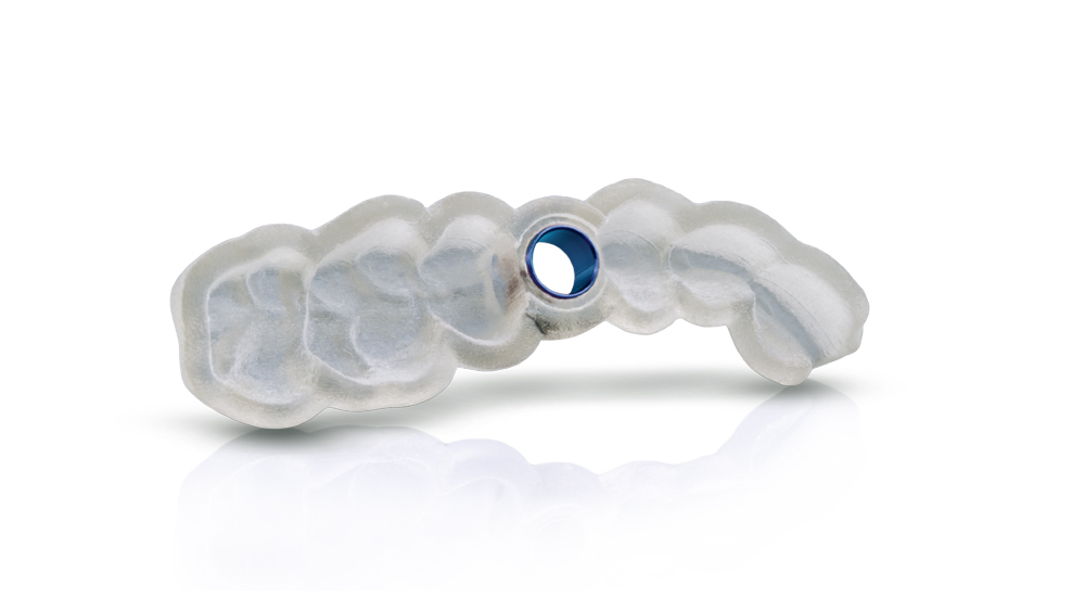
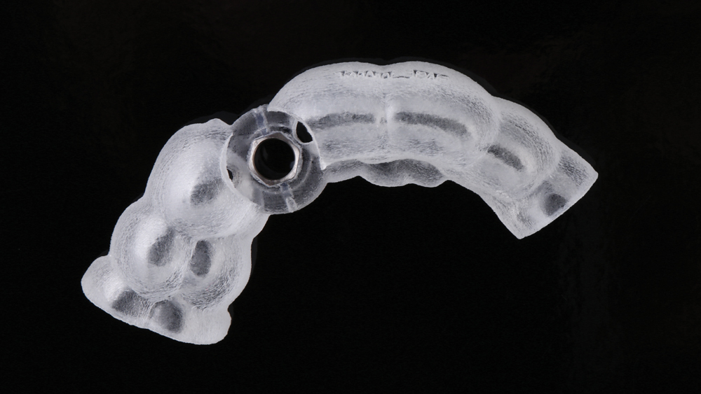
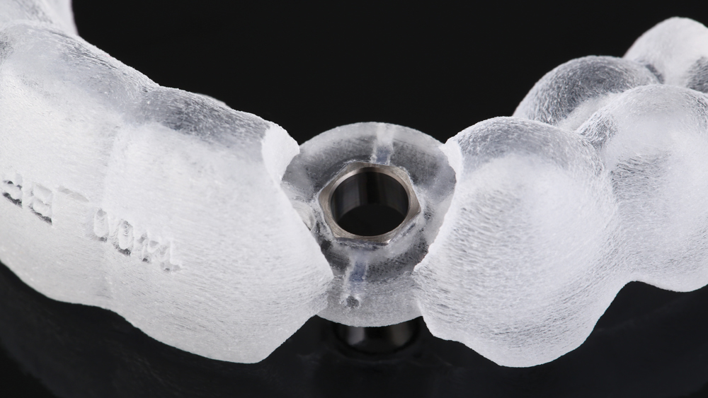
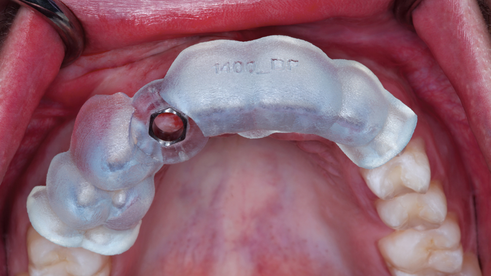
Figures 8a, 8b: A tooth-supported surgical guide was fabricated to precisely control the depth, angulation, and mesial-distal and buccal-lingual location of the implant osteotomy in accordance with the approved digital treatment plan. The titanium sleeve was secured within the surgical guide. Note that the Hahn Tapered Implant Guided Surgery System does not require drill keys, as the surgical kit includes diameter-specific drills for each available implant size.
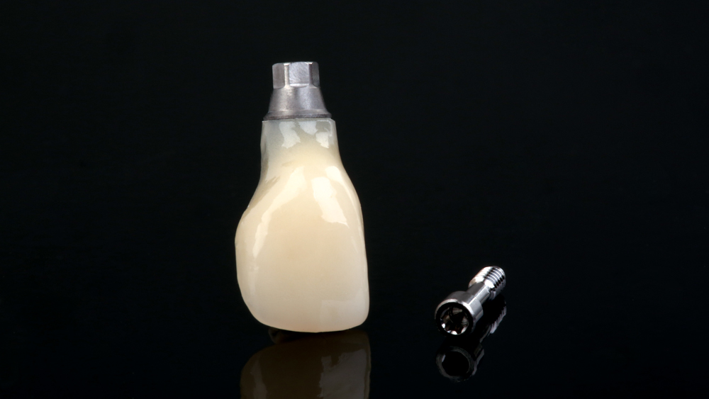
Figure 9: An immediate screw-retained BioTemps® Provisionals crown (Glidewell Laboratories; Newport Beach, Calif.) was designed and fabricated in advance of the surgical appointment based on the digitally determined implant position, providing a temporary restoration to contour the soft tissue, interdental papillae and emergence profile during the healing phase for an ideal prosthetic outcome.
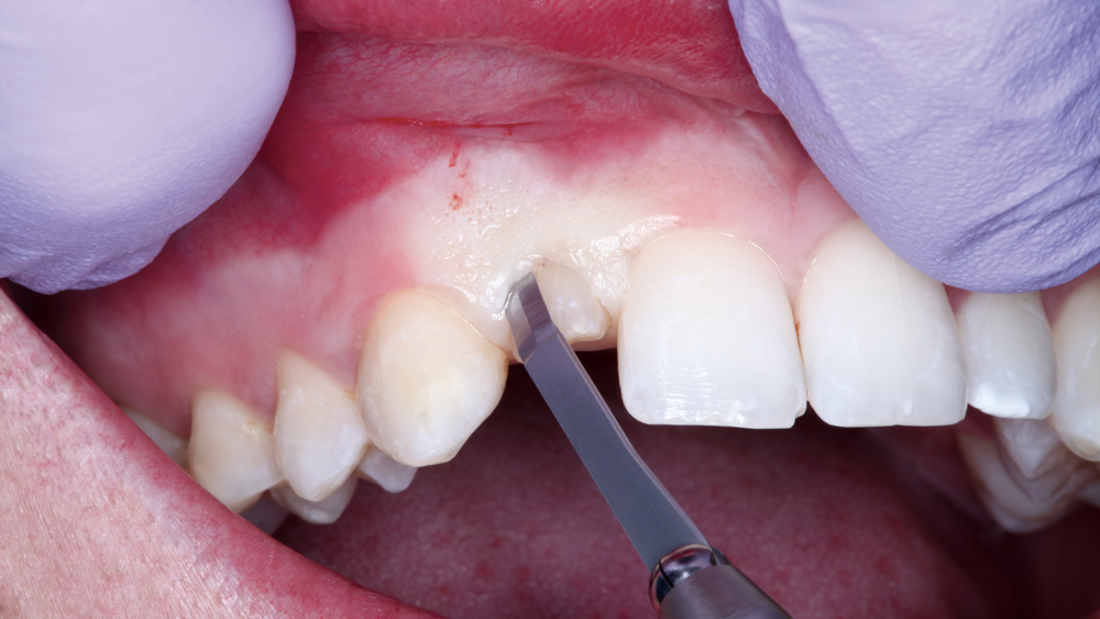
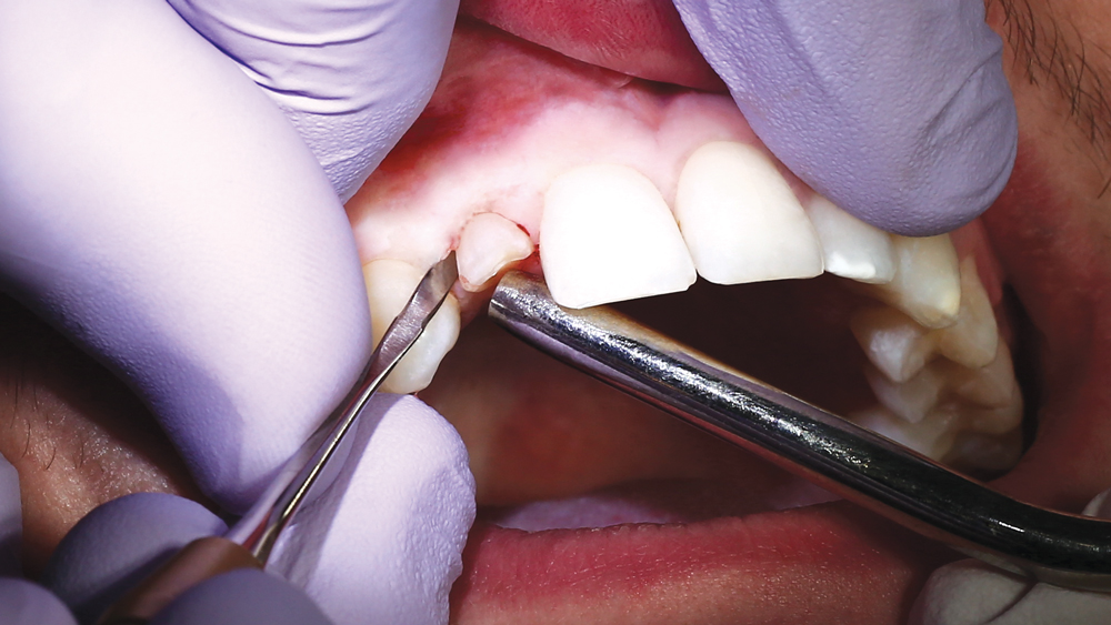
Figures 10a, 10b: At the surgical appointment, after tracing around the circumference of the crown with a microblade to separate the tooth from the periodontal ligament, the deciduous tooth was elevated using periosteal elevators.
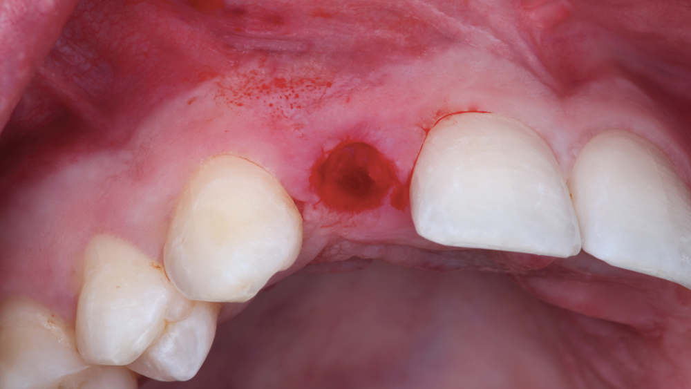
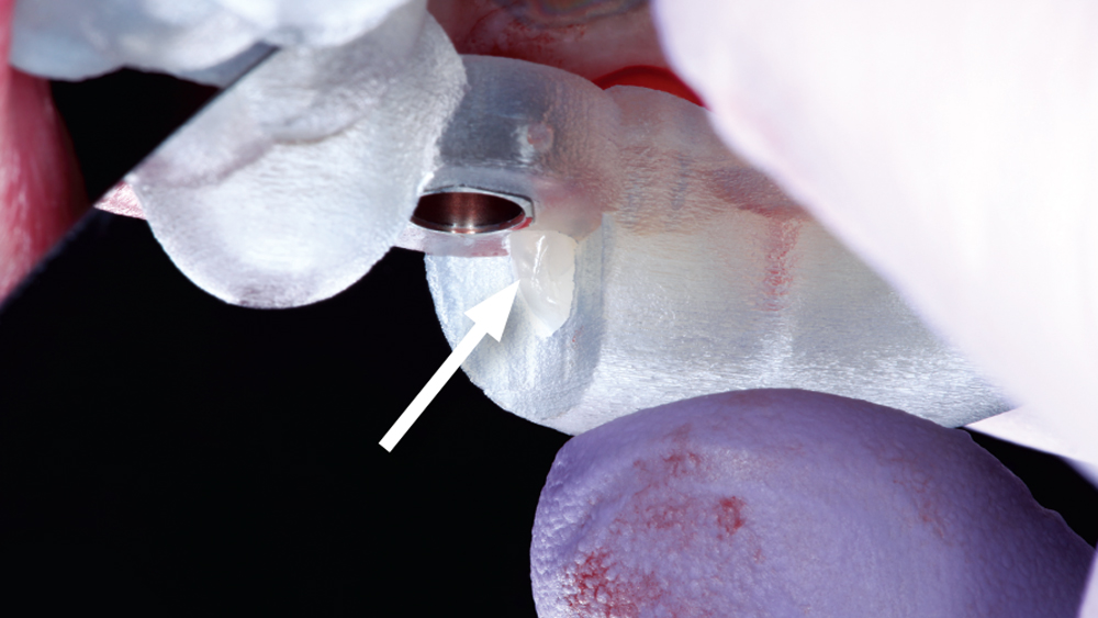
Figures 11a–11c: After extracting the tooth, the surgical guide was tried-in and exhibited a precise fit. Note the window of the surgical guide (11c), which is incorporated in the design to ease verification of full seating of the guide against the teeth.
Figure 12: The implant osteotomy was created using the Hahn Guided Surgical Kit, which includes an easy-to-follow drill sequence, color-coded instrumentation, and diameter-specific drills with built-in depth stops.
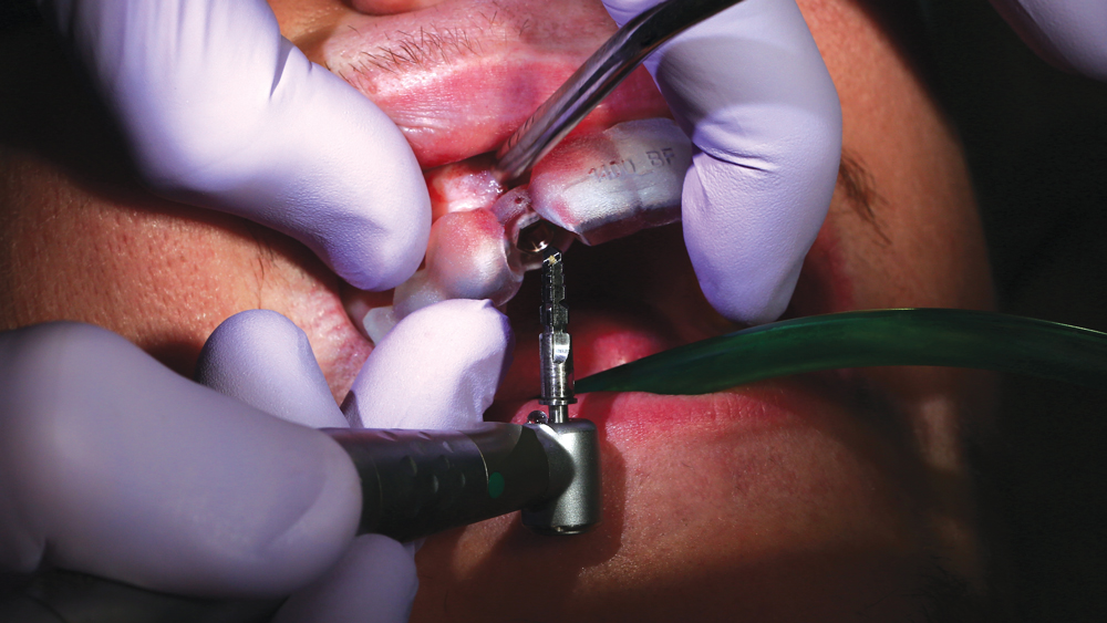
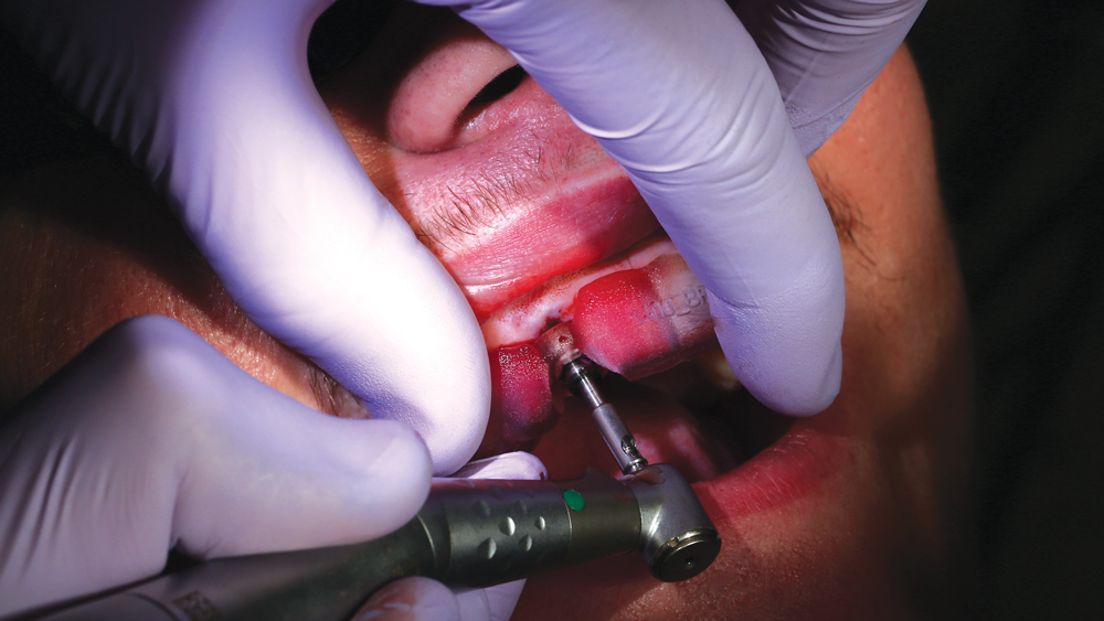
Figures 13a, 13b: The implant osteotomy was created using the Hahn Guided Surgery System, following a streamlined surgical protocol. After using the alignment and pilot drills to initiate the proper location and trajectory of the osteotomy, the shaping drills were utilized to establish the precise diameter and length of the prescribed 3.5 mm x 13 mm Hahn Tapered Implant.
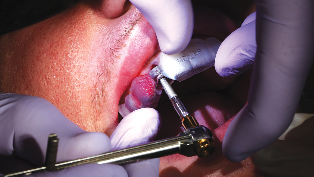
Figure 14: After flushing the site with sterile water to ensure the osteotomy was free of bone debris, the implant was placed directly through the guide utilizing a torque wrench.
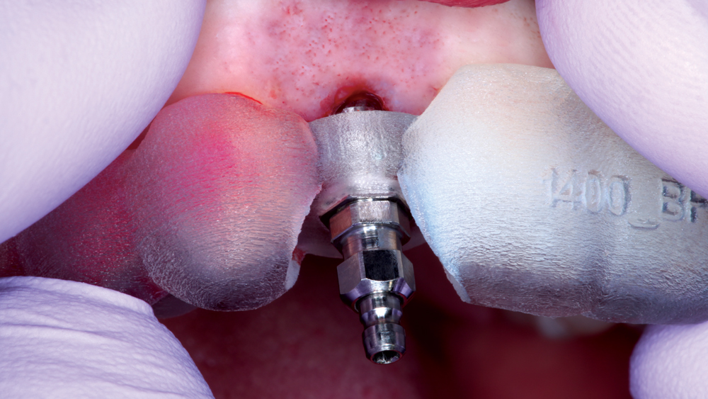
Figure 15: With the implant fully seated, the hex flange of the implant mount was flush with the hex of the guide sleeve. This confirmed proper orientation of the implant platform, which was important for proper fit and alignment of the BioTemps screw-retained crown and, eventually, the final restoration.
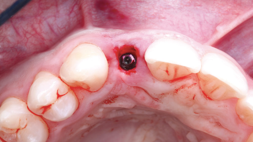
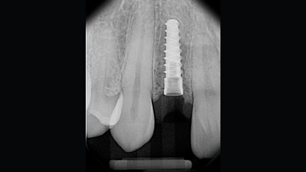
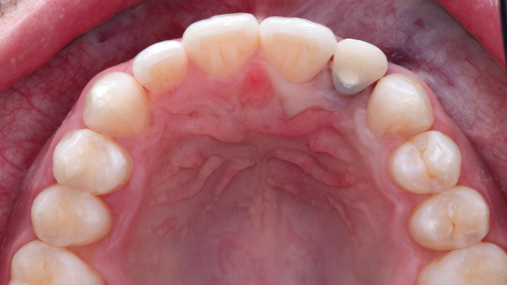
Figures 16a, 16b: Postoperative occlusal and radiographic views show the implant in the exact position determined by the digital treatment plan. The deep, sharp threads of the Hahn Tapered Implant helped establish the high primary stability that is essential for immediate placement cases and necessary for immediate provisionalization.
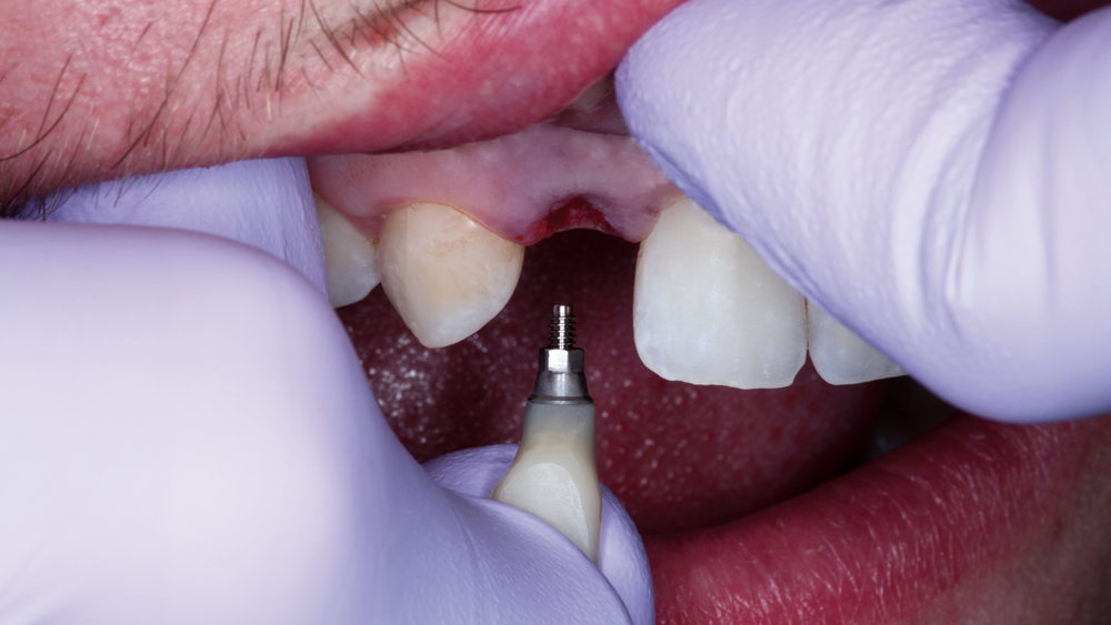
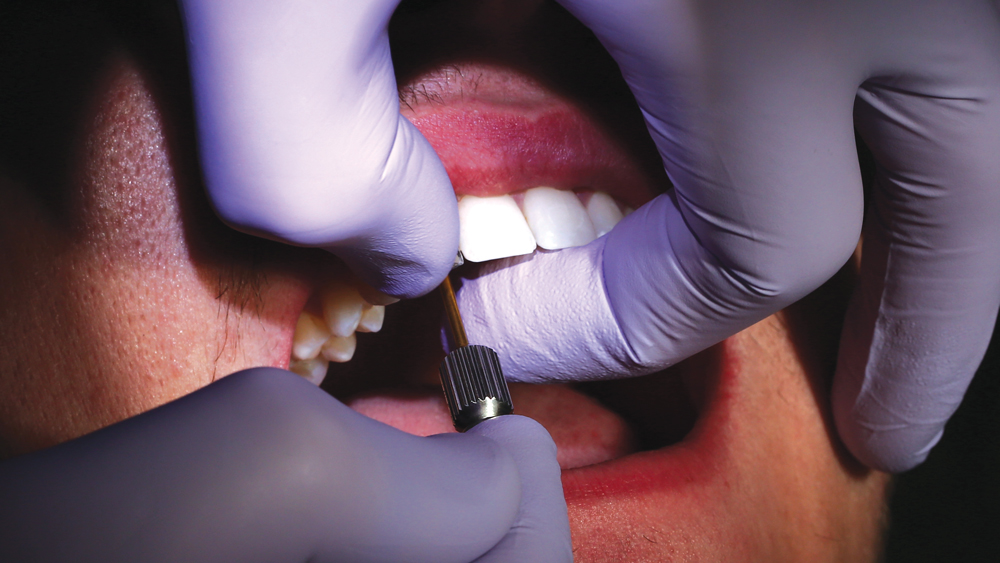
Figures 17a, 17b: The BioTemps screw-retained crown was delivered using a hand driver. Because the temporary crown was fabricated based on the digitally determined implant position, the BioTemps provisional exhibited a precise fit.
Figures 18a, 18b: The occlusion of the BioTemps provisional was checked, and slight alterations were made to the crown contours for esthetics and to avoid occlusal contact during function. The patient was very pleased with the esthetics of the delivered BioTemps crown.
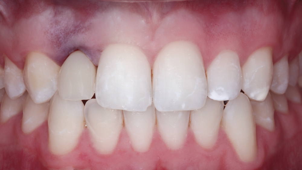
Figure 19: At the one-month follow-up appointment, the patient had healed well. Facial view shows emergence profile and interproximal papilla development fostered by the provisional crown. Oral hygiene habits were encouraged for the patient to address the slight inflammation of the soft tissue.
Figure 20: After four months of healing, an Inclusive® Scanning Abutment (Glidewell Direct) was connected to the implant, and a digital impression was created using the TRIOS intraoral scanner for the fabrication of the final restoration.
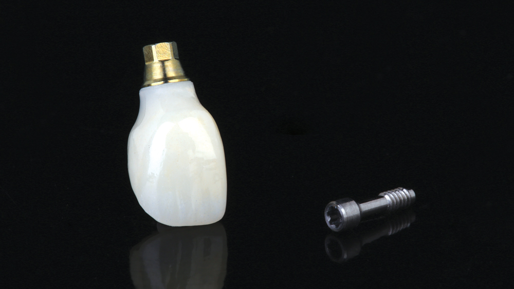
Figures 21a, 21b: The final crown was designed using dental CAD software to mimic the contours of the contralateral tooth. A screw-retained restoration was selected for retrievability and to create an ideal emergence profile.
Figure 22: The final screw-retained crown was fabricated from BruxZir® Anterior Solid Zirconia, which was chosen for its combination of strength and lifelike esthetics.
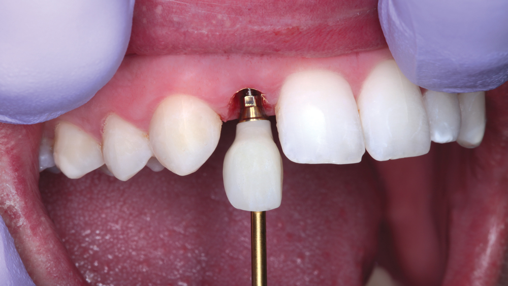
Figure 23: At the final delivery appointment, the temporary crown was removed. Note the papilla development resulting from the use of the BioTemps provisional restoration.
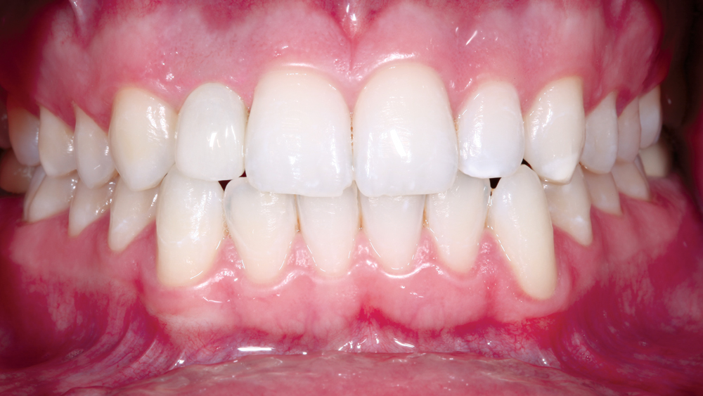
Figures 24a, 24b: The final BruxZir Anterior crown was delivered. The restoration fit well, met the desired esthetic requirements, and did not require any chairside adjustments. The accurate fit and natural emergence profile were predictable outcomes due to the precise digital process used to produce the treatment plan, surgical guide, provisional crown and final restoration.
Figure 25: After confirming that the tooth was slightly out of occlusion during function and maximum intercuspation, the screw access channel of the crown was sealed using Camouflage® NanoHybrid Composite (Glidewell Direct).
Figures 26a, 26b: The patient was extremely happy with the final restoration, as well as the efficient, minimally invasive treatment protocol that helped ensure such a predictable, esthetic outcome.
- 1
CONCLUSION
With the release of the Hahn Guided Surgery System, clinicians now have an efficient option for the digitally guided surgical placement of Hahn Tapered Implants. With simplified instrumentation, a streamlined surgical protocol, and support from a dental lab that ensures a restorative-driven approach is maintained throughout the course of treatment, the Hahn Guided Surgery System is a valuable tool for novice and experienced implant practitioners alike.