Removable and Fixed Solutions for the Edentulous Patient
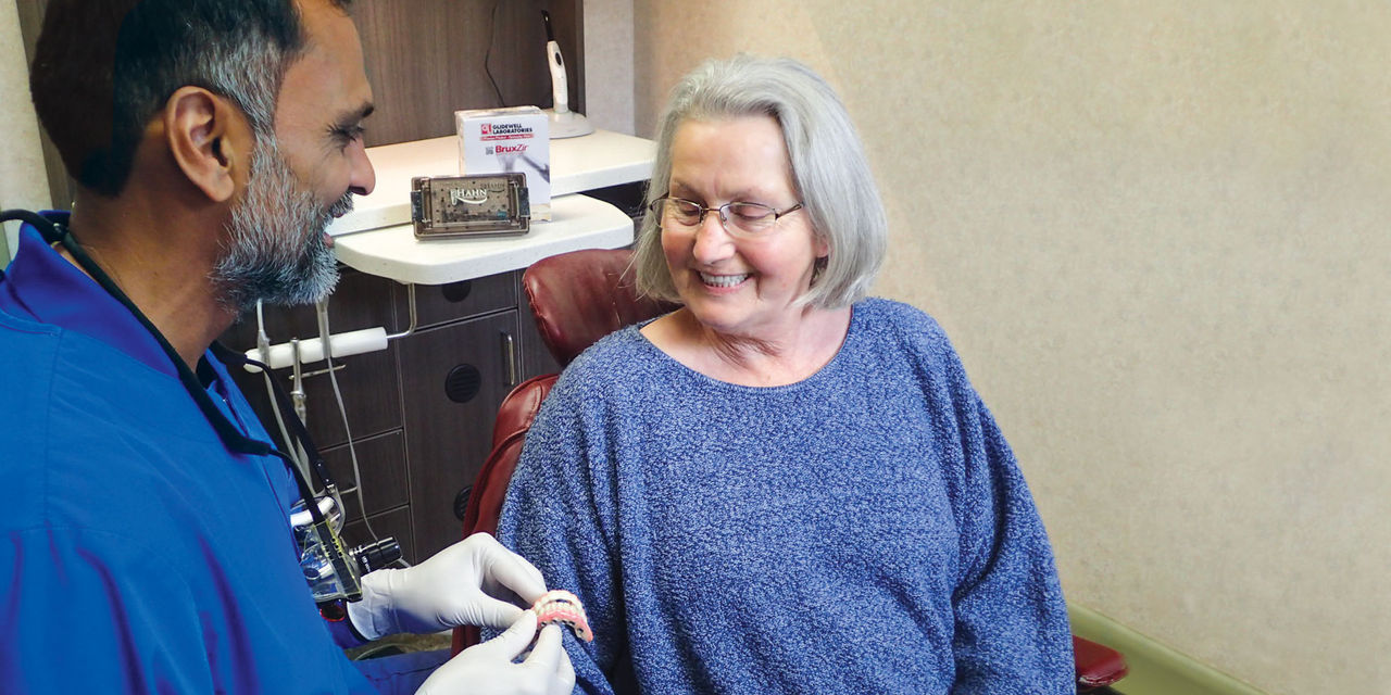
The fully edentulous patient faces many challenges, including diminished chewing capability and compromised speech function, as well as social and psychological issues, that combine to create a negative impact on quality of life.1 Thankfully, modern dentistry affords the clinician a range of options for successful treatment of the edentulous patient, from the traditional complete denture to the fixed monolithic zirconia full-arch implant restoration. Depending on the oral condition and financial circumstances of the patient, a treatment plan can be developed that addresses the unique challenges of each case. While some cases require a fixed or removable implant solution to effectively restore function, a well-designed complete denture can meet the needs of others.
Studies have shown that fixed full-arch implant restorations offer the highest levels of prosthetic stability, function, satisfaction and quality of life for the edentulous patient.2,3,4 However, patients with favorable anatomical features can often be provided with a relatively stable conventional denture, particularly in the maxilla. Upper dentures with palatal coverage can usually achieve sufficient retention and function.5 When a patient requires full-mouth rehabilitation but only can afford implant therapy for one arch, an implant solution is typically recommended for the mandible, where it’s more difficult to establish a stable and retentive complete denture.
By comprehensively assessing the patient’s chief complaint, bone volume, anatomical landmarks, and the fit, stability and comfort of any existing oral appliances, clinicians can confidently determine whether a traditional complete denture or an implant-supported prosthesis is indicated. After presenting the treatment options, doctors can work with the patient in developing a solution that is both affordable and effective.
CASE REPORT
This patient was unhappy with the esthetics of her existing upper denture as well as the discomfort and instability caused by her loose-fitting mandibular partial denture. Although her concerns with the maxillary denture could be addressed with a new, more esthetic appliance, she preferred implant treatment for her mandible, where a fixed solution was needed to adequately restore function, stability and comfort.
Treatment plan:
- Upper: Full denture
- Lower: Fixed BruxZir® Full-Arch Implant Prosthesis
The patient’s remaining mandibular teeth were extracted followed by immediate implant placement and delivery of a fixed provisional appliance. After integration of the implants, the prosthetic designs for the new upper denture and lower implant restoration were determined in tandem. Ultimately, the dual-arch restorations addressed the functional and esthetic challenges of the case while staying within the financial means of the patient, demonstrating the benefits of a flexible, multifaceted approach to restorative dentistry.
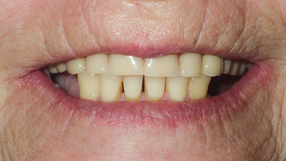
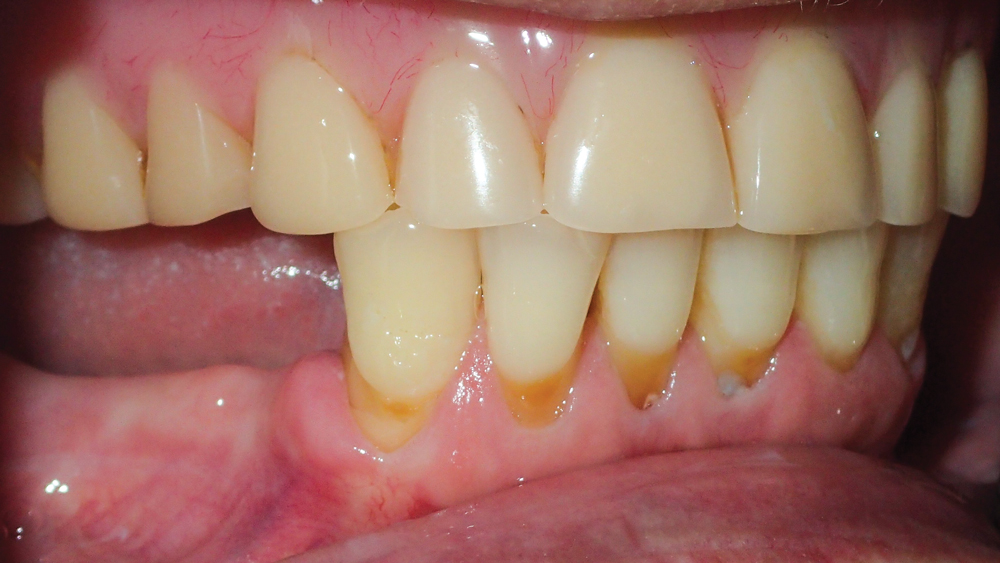
Figure 1: The patient presented with a heavily worn, unesthetic upper denture and a lower partial denture that was functioning poorly and was no longer utilized. She requested an affordable solution that would give her the esthetics she desired for her maxilla while restoring comfort and chewing capability for her lower arch.
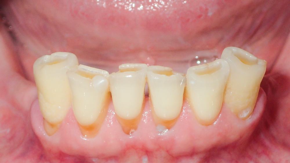
Figure 2: Preoperative condition of the mandibular arch. The patient’s remaining lower teeth were painful and exhibited periodontal involvement and loss of attached tissue. The patient did not wish to spend money salvaging her remaining mandibular teeth, which had an unfavorable long-term prognosis.
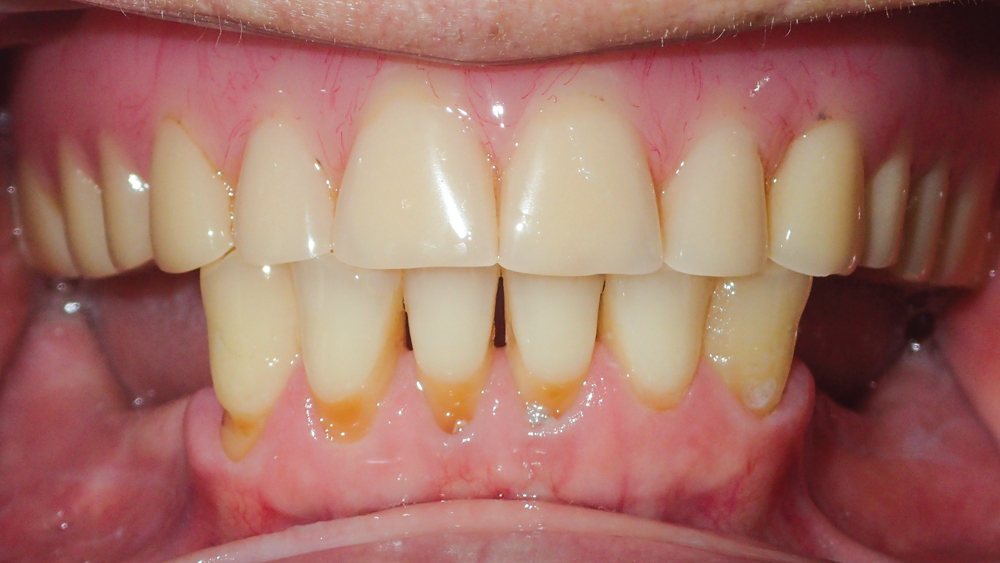
Figure 3: While the patient’s existing upper denture provided sufficient retention, it did not function properly and the incisal edges in the anterior were subjected to excessive wear due to the lack of posterior occlusion. The patient’s maxillary arch exhibited the anatomical landmarks needed for a stable complete denture, including ridge thickness, a favorable U-shaped palate for a strong seal, and firm, keratinized soft tissue.
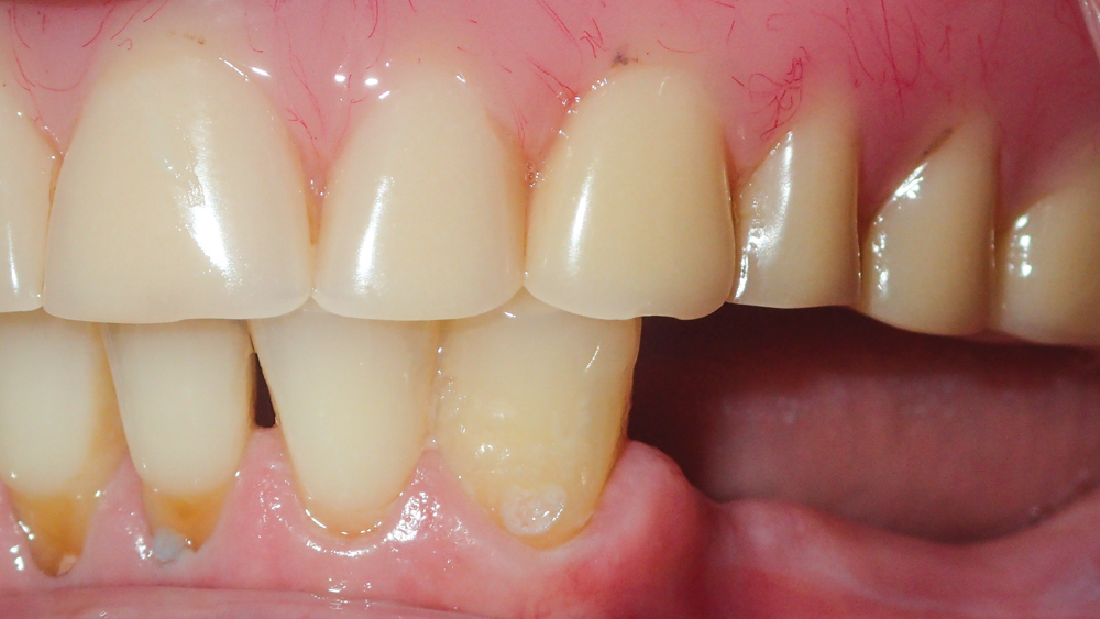
Figures 4a, 4b: Lateral views illustrate ridge resorption and thin soft tissue in the posterior areas of the mandible, which caused the patient pain, discomfort, and inadequate retention and function when she wore the existing partial appliance. A fixed implant solution was ideal for the mandible, as a removable, tissue-supported appliance could not achieve the stability and chewing capability the patient desired. Despite the bone loss in the posterior, the ridge width in the premolar areas was sufficient for a 3.5 mm implant on each side of the arch, which, in combination with four dental implants situated between the mental foramina, would provide the A-P spread needed for a fixed full-arch implant prosthesis.
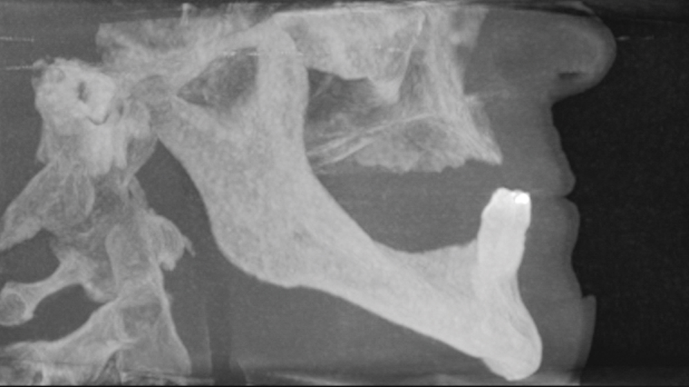
Figure 5: CBCT scanning confirmed sufficient bone volume for the placement of six implants in the mandible. The 3-D imaging also helped determine the proper diameters and lengths of implants to place while avoiding the inferior alveolar nerve and mental foramina. Treatment options were presented to the patient, using case examples and models to show the differences between a traditional denture, an implant overdenture and a fixed full-arch implant restoration.
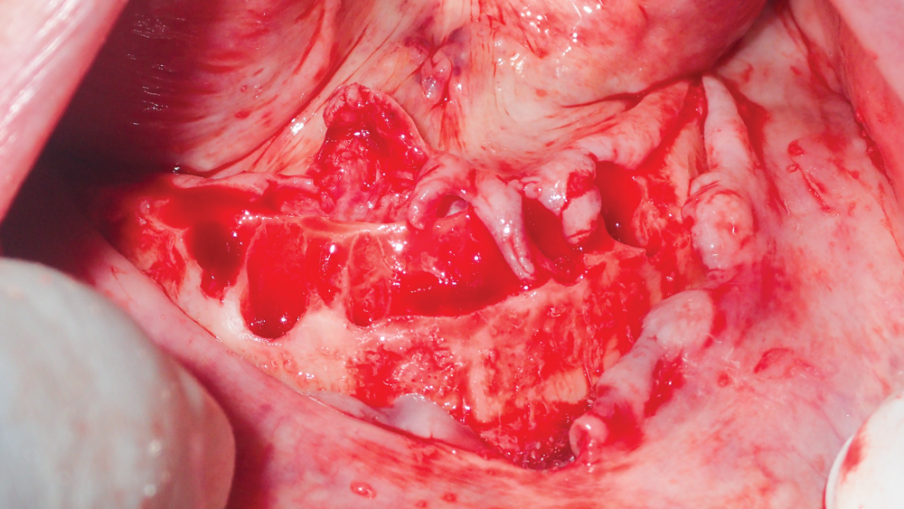
Figure 6: The patient, who desired but could not afford implant therapy for both arches, agreed to a treatment plan in which her existing maxillary appliance would be replaced with a new complete denture, with implants placed in the mandible to support a fixed restoration. The patient opted for extraction with immediate implant placement — an approach with success ratios that compare favorably to extraction with delayed implant placement.6,7 Many patients find this approach to implant therapy appealing due to the reduced number of surgical appointments, shortened treatment time, and opportunity for an immediate fixed restoration. To begin the procedure, a full-thickness flap was reflected and the anterior teeth of the mandible were extracted atraumatically using periotomes.
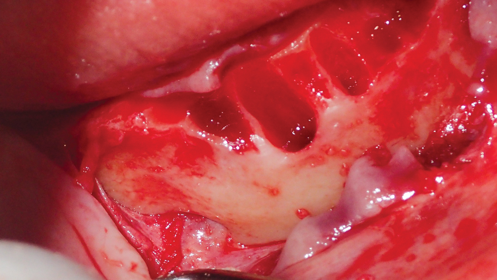
Figure 7: Lateral view of the mandible following completion of the bone-leveling procedure, which was performed in the anterior using a straight surgical handpiece with acrylic bur to create a uniform prosthetic platform for the implants. Note the location of the mental foramen, the visualization of which confirmed proper length of the 3.5 mm x 8 mm implants selected for the premolar areas.
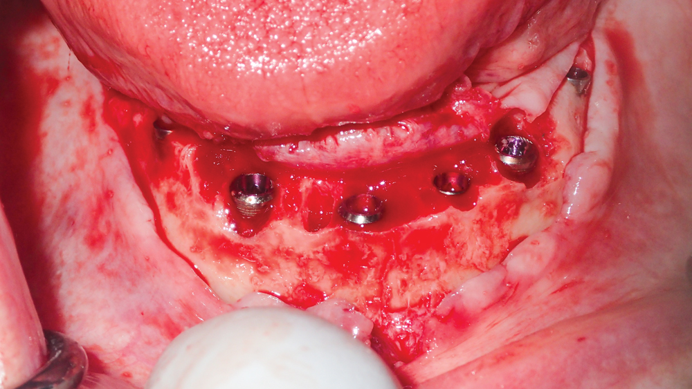
Figure 8: Hahn™ Tapered Implants (Glidewell Direct; Irvine, Calif.) were selected for the surgical procedure because of this system’s ability to maximize primary stability and facilitate positioning within the limited bone volume of the residual ridge. After the osteotomies were created following a user-friendly drilling protocol, the implants were threaded into place, including 4.3 mm x 13 mm implants in the areas of teeth #22, #25 and #27, a 3.5 mm x 13 mm implant in the area of #23, and 3.5 mm x 8 mm implants in the areas of #20 and #29. The deep, sharp threads of the Hahn Tapered Implant eased positioning within the extraction sockets by engaging the lingual wall during insertion, helping maintain a safe distance from the facial plate.
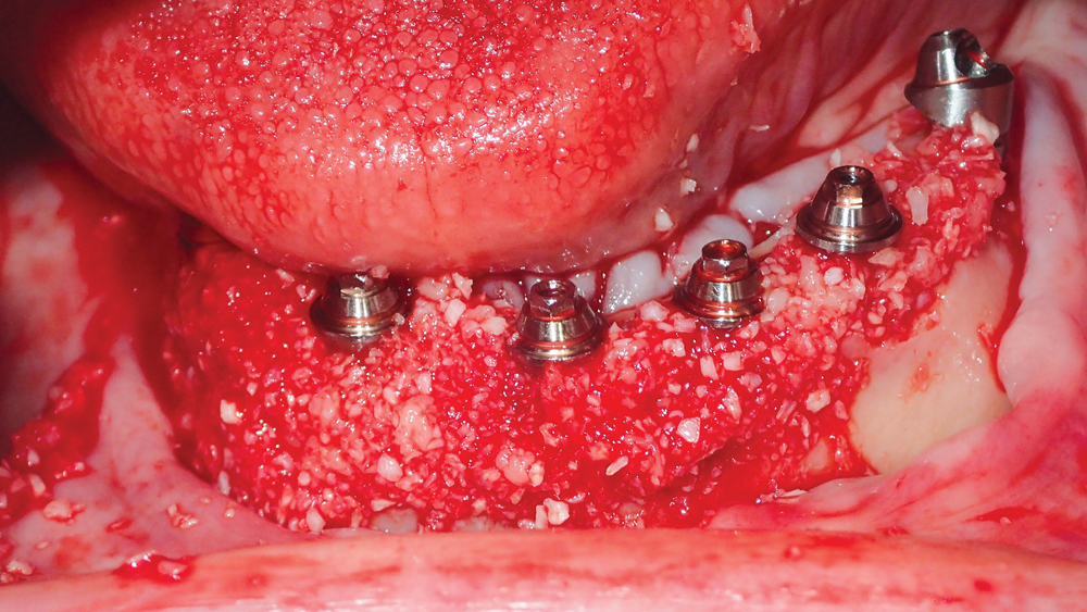
Figure 9: Inclusive® Multi-Unit Abutments (Glidewell Direct) were connected to the implants to establish a consistent, tissue-level prosthetic platform. Newport Biologics™ Mineralized Cortico/Cancellous Allograft Blend (Glidewell Direct) was placed in the gaps between the implants and the extraction sockets and was layered on top of the exposed cortical plate. By providing a scaffold for cell in-growth and consistent bone regeneration around the implants, the cortico/cancellous allograft material helped ensure a predictable outcome.
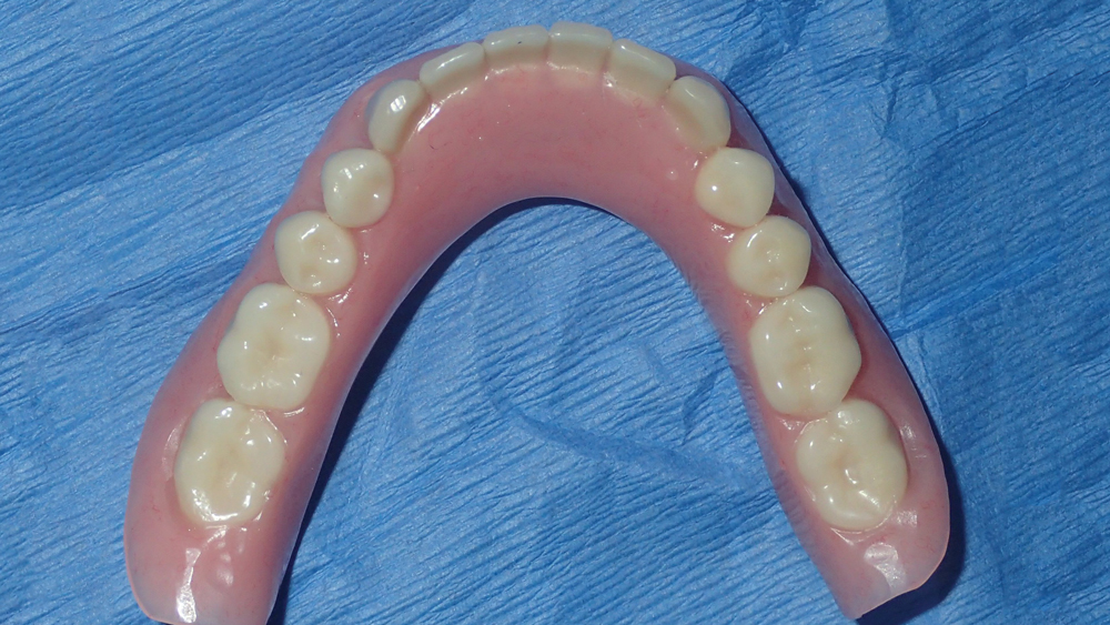
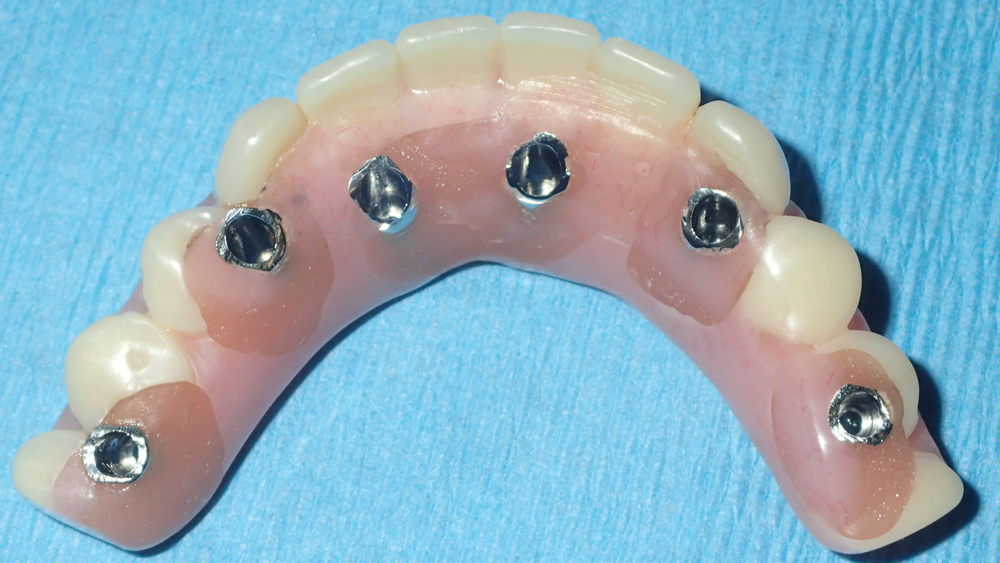
Figure 10: An immediate denture was fabricated in advance of the surgical appointment. The high primary stability established by the Hahn Tapered Implants allowed for conversion of the denture into a fixed screw-retained temporary appliance for immediate loading.
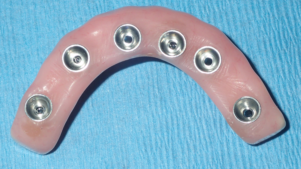
Figures 11a, 11b: The immediate denture was modified to serve as a fixed provisional prosthesis by adding and trimming temporary cylinders and removing the cantilevers in the molar areas of the prosthesis, which avoided the application of excess forces on the implants during healing.
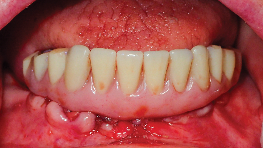
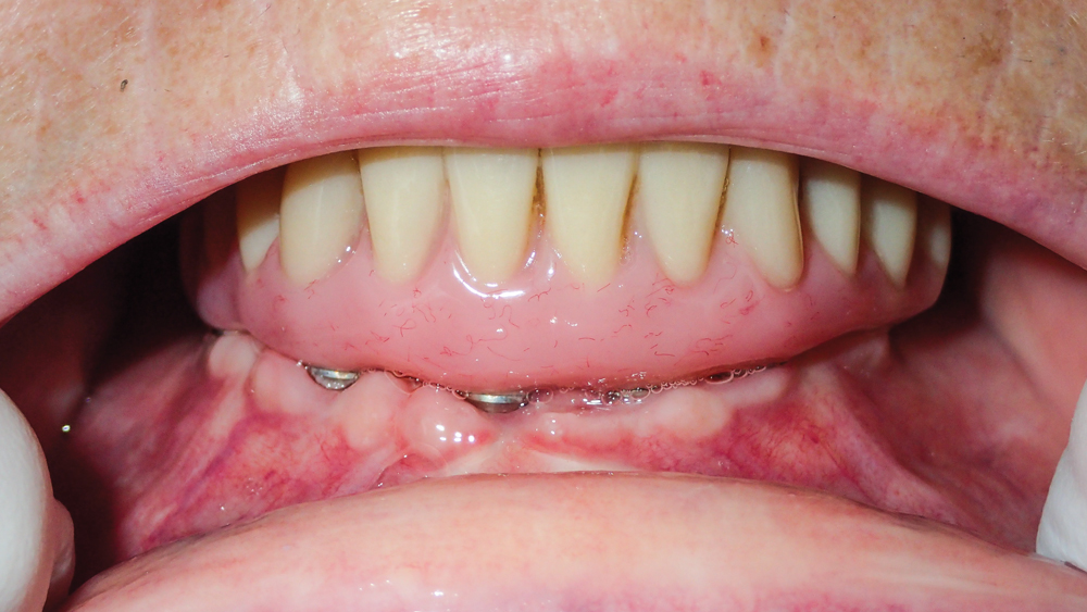
Figure 12: The converted denture was connected to the implants on the day of surgery, extending from premolar to premolar. The screw-retained provisional appliance immediately restored form and function and allowed the patient to leave the surgical appointment with a fixed prosthesis in place. The patient was extremely pleased to avoid wearing a removable appliance throughout the course of treatment.
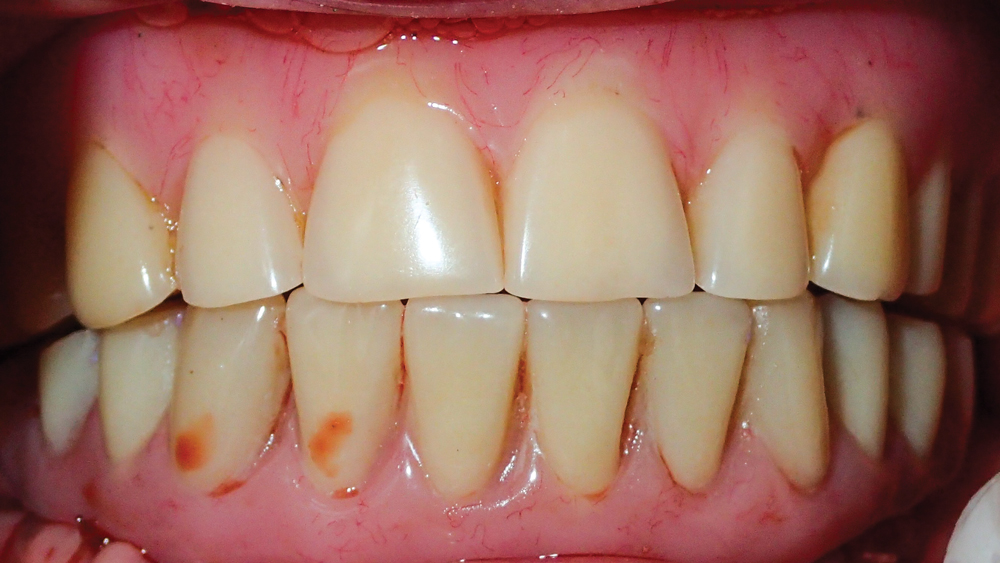
Figure 13: The occlusion of the provisional implant prosthesis was verified against the patient’s existing upper denture, which was left in place until a new prosthetic design could be determined in concert with the lower full-arch implant restoration following integration of the implants.
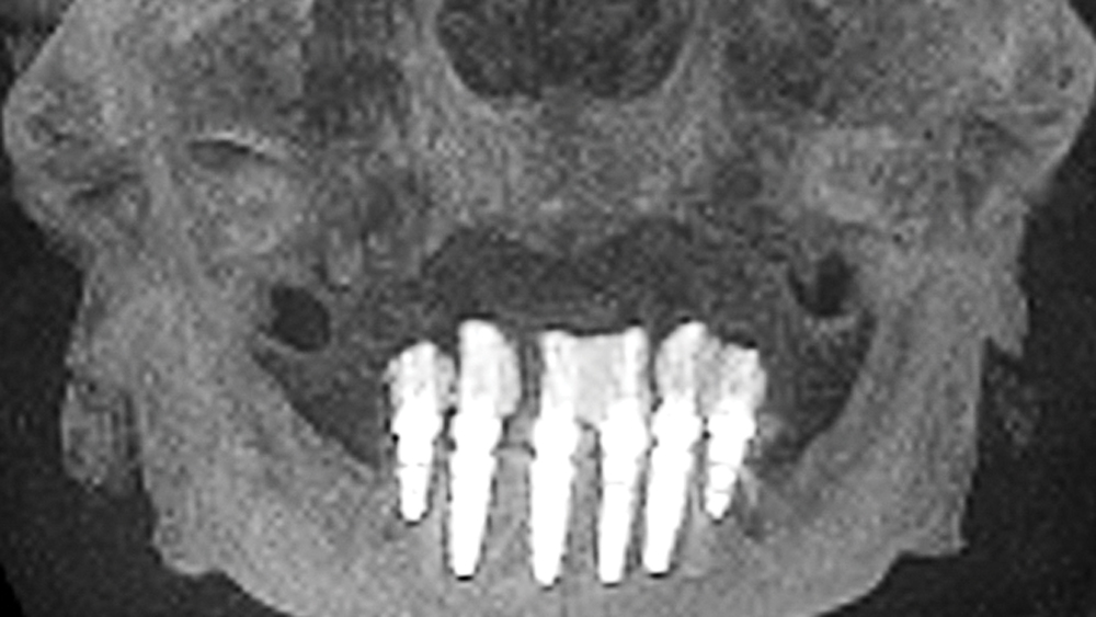
Figures 14a, 14b: Postoperative CBCT scans illustrate the six axially placed Hahn Tapered Implants as well as the temporary cylinders of the immediate provisional implant prosthesis.
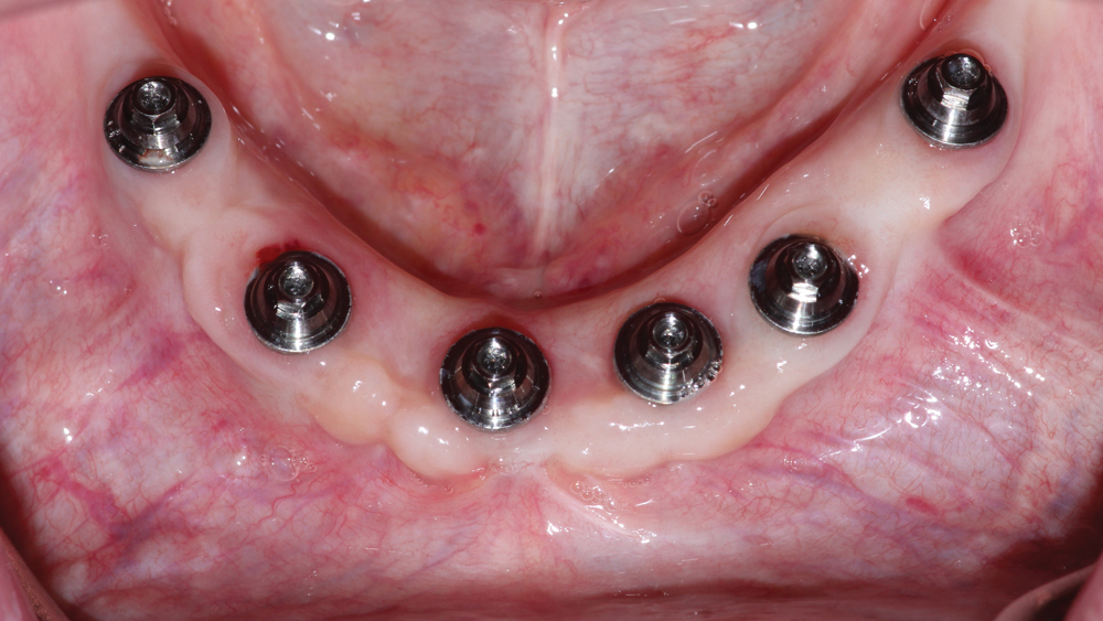
Figures 15a, 15b: After five months of healing, the patient exhibited excellent soft-tissue health. The patient’s appliances were removed, integration of the implants was confirmed, and impressions were taken of both arches to begin the fabrication process for the mandibular full-arch implant restoration and the new maxillary complete denture. To meet the patient’s desire for a fixed restoration while ensuring the best possible long-term prognosis, the patient was prescribed a BruxZir Full-Arch Implant Prosthesis, which eliminates any concerns with wear and breakage at a cost that compares favorably with screw-retained hybrid dentures. Further, the opportunity to test-drive a PMMA provisional implant prosthesis addressed the patient’s concern with achieving an accurate fit for the final restoration.
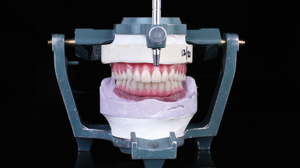
Figure 16: After obtaining jaw relation records, wax setups were produced and tried in to evaluate function, lip support, smile line, esthetics and occlusion. The conversion denture delivered at the time of surgery served as the basis for the lower restoration, and a new design was determined for the maxillary denture. The final wax setup and verified master model were submitted to Glidewell Laboratories for copy-milling of the BruxZir Full-Arch Implant Prosthesis, significantly reducing the cost of the final restoration.
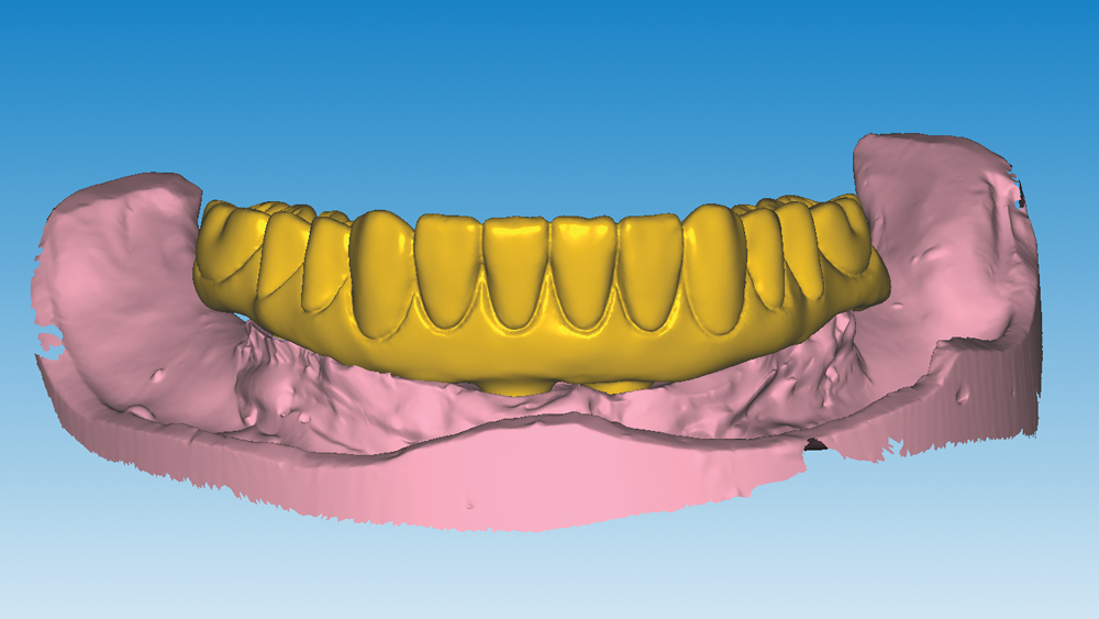
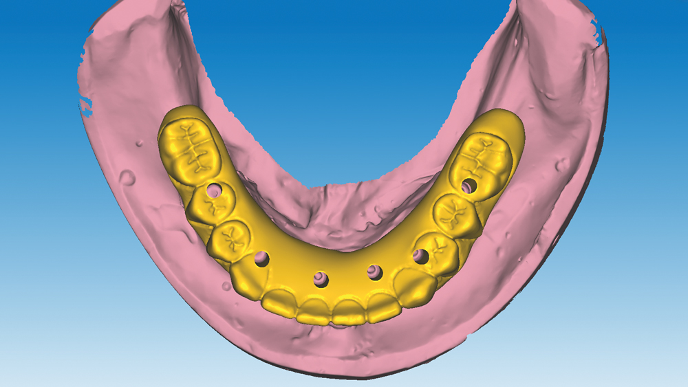
Figures 17a, 17b: After scanning the final wax setup and master model, Glidewell Laboratories created the digital design for the monolithic zirconia implant restoration using advanced CAD/CAM software. Because this case was submitted as a copy-mill restoration, the digital design represents a precise replica of the final wax setup; adjustments were made only to the openings through which titanium connections and screws are used to attach the prosthesis to the implants.
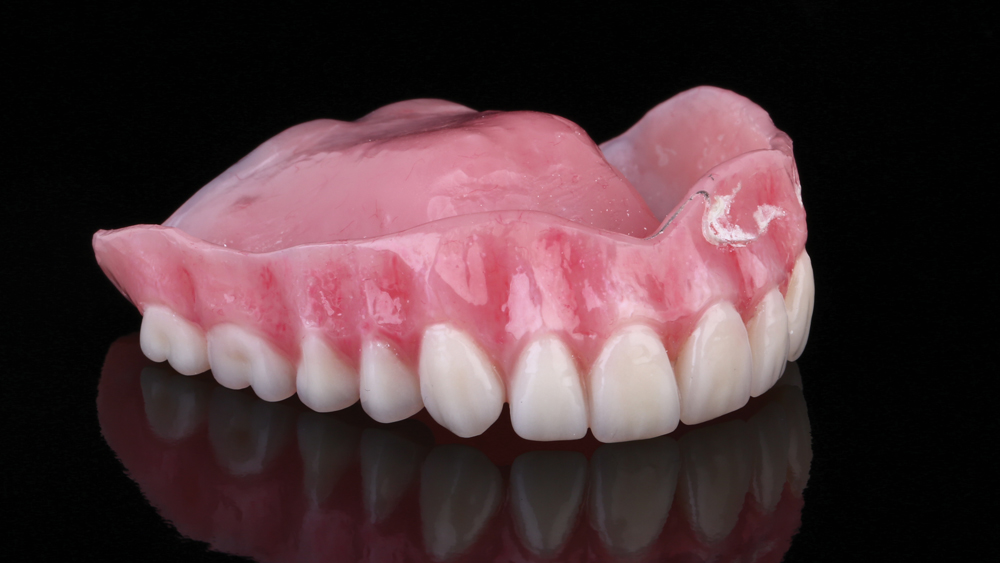
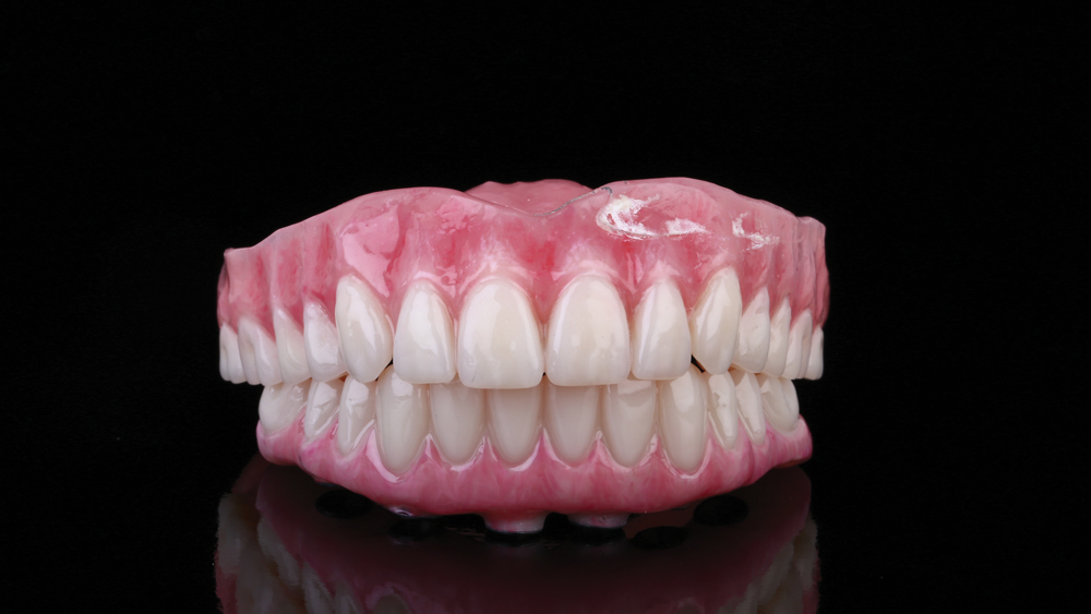
Figure 18: After fine-tuning the function, occlusion, and teeth shape and size of the upper denture with wax setups, a new maxillary denture was produced, significantly improving upon the esthetics of the patient’s existing appliance.
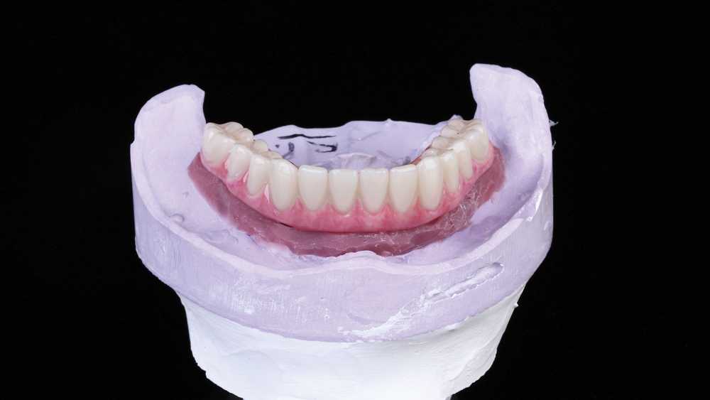
Figure 19: The lab fabricated a PMMA provisional implant prosthesis, which is recommended by the lab for copy-mill cases to ensure the design is completely accurate before the final restoration is milled from monolithic zirconia. The provisional implant prosthesis was attached to the master model to confirm a passive fit.
Figures 20a, 20b: Advanced staining techniques were used to achieve consistent, lifelike gingival and tooth shades for the denture and implant prosthesis.
Figure 21: The provisional implant prosthesis was tried in and achieved an accurate fit due to the precision of the CAD/CAM copy-mill process. Screws were then threaded through the access openings to secure the prosthesis to the implants, and the screw access holes were filled with Camouflage® NanoHybrid Composite.
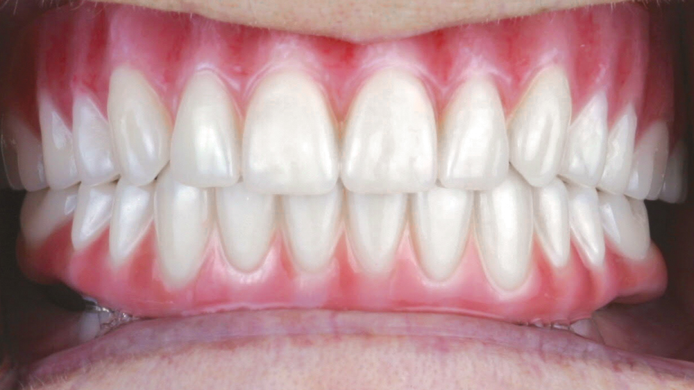
Figure 22: The final upper denture was seated and the esthetics, function and occlusion were confirmed with both appliances in place. Minor occlusal adjustments were made to the denture and provisional appliance. After three weeks of wear, the patient was satisfied with the design of the implant prosthesis, so the provisional was returned to the lab with the master cast for fabrication of the final restoration.
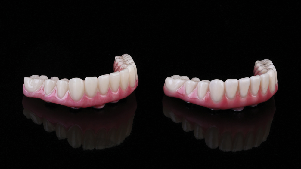
Figure 23: The final mandibular restoration was milled from BruxZir Full-Strength Solid Zirconia. As an exact replica of the patient-confirmed PMMA provisional implant prosthesis (left), the final full-arch BruxZir restoration (right) aligned with the precise expectations of the patient.
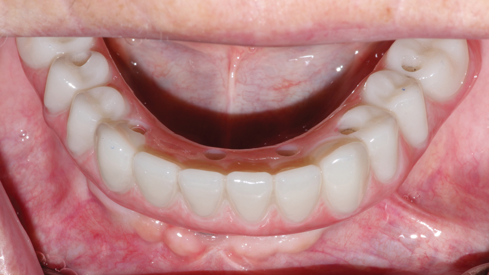
Figure 24: The final BruxZir Full-Arch Implant Prosthesis was delivered with relative ease. As a result of verification with the PMMA provisional appliance, the final restoration fit perfectly and did not require any alterations.
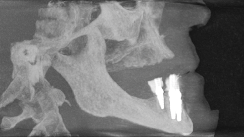
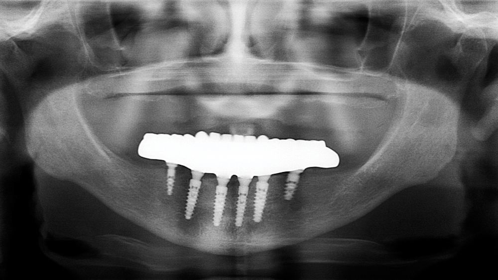
Figure 25: Final CBCT scan illustrates the BruxZir Full-Arch Implant Prosthesis fully seated over the Hahn Tapered Implants.
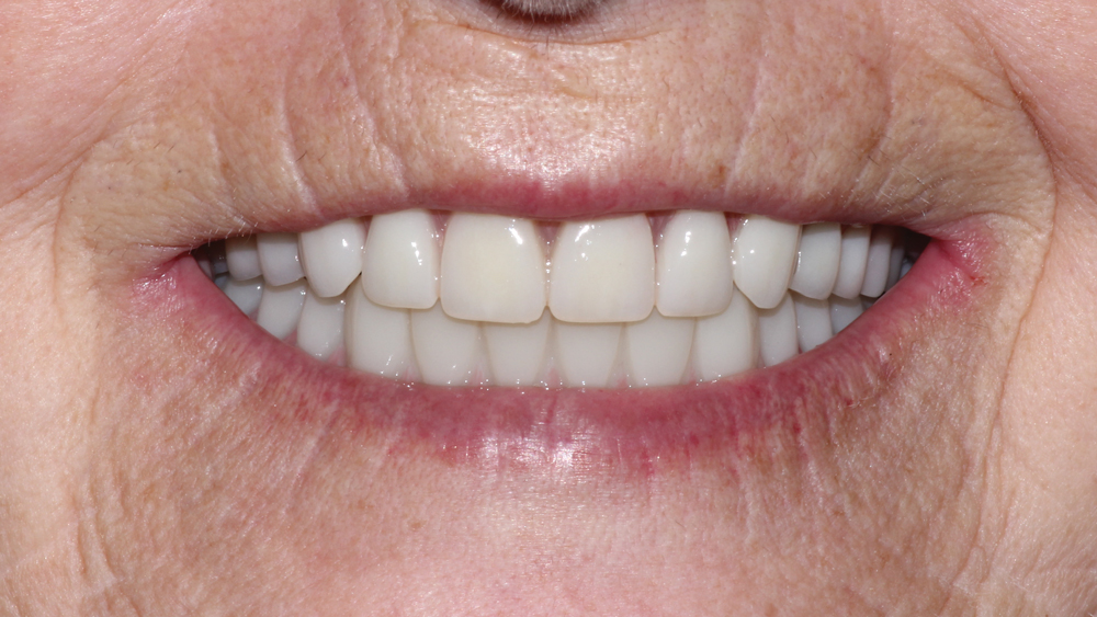
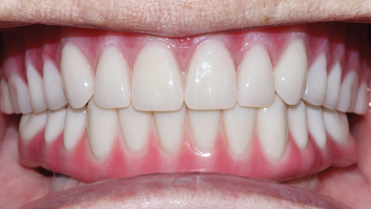
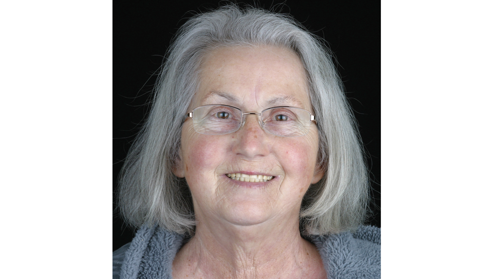
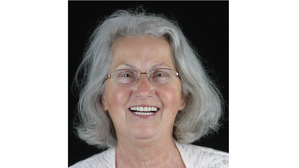
Figures 26a, 26b: Before and after images illustrate the transformation provided by the new maxillary complete denture and the mandibular full-arch BruxZir restoration — a combination that met the functional and esthetic needs of the patient, who now enjoys substantially improved chewing capability, comfort, confidence and quality of life.
- 1
- 2
- 3
- 4
- 5
- 6
- 7
- 8
- 9
- 10
- 11
- 12
- 13
- 14
- 15
- 16
- 17
- 18
- 19
- 20
- 21
- 22
- 23
- 24
- 25
- 26
- 27
- 28
- 29
- 30
- 31
- 32
- 33
The dual-arch restorations addressed the functional and esthetic challenges of the case while staying within the financial means of the patient.
CONCLUSION
When providing treatment for the edentulous arch, the ability to offer traditional removable dentures as well as implant-supported prostheses affords the flexibility required to address the various needs of patients. By carefully considering the oral health, anatomy and financial circumstances of each case, clinicians can devise a treatment plan that effectively restores form and function within the budget of the patient.
Available CE Course
References
- ^Zembic A, Wismeijer D. Patient-reported outcomes of maxillary implant-supported overdentures compared with conventional dentures. Clin Oral Implants Res. 2014 Apr;25(4):441-50.
- ^Turkyilmaz I, Company AM, McGlumphy EA. Should edentulous patients be constrained to removable complete dentures? The use of dental implants to improve the quality of life for edentulous patients. Gerodontology. 2010 Mar;27(1):3-10.
- ^Brennan M, Houston F, O’Sullivan M, O’Connell B. Patient satisfaction and oral health-related quality of life outcomes of implant overdentures and fixed complete dentures. Int J Oral Maxillofac Implants. 2010 Jul-Aug;25(4):791-800.
- ^Preciado A, Del Río J, Lynch CD, Castillo-Oyagüe R. Impact of various screwed implant prostheses on oral health-related quality of life as measured with the QoLIP-10 and OHIP-14 scales: a cross-sectional study. J Dent. 2013 Dec;41(12):1196-207.
- ^Misch CE. Contemporary implant dentistry. 3rd ed. St. Louis: Mosby; 2007. p. 367.
- ^Colomina LE. Immediate loading of implant-fixed mandibular prostheses: a prospective 18-month follow-up clinical study–preliminary report. Implant Dent. 2001;10(1):23-9.
- ^Najafi H, Siadat H, Akbari S, Rokn A. Effects of immediate and delayed loading on the outcomes of all-on-4 treatment: a prospective study. J Dent (Tehran). 2016 Nov;13(6):415-22.