Chairside CAD/CAM

July 6, 2026
Chairside CAD/CAM

When chairside CAD/CAM was pioneered in the 1980s by Swiss dentist Dr. Werner Mörmann and colleagues, the system held the potential for transforming restorative dentistry by replacing multi‑appointment, lab‑dependent workflows with same‑day production. But technological hurdles prevented widespread adoption of the procedure.

Advances in intraoral scanning, computing power, materials science, and precision manufacturing have made chairside CAD/CAM far more reliable and accessible over time. One of the fastest areas of digital adoption has occurred in digital scanning, which now accounts for over 70% of impressions received by Glidewell dental lab. Still, adoption is not universal, because some chairside CAD/CAM systems remain costly, complex, or difficult to integrate with existing clinical and laboratory processes.

Improved versions of chairside CAD/CAM, such as the glidewell.io In-Office Solution, reflect a broader shift toward simplifying clinical production. Rather than treating chairside milling as a standalone tool, the latest systems seek to combine scanning, design, and fabrication into cohesive digital ecosystems that draw inspiration from large‑scale manufacturing principles.

Designed to ease the transition from traditional lab workflows to streamlined in‑office production, glidewell.io is informed by a laboratory perspective. It makes it possible to scan, design with AI, and mill using a single, intuitive process shaped around how clinicians diagnose and plan care, reducing the need to engage with complex software.

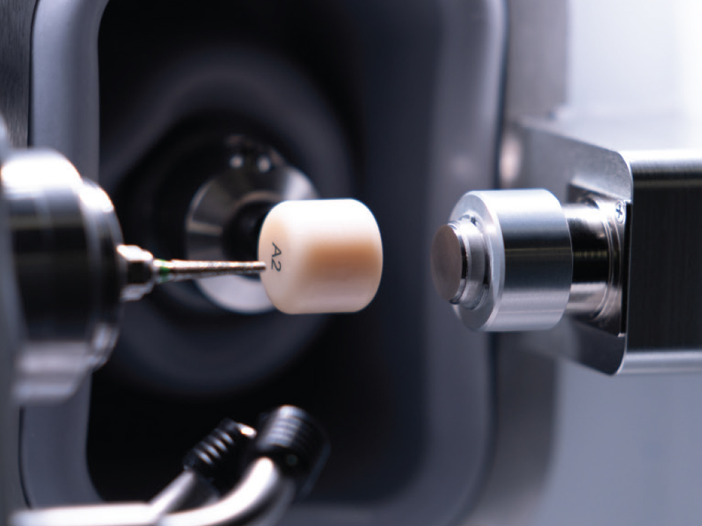
At the center of this ecosystem is a compact, high‑speed in‑office mill supported by design software that guides clinicians from scan to proposal to final restoration through a structured, user‑friendly workflow.

As materials advance and software becomes even more intuitive, the line between operatory and laboratory continues to blur. Practices can increasingly choose when to produce restorations in‑office or rely on outside fabrication, a hybrid approach that reflects the future of restorative dentistry.
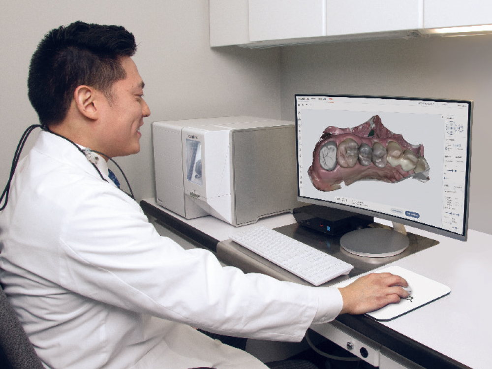
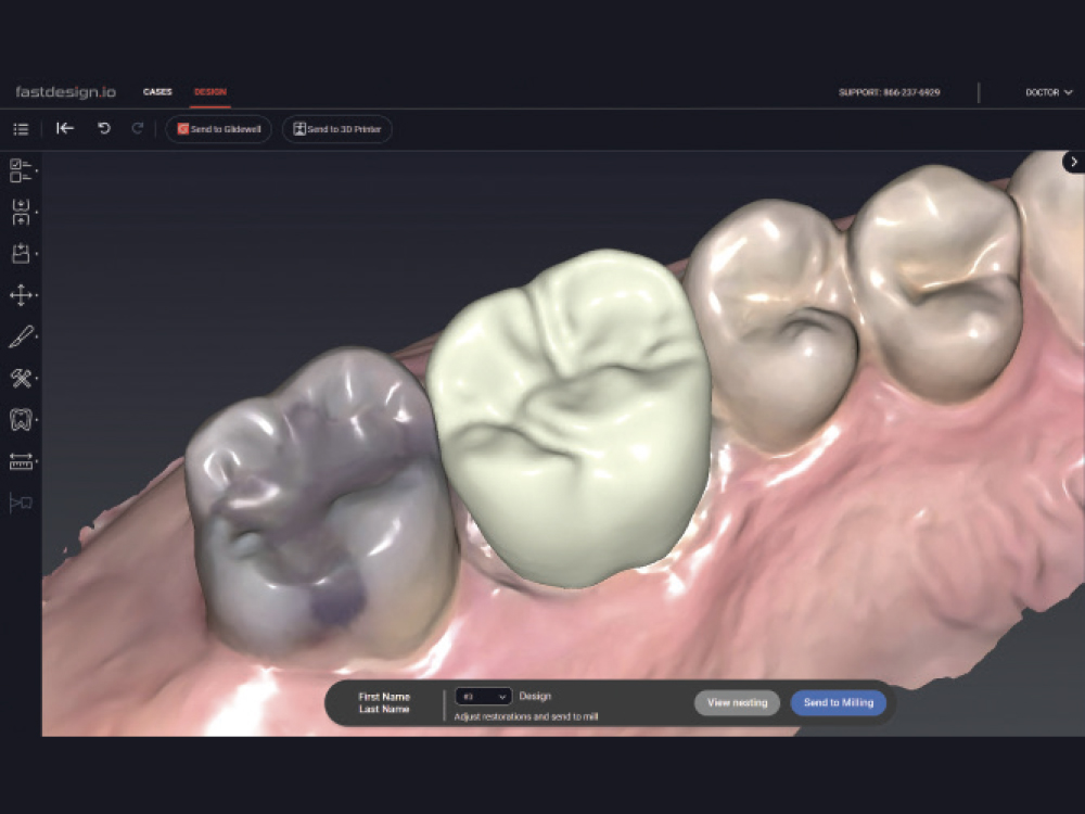
The glidewell.io solution is informed by a laboratory perspective
At the center of this ecosystem is a compact, high‑speed in‑office mill supported by design software
Practices can increasingly choose when to produce restorations in‑office or rely on outside fabrication
Same-Day, Multiple Restorations

Same-Day, Multiple Restorations

Dr. Chi

by Justin Chi, DDS, CDT

Director of Clinical Technologies, Glidewell

Irvine, California

CASE REPORT

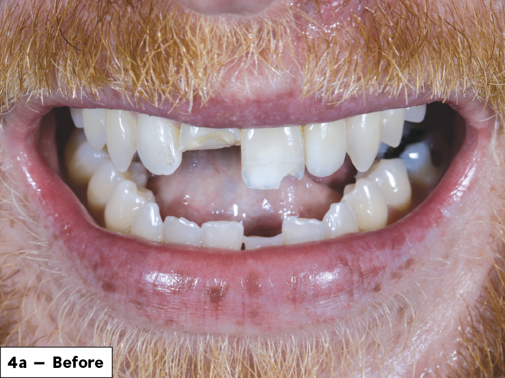
Gregory came to our practice following facial trauma sustained during an altercation. Clinical examination revealed multiple anterior tooth fractures involving both maxillary and mandibular anterior teeth. Comprehensive treatment of Gregory’s injuries required a digital approach to manage multiple restorations, including a BruxZir® Esthetic NOW crown for tooth #8 and CAMouflage® NOW incisal veneer on #24. The smaller incisal fractures on #9, #23, and #26 were restored using direct composite resin. By starting treatment with the direct restorations, I was able to utilize digital copy functions on the design software to mirror contralateral anatomical contours for the full crowns.
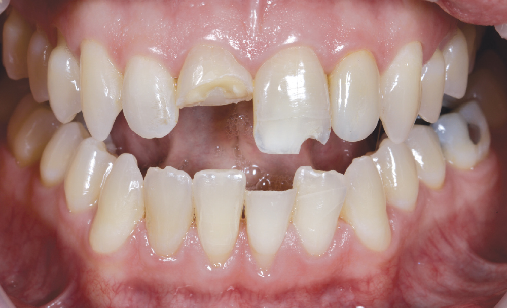
he initial consultation determined that tooth #8 exhibited a horizontal crown fracture involving approximately 50% of the clinical crown. Tooth #9 demonstrated a distoincisal fracture with rounded enamel-dentin involvement and loss of incisal edge continu

Figure 1: The initial consultation determined that tooth #8 exhibited a horizontal crown fracture involving approximately 50% of the clinical crown. Tooth #9 demonstrated a distoincisal fracture with rounded enamel-dentin involvement and loss of incisal edge continuity.

In the mandibular anterior region, tooth #24 presented with a horizontal fracture of the incisal third. Minor enamel fractures were found on the incisal edges on teeth #23 and #26. No obvious displacement of the teeth was observed clinically, and soft-tissue examination revealed no active lacerations.

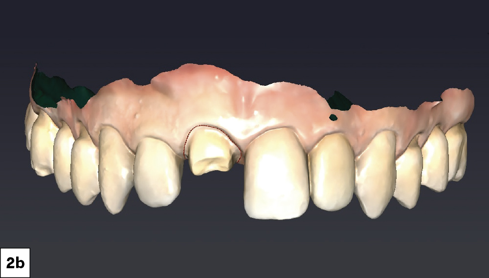
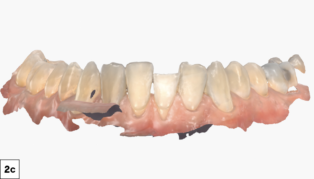
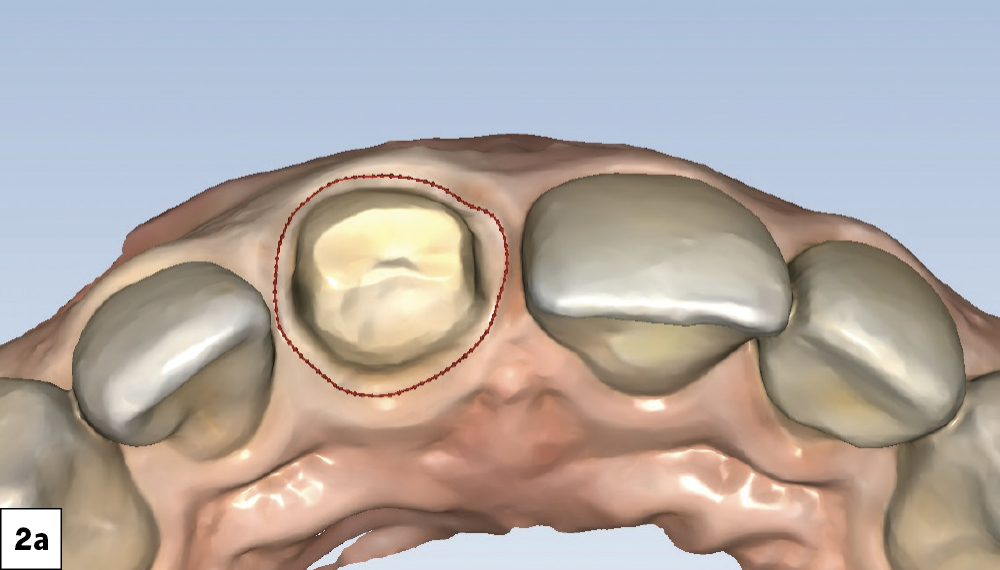
 After eliminating undercuts, ensuring optimal clearance from the opposing teeth, and rounding the internal line angles, the case was ready for intraoral scanning
Full-arch digital impressions were obtained using the fastscan.io™ Scanning Solution featuring Medit® i900.
The objective was to capture contaminant-free scans with clearly exposed finish lines while fully capturing neighboring teeth and proximal contact areas.

Figures 2a–2c: After eliminating undercuts, ensuring optimal clearance from the opposing teeth, and rounding the internal line angles, the case was ready for intraoral scanning. Full-arch digital impressions were obtained using the fastscan.io Scanning Solution featuring Medit® i900®. The objective was to capture contaminant-free scans with clearly exposed finish lines while fully capturing neighboring teeth and proximal contact areas.

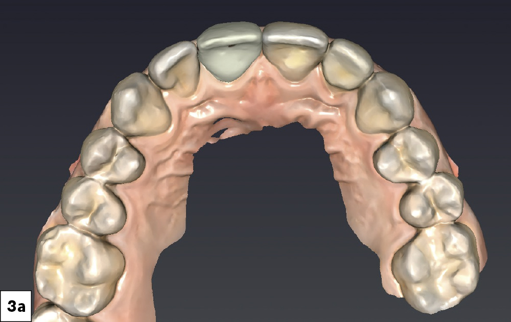
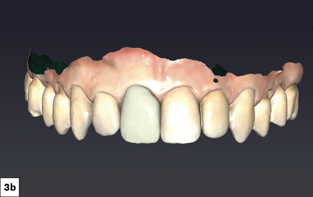
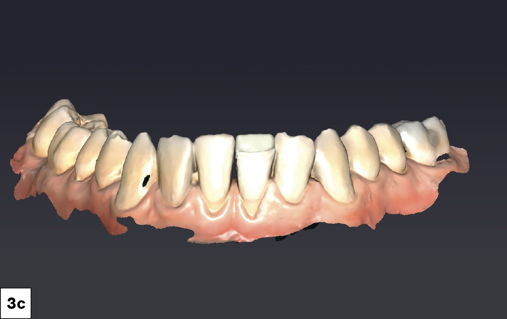
I utilized the fastdesign.io mirror function to create restorative designs that establish symmetrical contours between teeth #8 and #9 as well as for #24 and #25. I also verified proximal contacts and occlusion prior to milling.

Figures 3a–3c: I utilized the fastdesign.io mirror function to create restorative designs that establish symmetrical contours between teeth #8 and #9 as well as for #24 and #25. I also verified proximal contacts and occlusion prior to milling.

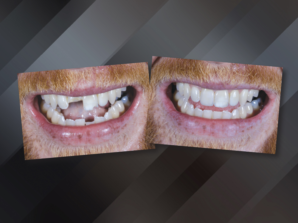
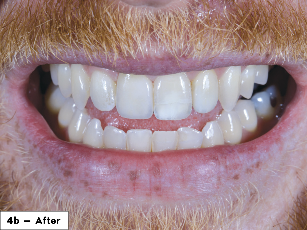
We were able to successfully restore Gregory’s smile in one visit

Figures 4a, 4b: I was able to provide a functional, esthetic solution quickly that completely satisfied the patient.

CONCLUSION

We were able to successfully restore Gregory’s smile in one visit with a combination of direct composite restorations and chairside milling that included a BruxZir Esthetic NOW crown and CAMouflage NOW veneer.
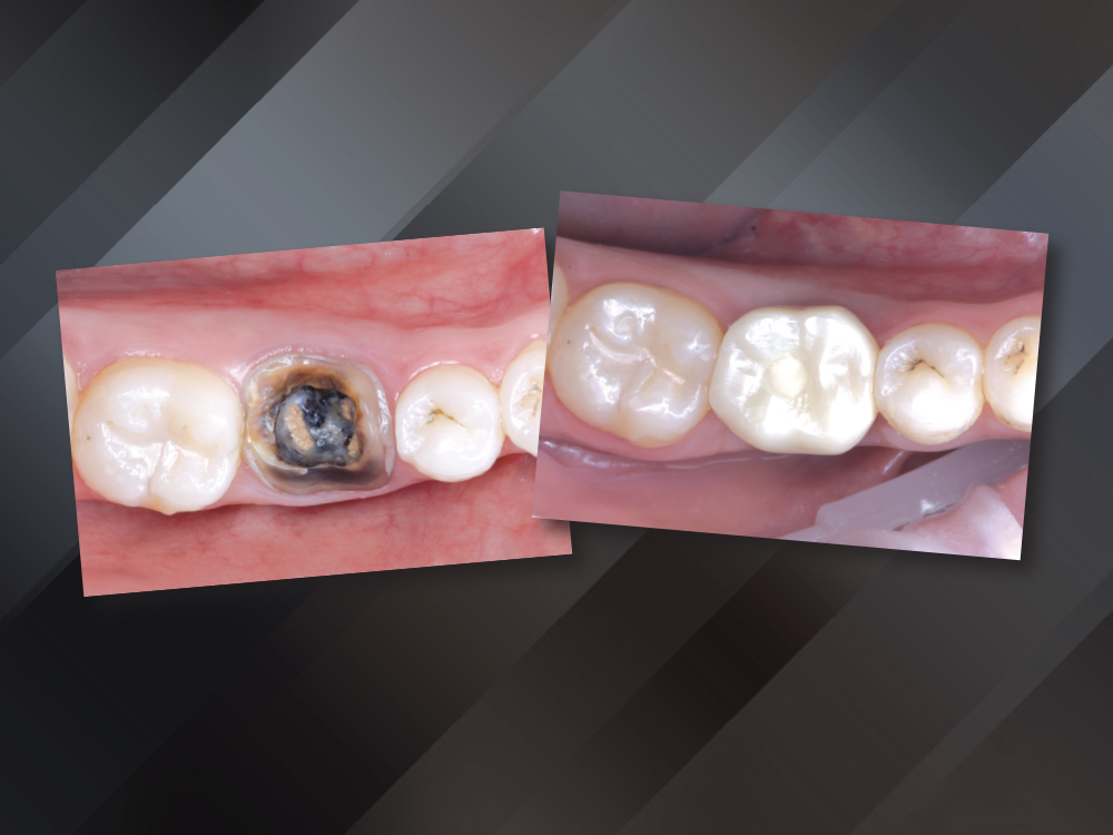
Extraction  and Immediate Implant Placement
Extraction and Immediate Implant Placement

CASE REPORT

Tan Nguyen presented with a fractured molar that was nonrestorable. The treatment was tailored to the patient’s need for a quick and long-lasting restoration, and involved extraction and immediate guided implant placement using a plan provided by the Glidewell Digital Treatment Planning (DTP) team.

APPOINTMENT #1 (RECORD COLLECTION)
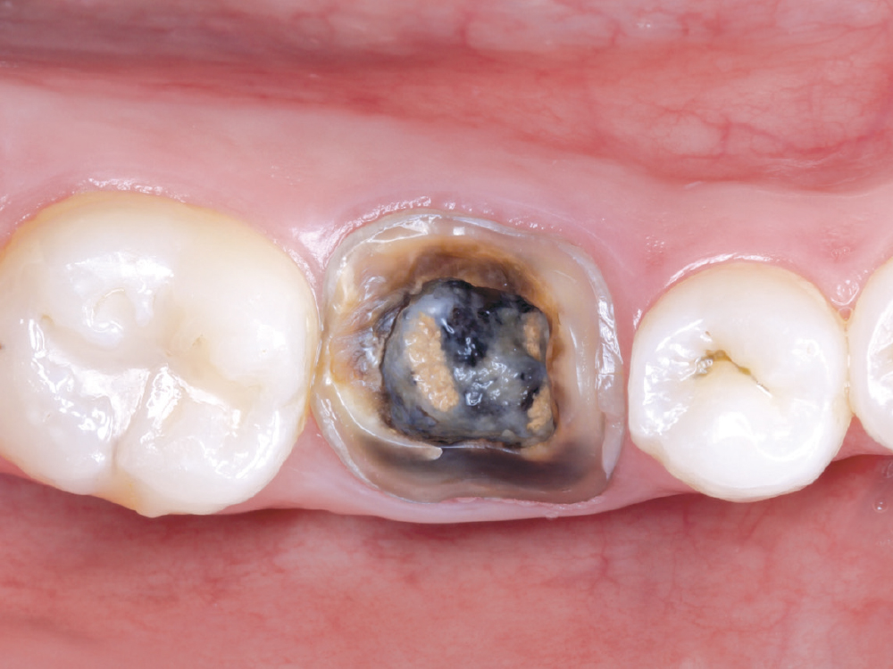
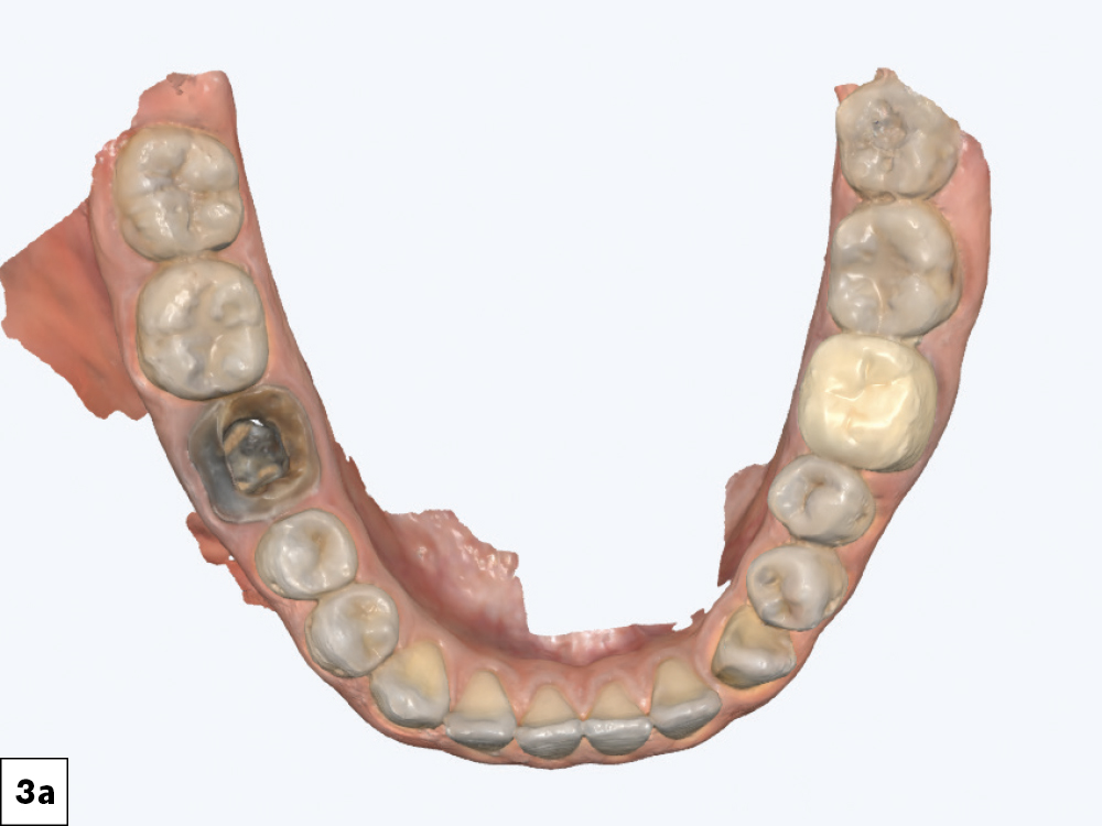
An intraoral scan and CBCT were performed.  Tooth #30 had a missing crown restoration, recurrent caries

Figure 1: An intraoral scan and CBCT were performed. Tooth #30 had a missing crown restoration, recurrent caries, as well as unresolved endodontic lesions. The tooth was deemed to be unrestorable.

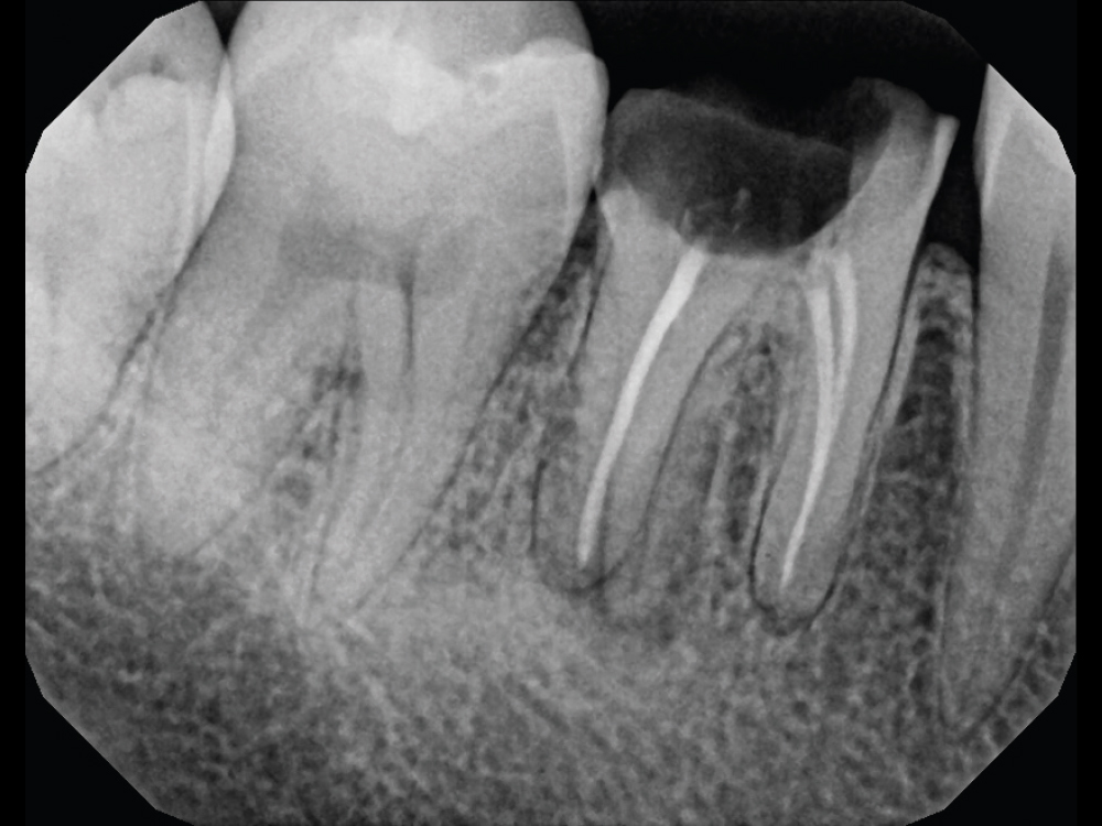
The PA radiograph revealed periapical radiolucency.

Figure 2: The PA radiograph revealed periapical radiolucency.

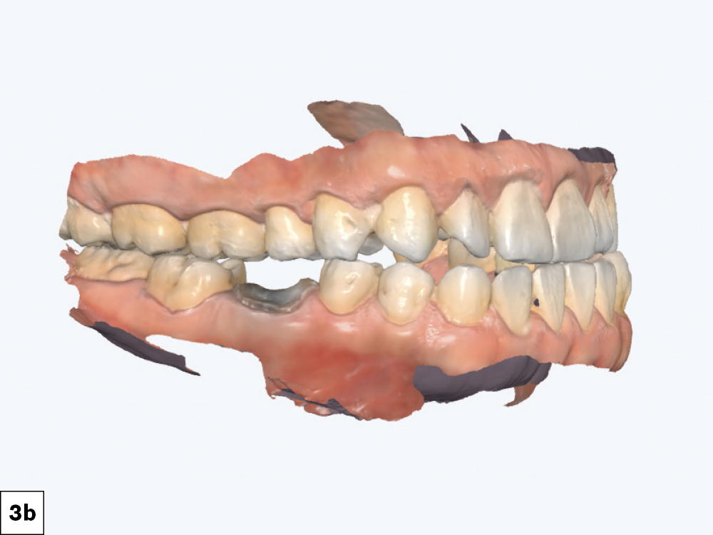
Intraoral scans were taken using the fastscan.io Scanning Solution featuring Medit i900
he intraoral scan and CBCT are required records for the DTP team who received the digital files along with the request for an immediate guided implant placement treatment plan.

Figures 3a, 3b: Intraoral scans were taken using the fastscan.io Scanning Solution featuring Medit i900. The intraoral scan and CBCT are required records for the DTP team who received the digital files along with the request for an immediate guided implant placement treatment plan.

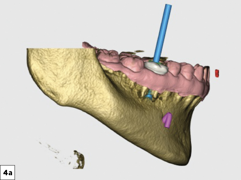
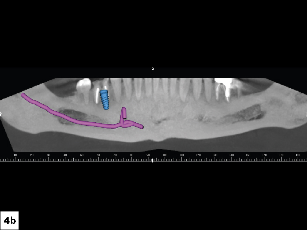
he approved digital treatment plan indicated exact implant location and confirmed there was adequate bone
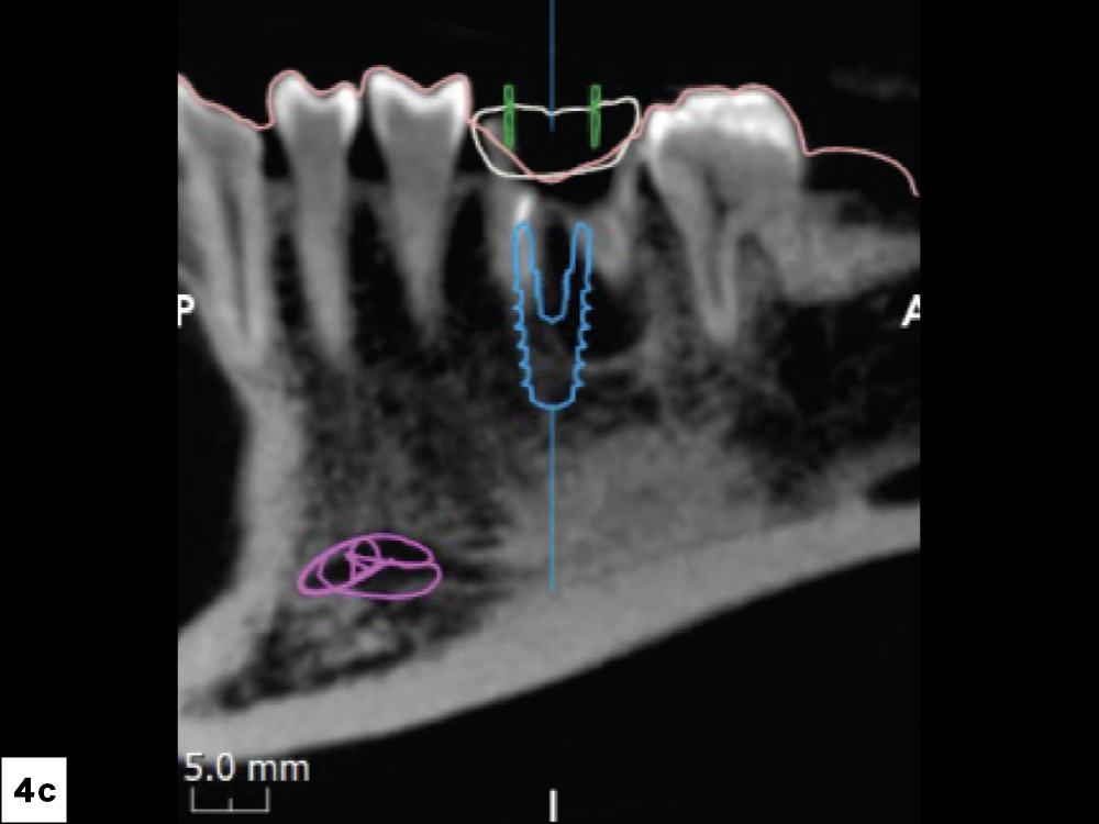
The approved digital treatment plan indicated exact implant location and confirmed there was adequate bone to place a  5 × 11.5 mm Glidewell HT™ Implant

Figures 4a–4c: The approved digital treatment plan indicated exact implant location and confirmed there was adequate bone to place a 5 × 11.5 mm Glidewell HT Implant (Glidewell Direct; Irvine, Calif.) immediately following extraction.

APPOINTMENT #2 (EXTRACTION AND IMMEDIATE IMPLANT PLACEMENT)
The surgical site was then grafted using Newport Biologics™ Mineralized Cortico/Cancellous Allograft Blend
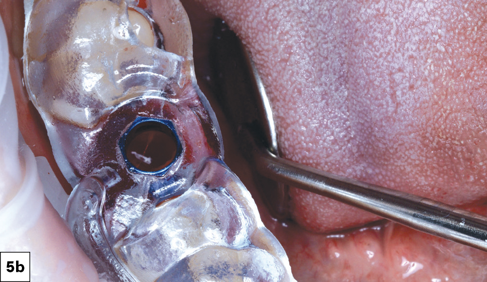
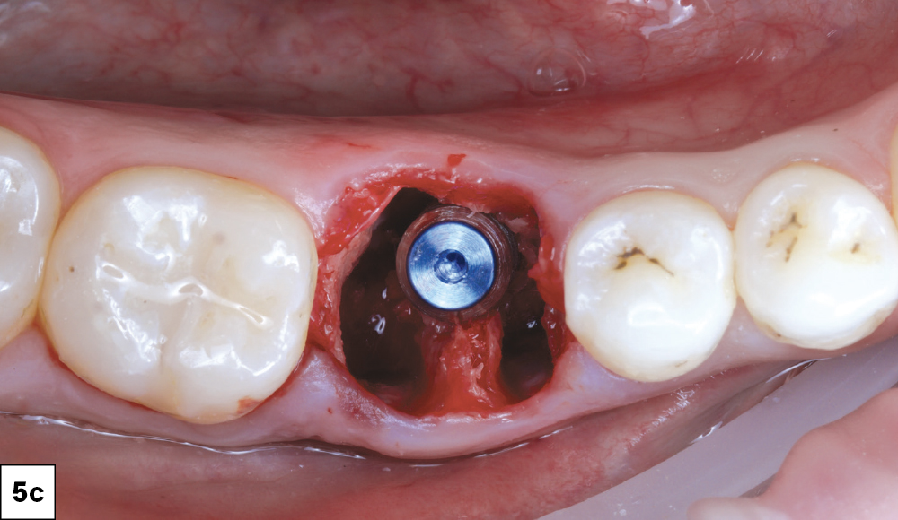
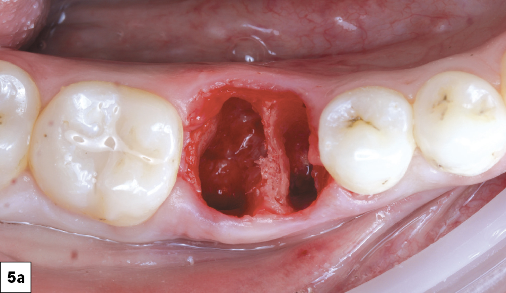
The tooth was carefully extracted and followed by socket debridement and curettage to remove all granulation tissue.
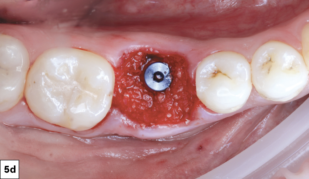
The surgical site was then grafted using Newport Biologics™ Mineralized Cortico/Cancellous Allograft Blend

Figures 5a–5d: The tooth was carefully extracted and followed by socket debridement and curettage to remove all granulation tissue. A surgical guide was used for implant placement, with adequate primary stability. The surgical site was then grafted using Newport Biologics™ Mineralized Cortico/Cancellous Allograft Blend (Glidewell Direct) to support optimal bone regeneration.

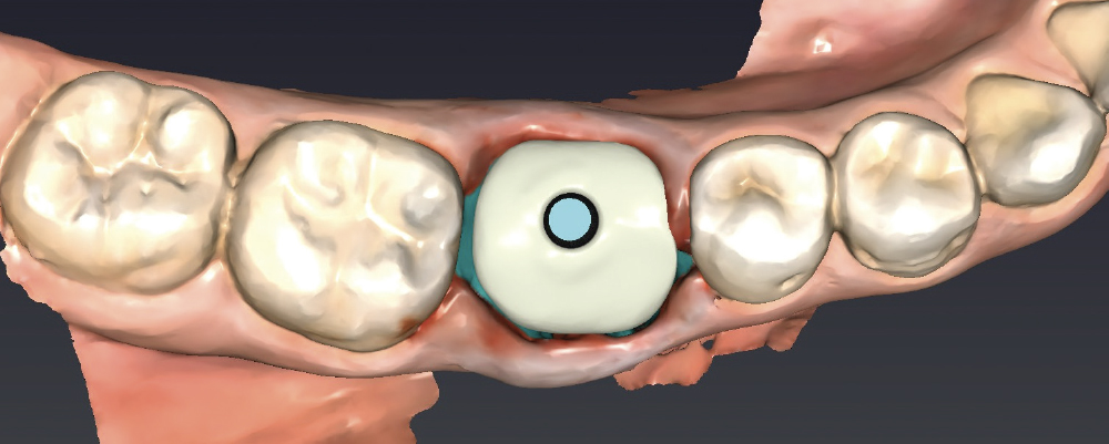
A custom healing abutment was created to mimic the gingival margins of the patient’s original tooth #30 using fastdesign.io™ Software and Design Station.

Figure 6: A custom healing abutment was created to mimic the gingival margins of the patient’s original tooth #30 using the fastdesign.io Software and Design Station.

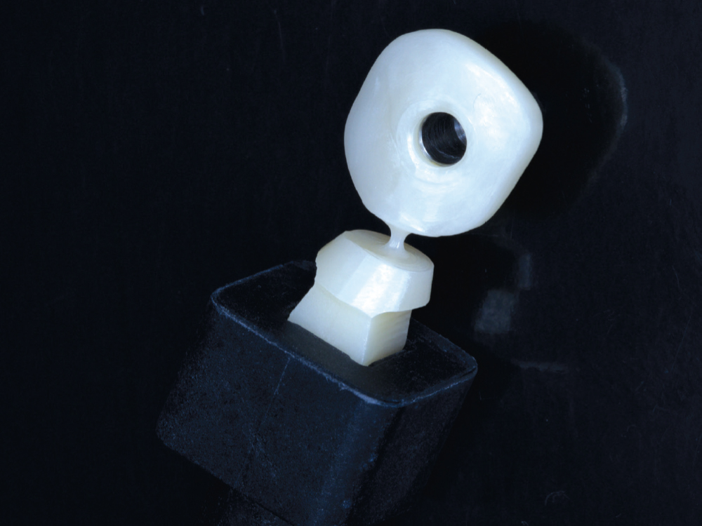
 The custom healing abutment was milled chairside in 30 minutes using a BruxZir® NOW Milling Block.

Figure 7: The custom healing abutment was milled chairside in 30 minutes using a BruxZir® NOW Milling Block.

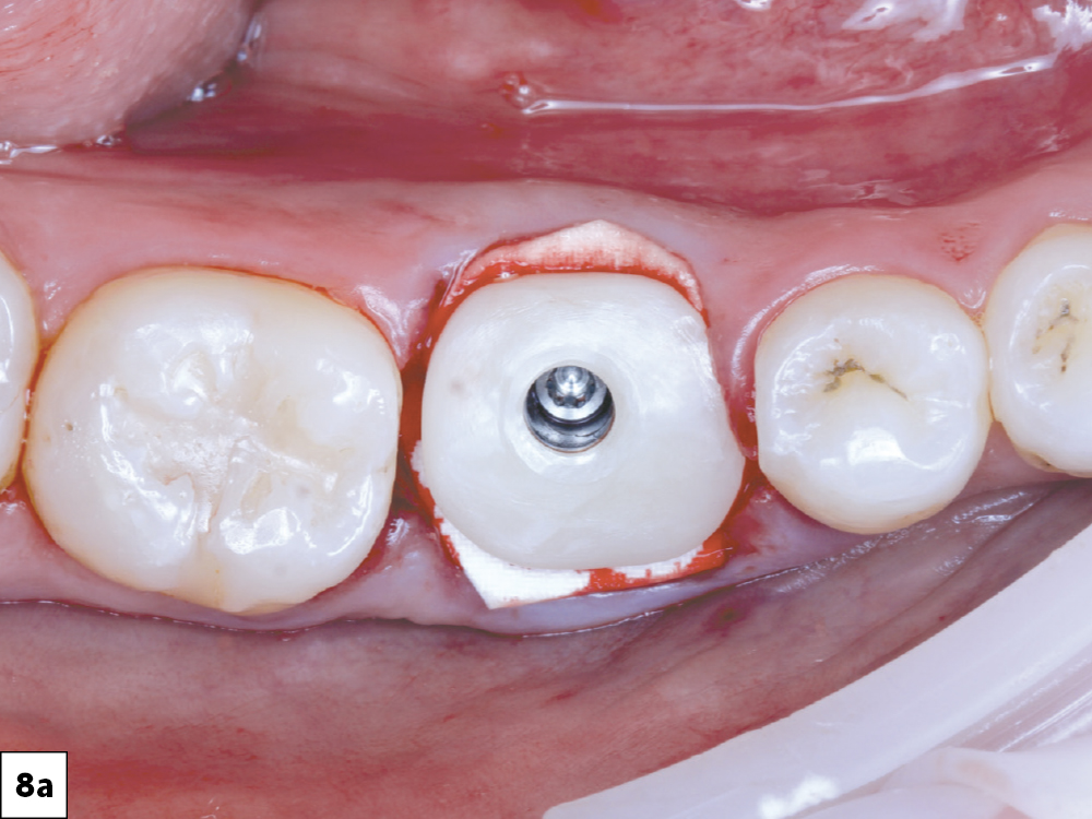
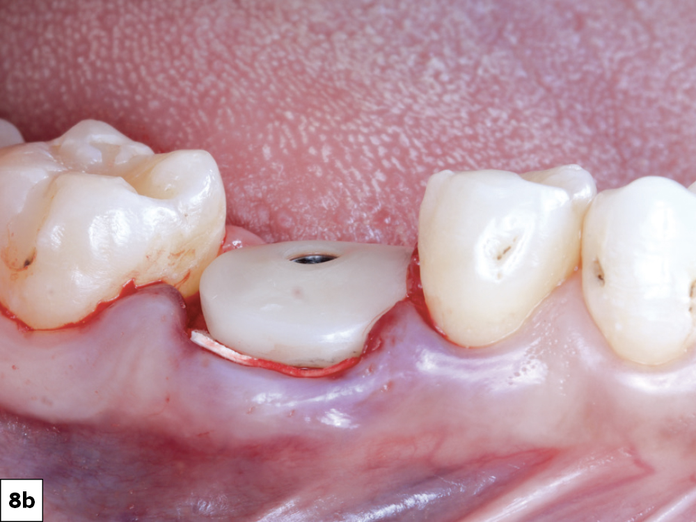
The custom healing abutment was seated after placing a layer of Newport Surgical Collagen Membrane 4–6 (Glidewell Direct) over the allograft.
This membrane is designed to remain effective for 4–6 months, preventing the migration of soft tissue into the healing implant site.

Figures 8a, 8b: The custom healing abutment was seated after placing a layer of Newport Surgical Collagen Membrane 4–6 (Glidewell Direct) over the allograft. This membrane is designed to remain effective for 4–6 months, preventing the migration of soft tissue into the healing implant site.

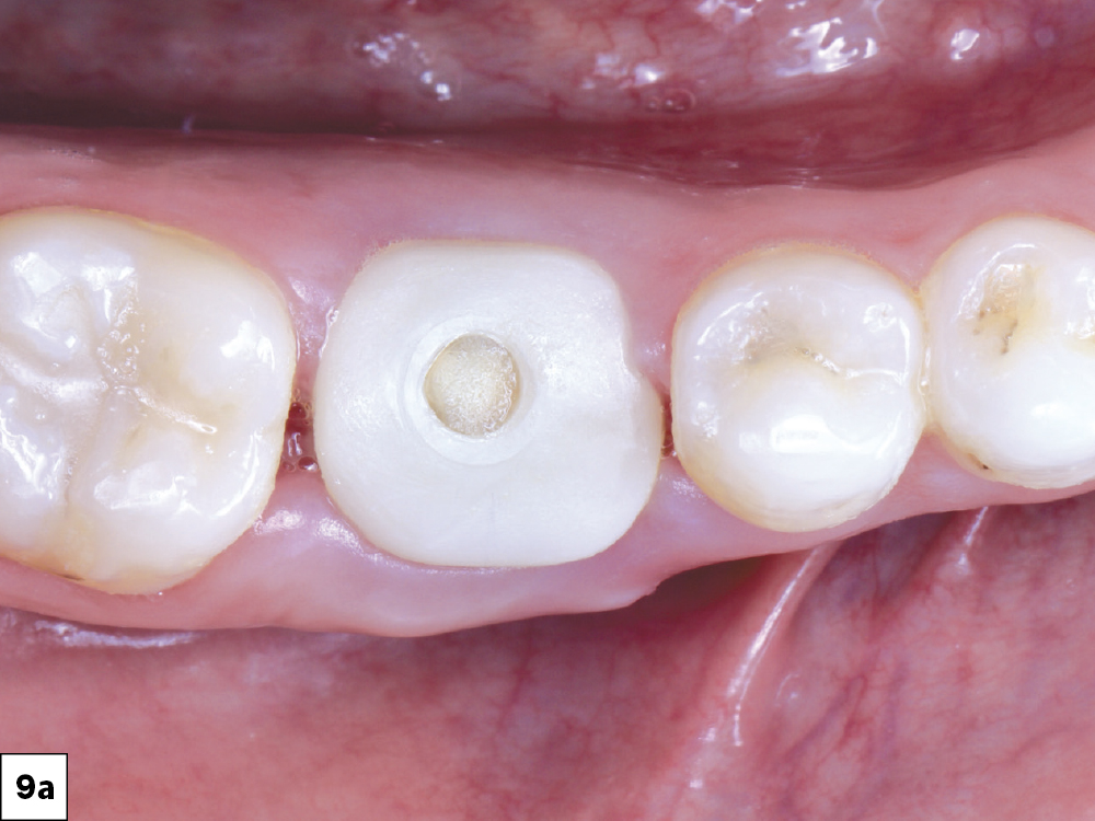
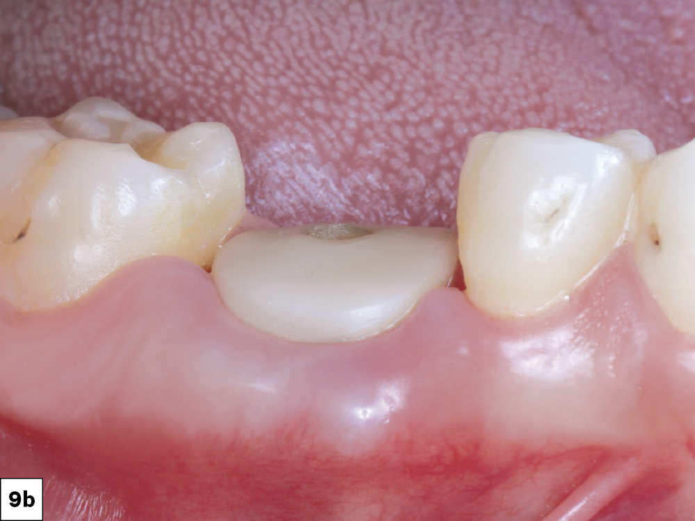
Two-week follow-up showed excellent resolution of the surgical site.

Figures 9a, 9b: Two-week follow-up showed excellent resolution of the surgical site.

APPOINTMENT #3 (FOUR MONTHS LATER)
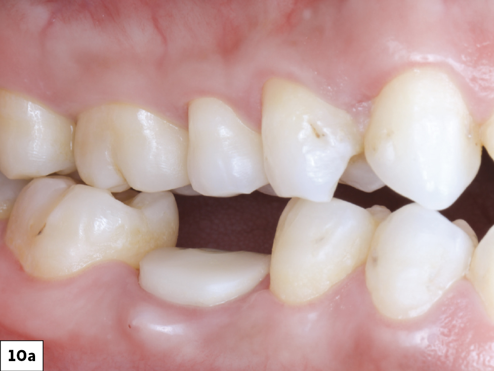
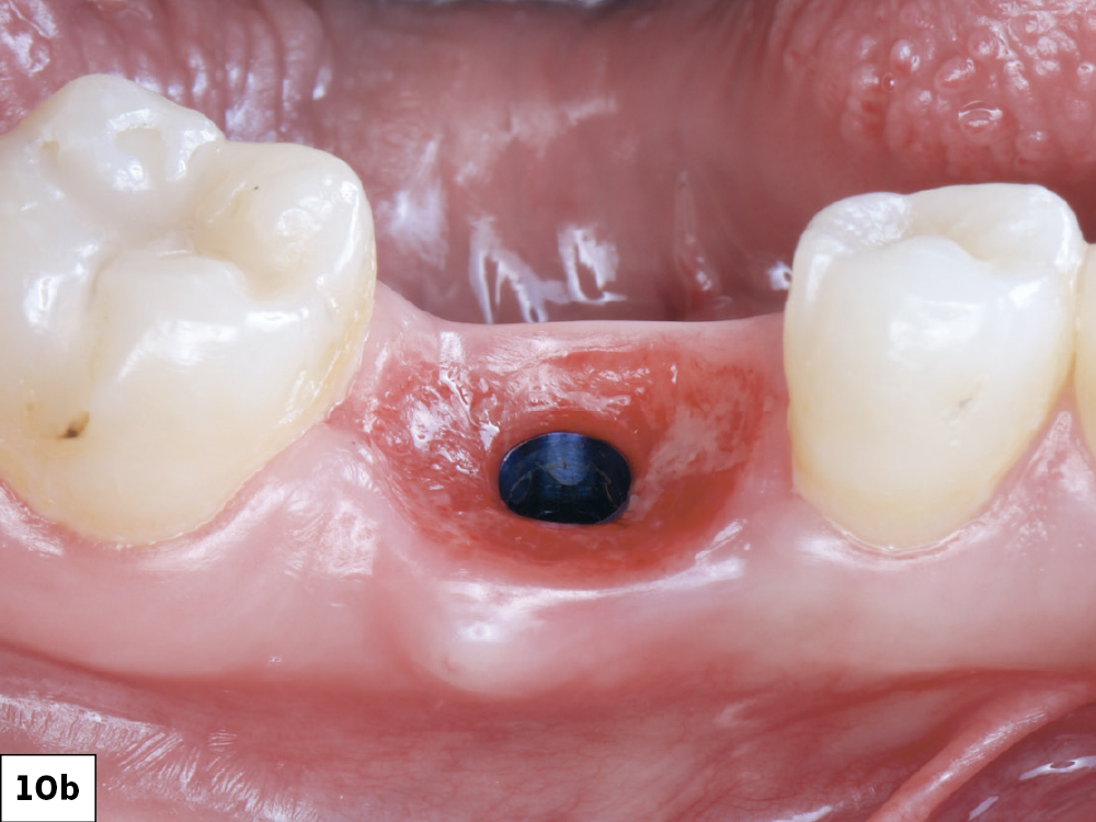
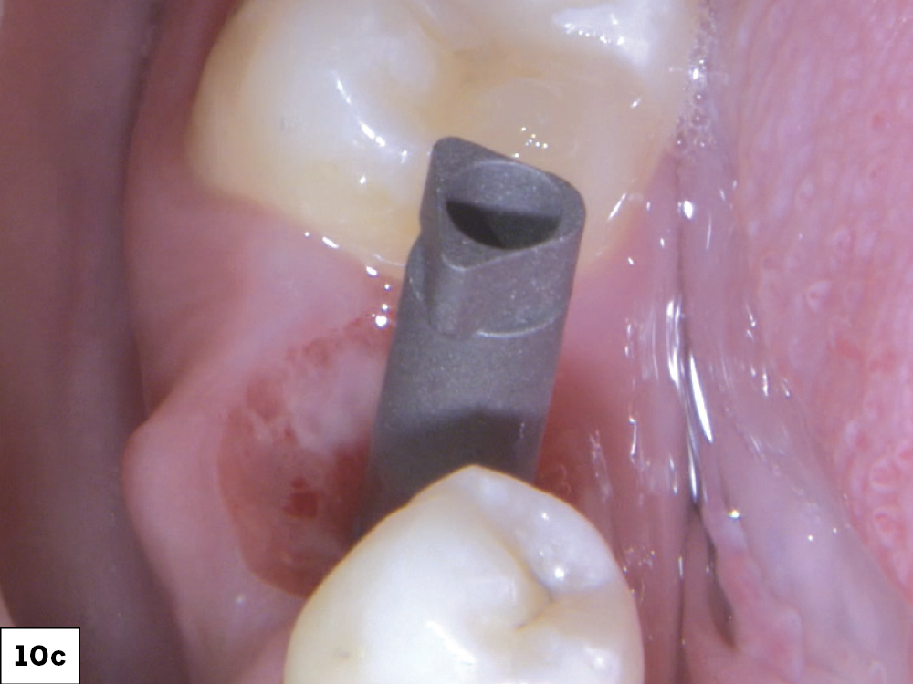
The healing abutment was removed, and a scan body was seated

Figures 10a–10c: The healing abutment was removed, and a scan body was seated; the favorable gingival architecture established by the custom healing abutment allowed for optimal emergence profile development in the restorative design.

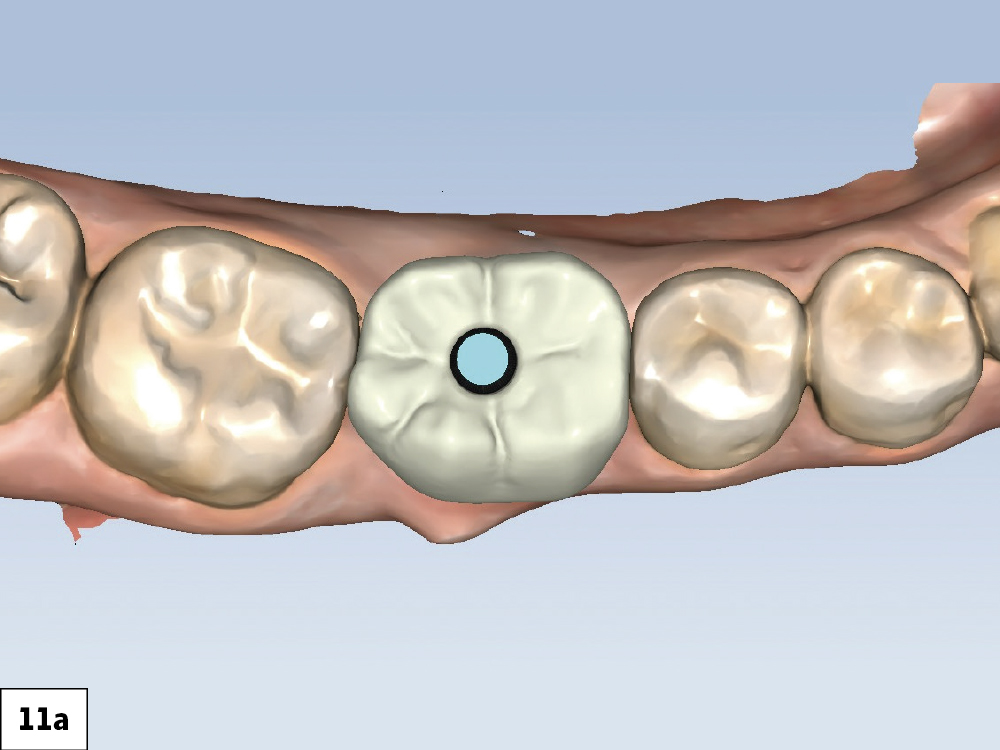
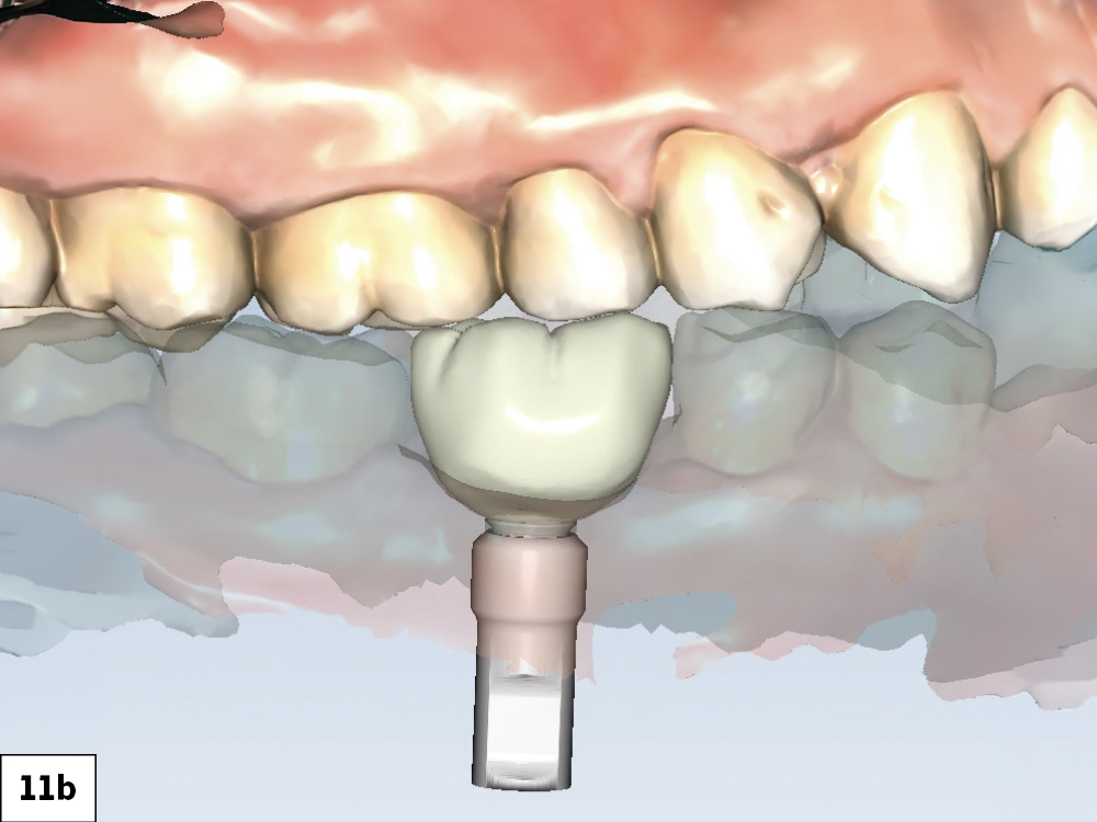
Chairside design using fastdesign.io software helped determine the ideal anatomy of the BruxZir NOW screw-retained crown prior to milling.

Figures 11a, 11b: Chairside design using fastdesign.io software helped determine the ideal anatomy of the BruxZir NOW screw-retained crown (SRC) prior to milling.

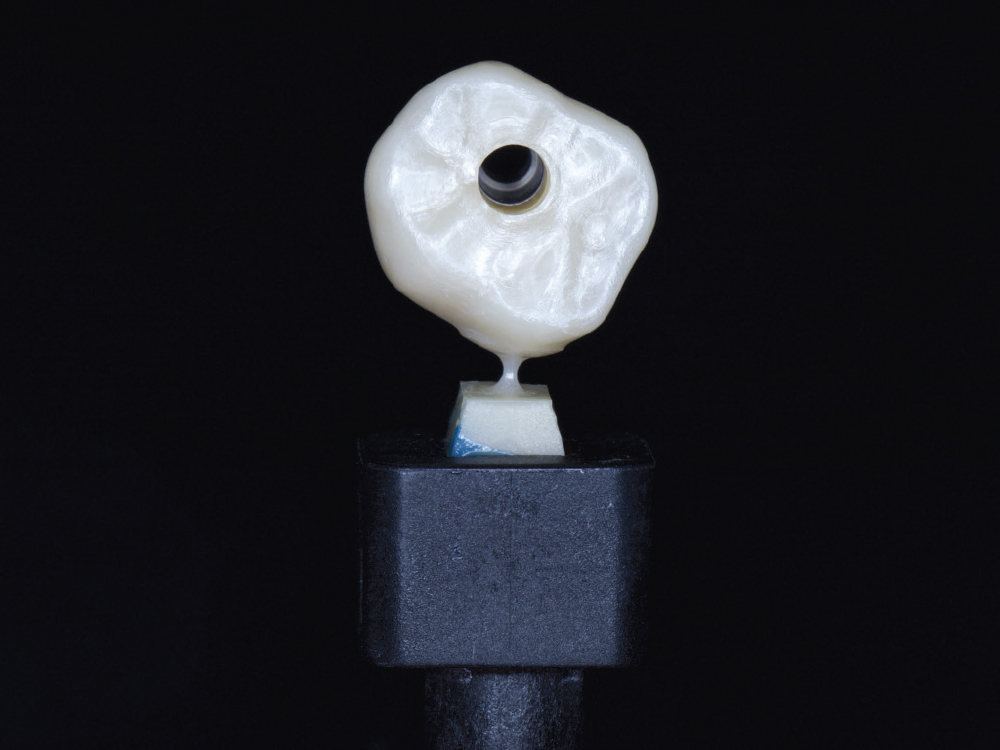
 Chairside-milled BruxZir NOW screw-retained crown.

Figure 12: Chairside-milled BruxZir NOW SRC.

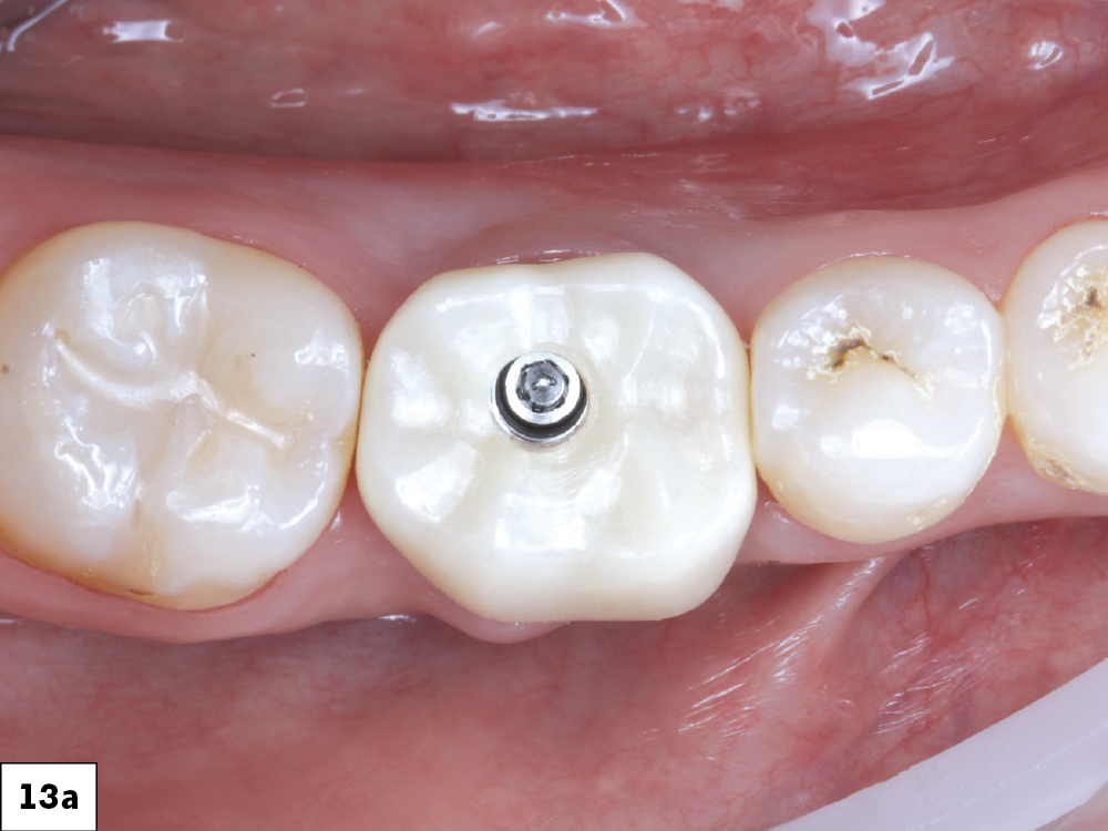
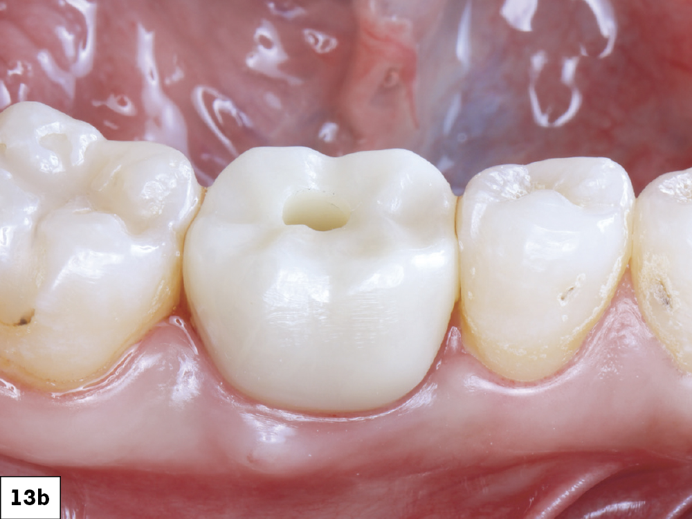
The BruxZir SRC was seated, torqued to 35 Ncm, and the screw access sealed with Teflon tape and composite resin.

Figures 13a, 13b: The BruxZir SRC was seated, torqued to 35 Ncm, and the screw access sealed with Teflon tape and composite resin.

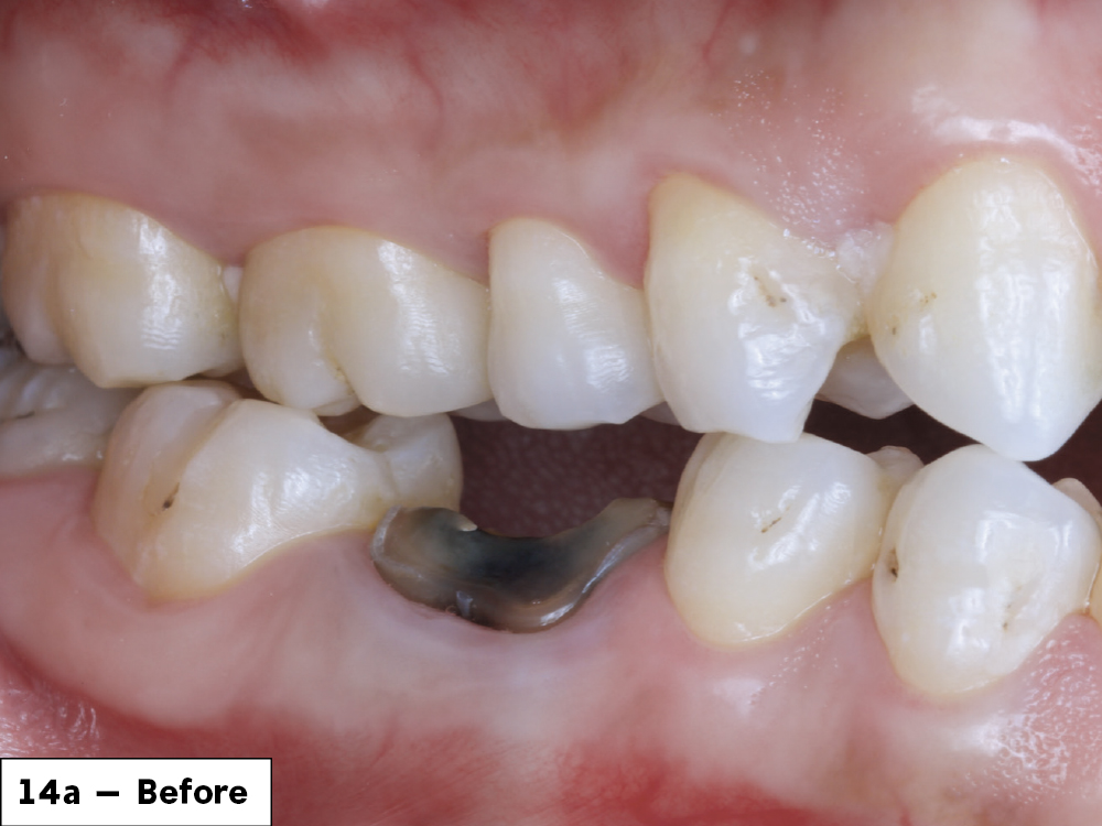
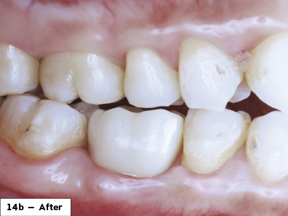
The patient left with a functional and esthetic tooth replacement and was extremely happy with the result.

Figures 14a, 14b: The patient left with a functional and esthetic tooth replacement and was extremely happy with the result.

CONCLUSION

The patient’s lower first molar was successfully replaced using modern digital workflows that included intraoral scanning, digital treatment planning, fastdesign.io software, and a custom healing abutment. The final BruxZir NOW SRC was milled and delivered in less than one hour.

Medit is a registered trademark of Medit Corporation.
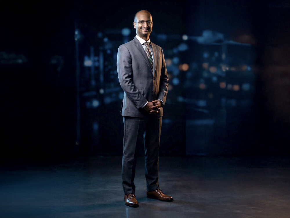
Prabhakar  Thirugnanasambandam

The Innovator

Prabhakar Thirugnanasambandam

VP of Materials & Hardware Manufacturing

A Bachelor’s in Mechanical Engineering, early experience in high‑precision luxury watch production, and a Master’s in Mechanical Engineering, Controls, and Automation from Utah State University helped Prabhakar build a foundation in accuracy and scalable output. He spent more than a decade developing advanced silicon nitride ceramic medical implants, and for the past 12 years at Glidewell, he has transformed complex ideas into reliable, high‑volume manufacturing systems, driving innovation from concept to production.