Materials Science R&D

July 6, 2026
MATERIALS SCIENCE R&D

The accelerating evolution of dental materials has been transformative for the practice of dentistry. Reliance on a limited number of metal‑based restorative materials has expanded into a cascade of ceramics, polymers, and hybrid composites, each engineered for strength, esthetics, biocompatibility, and longevity.

The materials science division at Glidewell, led by Vice President of Materials R&D, Raj Malyala, Ph.D., operates at the intersection of chemistry, engineering, biology, and clinical reality. As Malyala explains, “Dentistry is fundamentally a materials‑driven discipline that directly determines how restorations look, feel, and perform in the mouth.” 

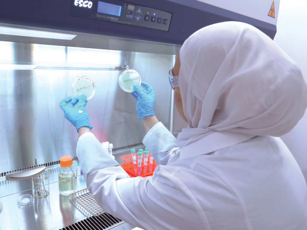
Materials undergo rigorous testing, from million‑cycle chewing simulations to microscopic surface analysis, ensuring every product meets strict standards for safety, durability, and biological compatibility. This scientific rigor is not academic excess; it’s essential because dental restorations reside in one of the body’s most complex microbiomes, where material composition plays a critical role in maintaining good oral health.

Advances in materials science translate directly into confidence and consistency. Stronger ceramics and optimized zirconia formulations mean restorations that resist fracture while maintaining lifelike esthetics. Equally important, good material design supports chairside efficiency. Fully sintered, mill‑ready ceramics and faster‑processing materials can shorten appointments, reduce remakes, and simplify workflows without sacrificing performance.

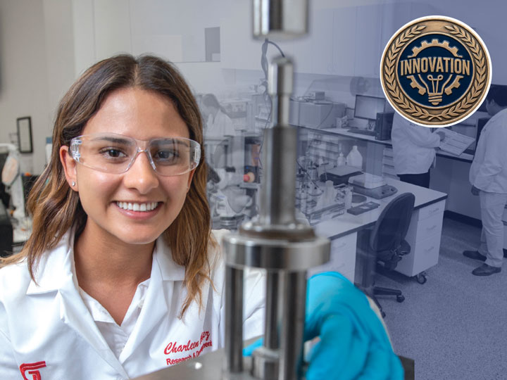
Powered by a multidisciplinary team of 60 scientists and engineers, including Ph.D.s in materials science, chemistry, and biomedical fields, Glidewell’s Materials R&D team pioneers preventive and restorative materials across the dental spectrum, from resins for 3D‑printed nightguards and dentures to advanced ceramics like BruxZir® Full-Strength, BruxZir Esthetic and BruxZir Fusion.
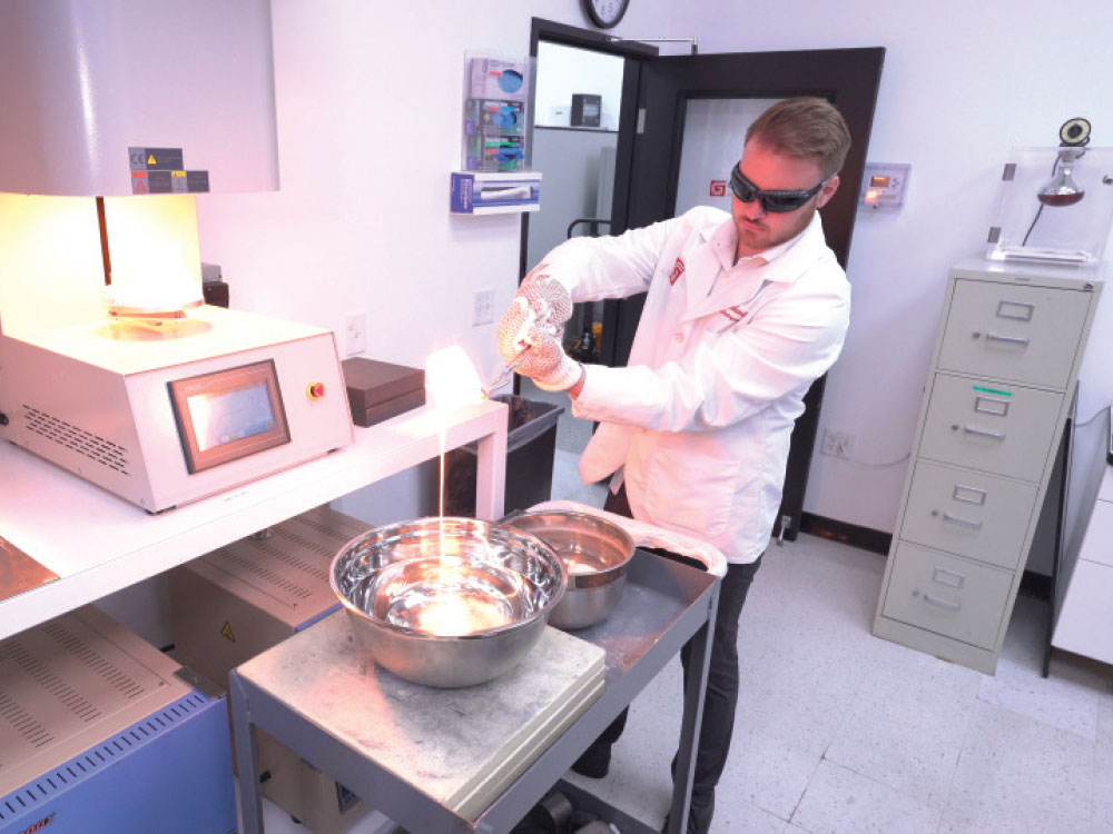
The materials science division at Glidewell
Powered by a multidisciplinary team of 60 scientists and engineers
Helping a Self-Conscious Patient Regain Her Smile

Helping a Self-Conscious Patient Regain Her Smile

Dr Taylor Manalili

by Taylor Manalili, DDS

Director of Clinical Prosthodontics, Glidewell

Irvine, California

CASE REPORT

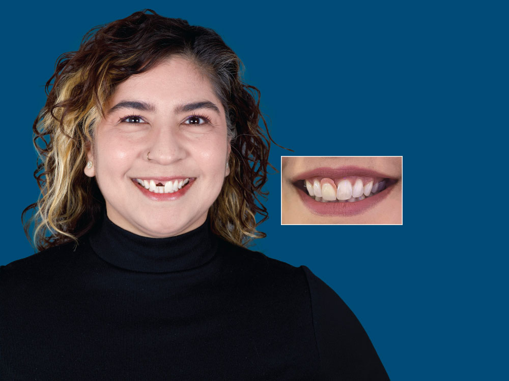
Melissa, a manager in Glidewell’s Lab Operations department, arrived at our clinic missing tooth #8. She was wearing an ill-fitting removable partial denture and expressed dissatisfaction with its appearance and lack of stability. Melissa was seeking a fixed, long-term solution to restore both the function of the missing tooth and confidence in her smile.
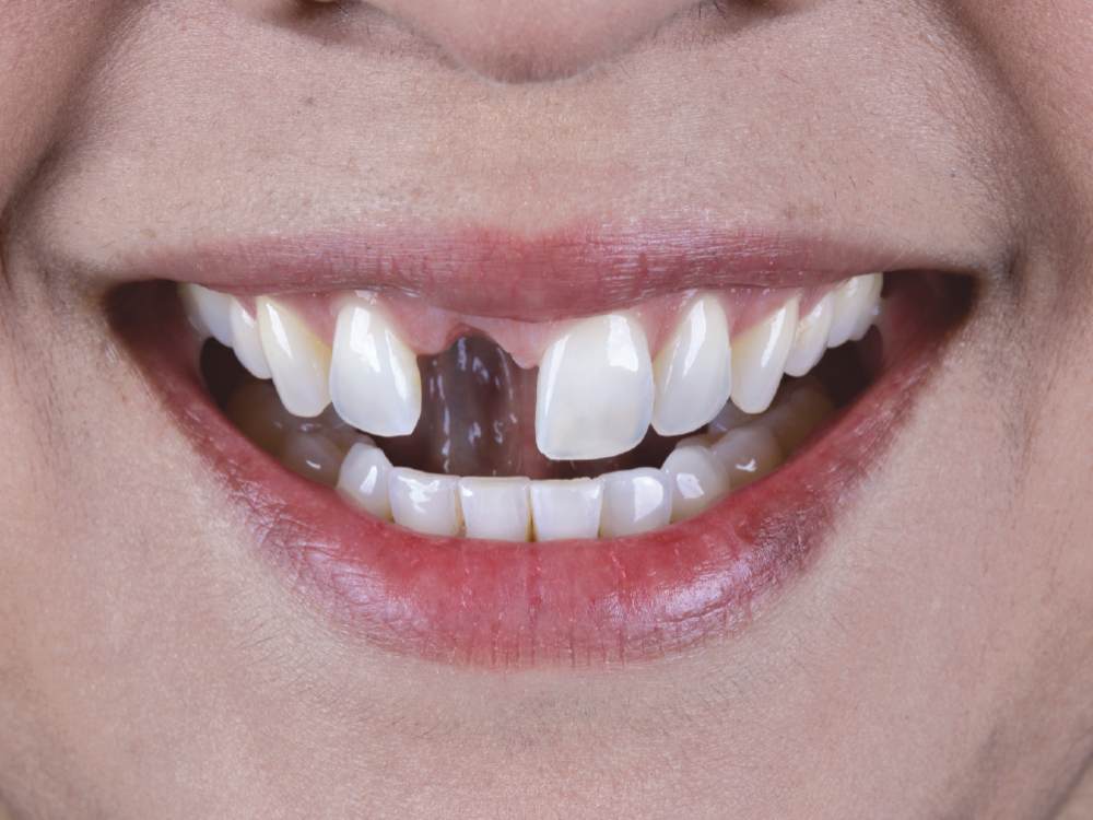
Patients with anterior esthetic concerns often control their smiles to reduce tooth display

Figure 1: Patients with anterior esthetic concerns often control their smiles to reduce tooth display. By eliciting the uninhibited Duchenne smile, we were able to determine maximum tooth and gingival exposure. This evaluation revealed an average lip line, with about 2 mm of gingiva visible above the margin when the patient attempted a maximum smile. 

often control their smiles to reduce tooth display
eliciting the uninhibited Duchenne smile

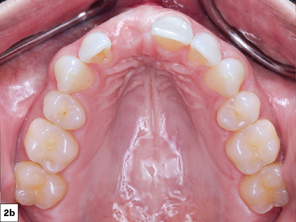
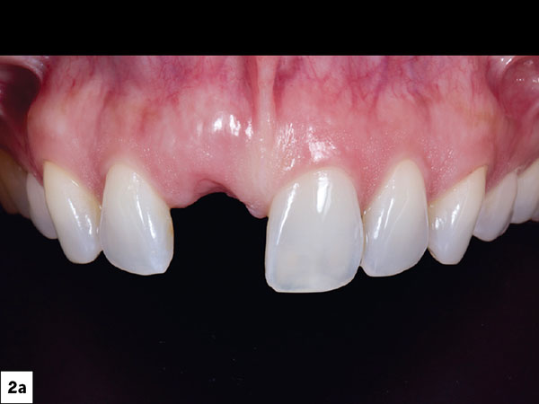
Figures 2a, 2b: The patient’s previous dentist had extracted #8 and performed a bone graft, leaving a favorable ridge for implant therapy. The patient exhibited a thick gingival biotype, providing additional treatment advantages. This tissue phenotype offers improved resilience against trauma and recession, supports long-term tissue stability, and contributes to more predictable esthetic outcomes.

The patient’s previous dentist had extracted #8
This tissue phenotype offers improved resilience against trauma and recession
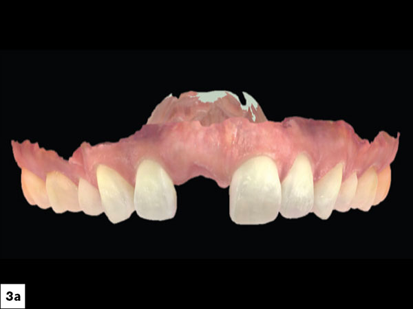
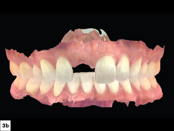
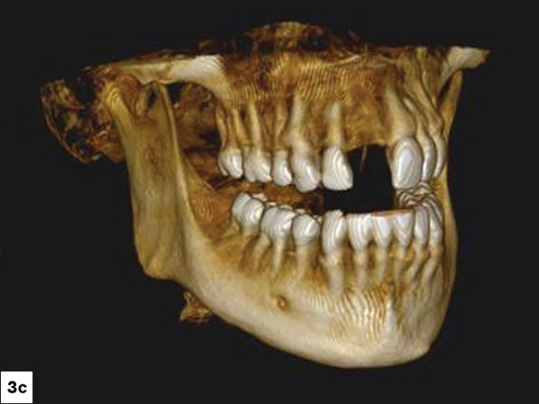
At the initial consultation, comprehensive digital records — including clinical photographs, intraoral scans, and CBCT imaging — were obtained

Figures 3a–3c: At the initial consultation, comprehensive digital records — including clinical photographs, intraoral scans, and CBCT imaging — were obtained.

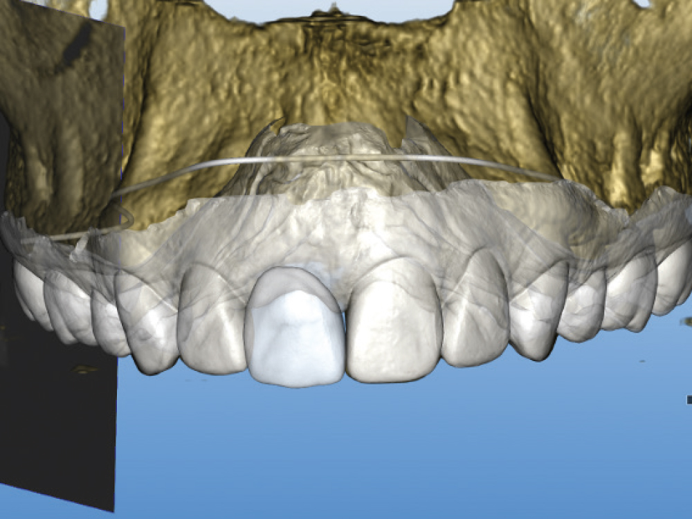
Combined, digital records enabled the design of a diagnostic prosthesis

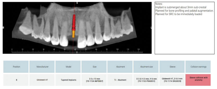
Figure 4: Combined, digital records enabled the design of a diagnostic prosthesis and precise implant planning — ensuring ideal positioning to support proper emergence profiles and restorative harmony.

IMPLANT PLANNING

This service provides significant value by coordinating both the surgical and restorative aspects of care

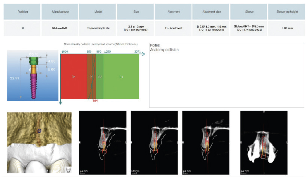
Figure 5: I collaborated with Glidewell’s Digital Treatment Planning (DTP) team. This service provides significant value by coordinating both the surgical and restorative aspects of care. The team assists in planning the implant position, fabricating a surgical guide, and designing a provisional screw-retained crown (SRC). Each case is returned to the clinician with a detailed surgical report, along with a video recording of the 3D surgical plan for visualization and approval prior to the procedure.

A Glidewell HT™ Implant was selected because its design is well-suited for immediate load situations

Figure 6: A Glidewell HT Implant was selected because its design is well-suited for immediate load situations due to its tapered body, sharp buttress threads, and self-tapping grooves. Its aggressive thread design promotes both placement efficiency and high primary stability — critical factors in the success of an immediate provisional restoration.

Glidewell tooth replacement

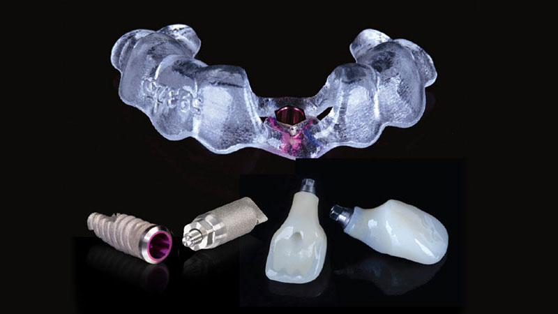
Figure 7: The Glidewell Tooth Replacement Solution (GTRS) for this case included a customized surgical guide, a 3.5 x 13 mm Glidewell HT Implant and a provisional SRC for use on the day of surgery, as well as a scan body to be used during the impression appointment for the final restoration.

SURGICAL DAY

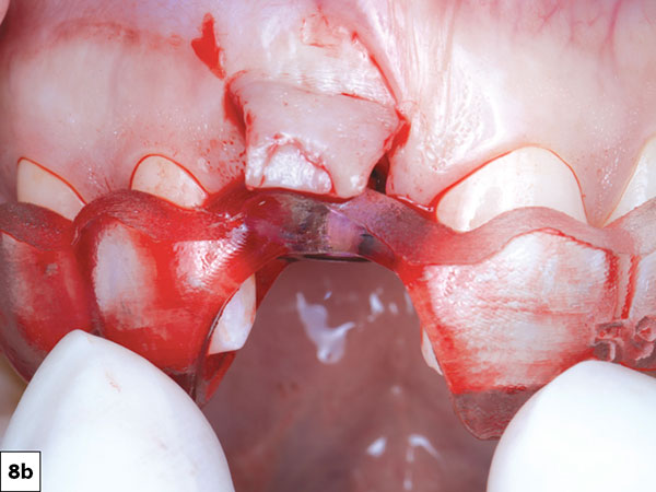
the surgical guide was verified for fit
Using the Glidewell HT™ Implant Guided Surgical Kit (Glidewell Direct; Irvine, Calif.), the osteotomy was prepared

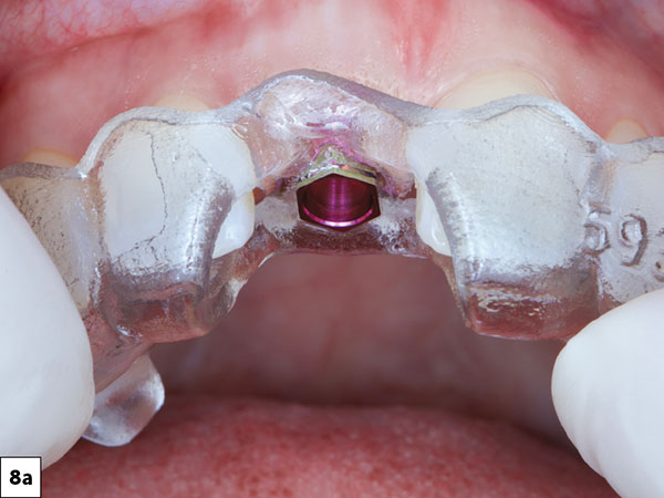
Figures 8a, 8b: On the day of surgery, the surgical guide was verified for fit, and a papilla-sparing flap was created to preserve keratinized soft tissue around the implant. Using the Glidewell HT Implant Guided Surgical Kit (Glidewell Direct; Irvine, Calif.), the osteotomy was prepared.

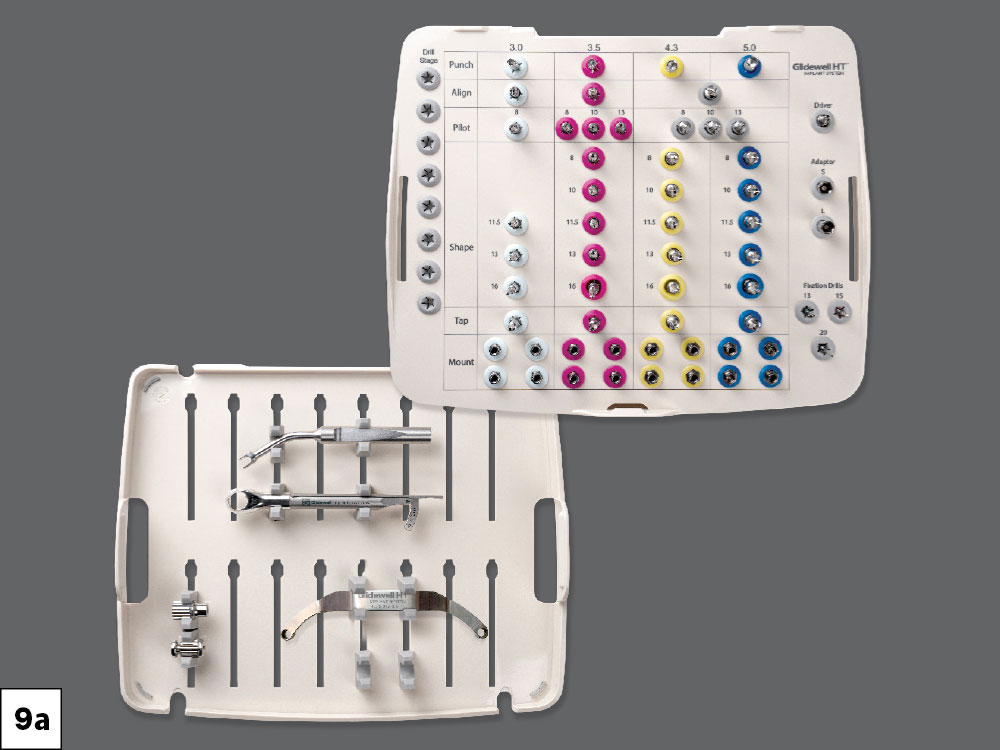
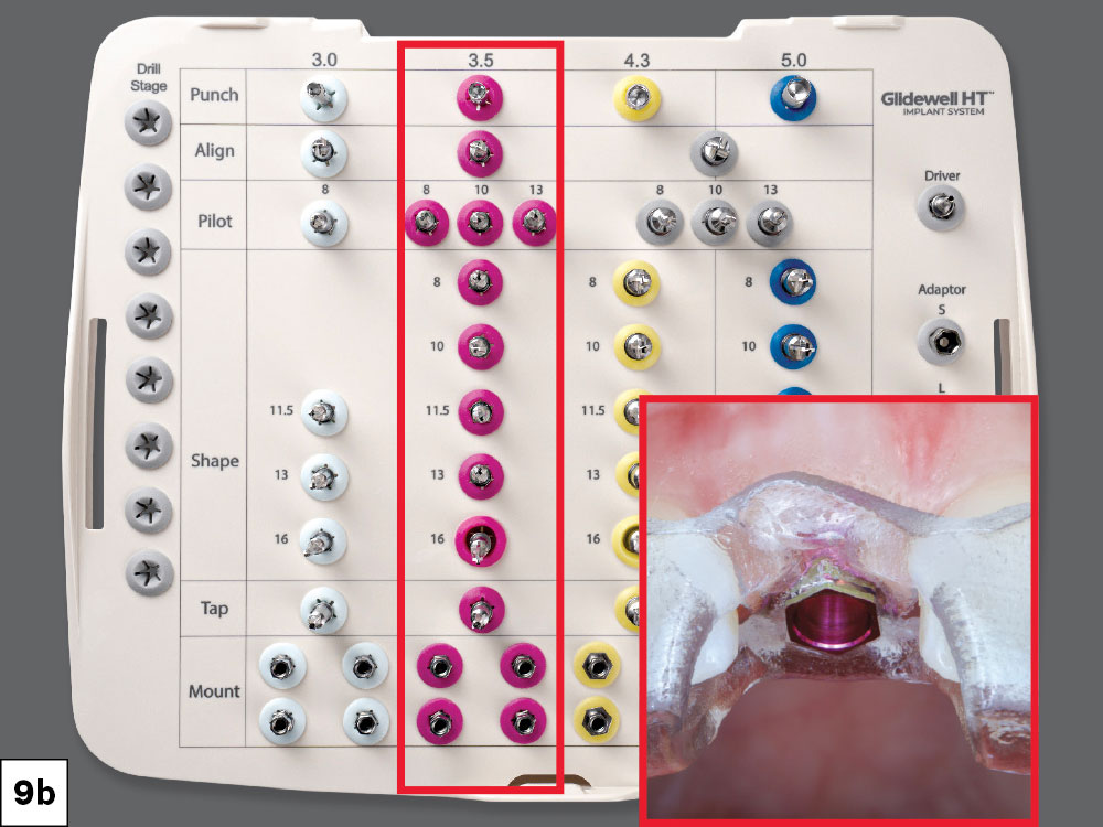
The cassette contains color-coded grommets, the drills are marked with corresponding color bands
The Glidewell HT Implant Guided Surgical Kit

Figures 9a, 9b: The Glidewell HT Implant Guided Surgical Kit features a fully color-coded system that makes it straightforward to follow intraoperatively. The cassette contains color-coded grommets, the drills are marked with corresponding color bands, and the guide sleeves are anodized to match. Each color represents a specific implant diameter in the Glidewell HT system, for ease of use during surgery.

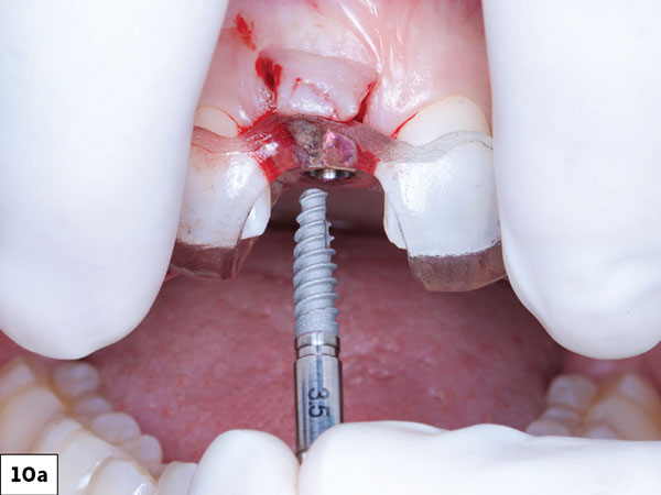
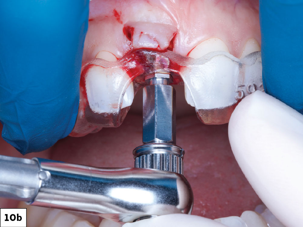
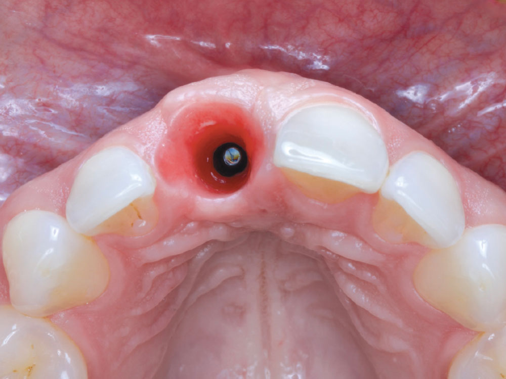
The implant was delivered through the guide, with orientation achieved by aligning the hex of the implant mount to the hex of the guide sleeve.

Figures 10a, 10b: The implant was delivered through the guide, with orientation achieved by aligning the hex of the implant mount to the hex of the guide sleeve.

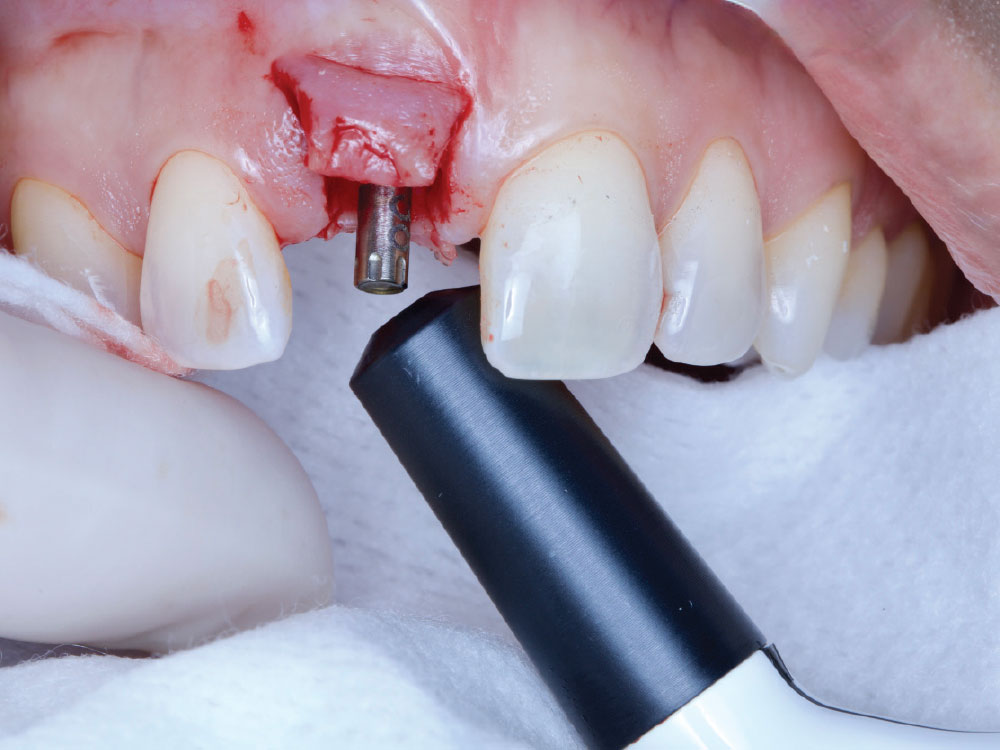
Once torqued into place, the stability of the implant was measured with a Penguin II device

Figure 11: Once torqued into place, the stability of the implant was measured with a Penguin II device (Glidewell Direct), which uses resonance frequency analysis to quantify stability with an Implant Stability Quotient (ISQ). An ISQ reading of 75 exceeded the minimum threshold of 70 for single-unit immediate loading.

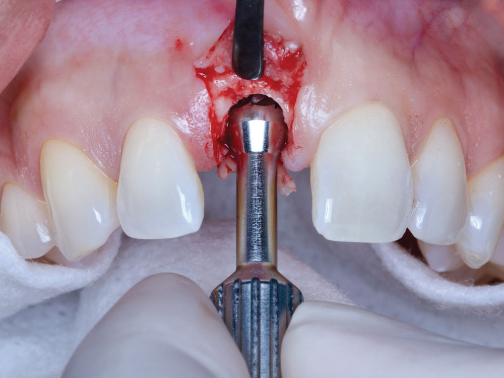
Following implant placement, a bone profiler was used to contour the crestal bone around the platform, creating a more favorable emergence profile. 10a 10b 66 chairsidemagazine.

Figure 12: Following implant placement, a bone profiler was used to contour the crestal bone around the platform, creating a more favorable emergence profile.

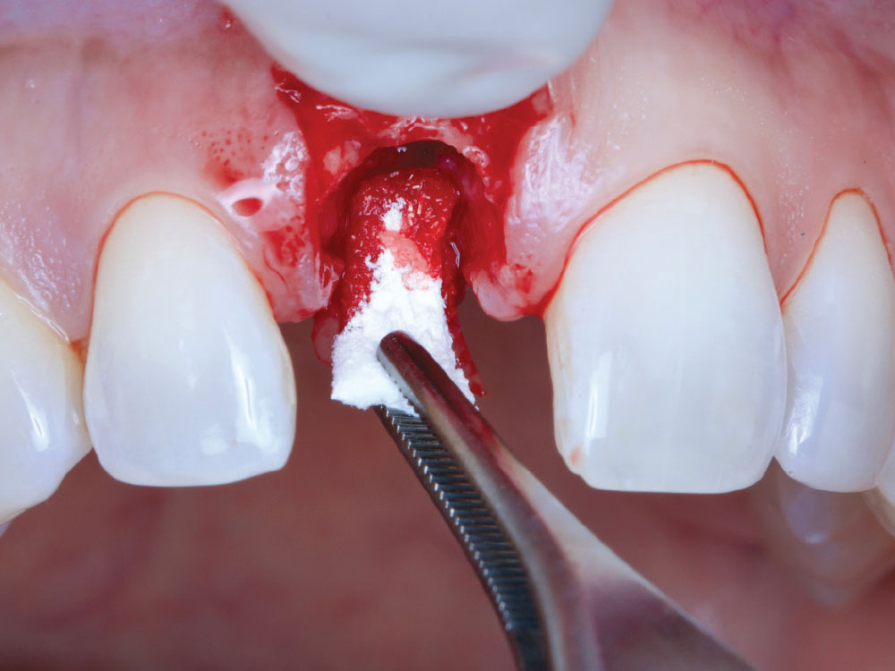
Given the implant positioning required to achieve a screwretained restoration

Figure 13: Given the implant positioning required to achieve a screw-retained restoration, additional grafting was performed on the palatal aspect using an OsteoGen® Plug (Glidewell Direct) to bulk out the ridge.

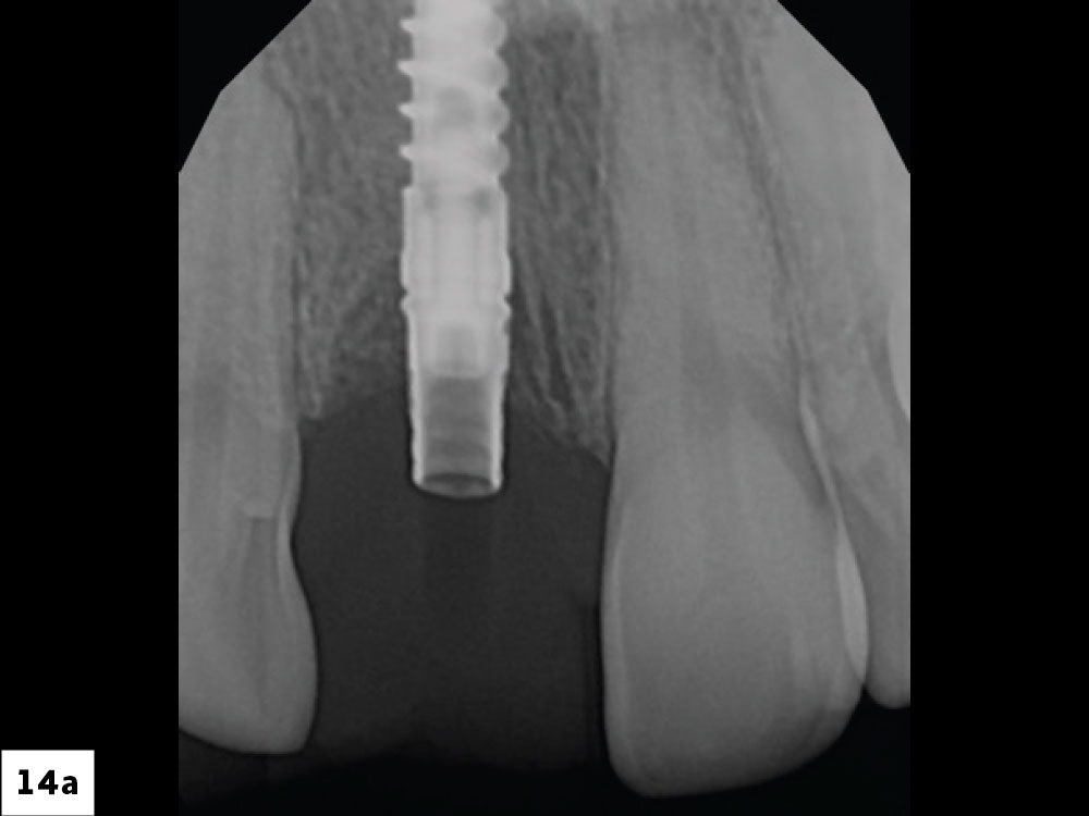
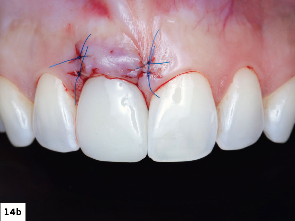
The prefabricated provisional SRC was seated and hand-tightened
A vertical bitewing was taken to confirm complete seating

Figures 14a, 14b: The prefabricated provisional SRC was seated and hand-tightened. A vertical bitewing was taken to confirm complete seating. The access was filled with Teflon tape and Telio® temporary material (Ivoclar Vivadent AG; Schaan/Liechtenstein). A gingival roll technique was performed with Perma Sharp® polypropylene sutures (Hu-Friedy Mfg. Co., LLC; Frankfurt, Germany) to further refine and support the soft tissues.

The patient was able to go home with a fixed provisional restoration that was out of occlusion

Figure 15: The patient was able to go home with a fixed provisional restoration that was out of occlusion.

THREE-MONTH FOLLOW-UP
The patient returned after three months for re-evaluation and final records.

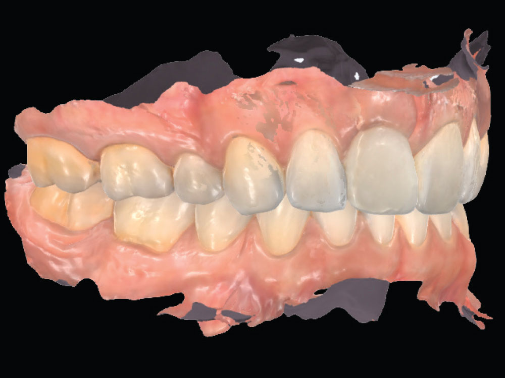
Figure 16: The patient returned after three months for re-evaluation and final records. At the beginning of the visit, an intraoral scan was performed, including a scan of the provisional in place, the opposing arch, and occlusion.

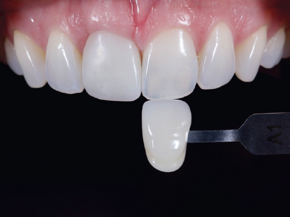
Shade mapping and supplemental photography were captured to guide the final restoration.

Figure 17: Shade mapping and supplemental photography were captured to guide the final restoration.

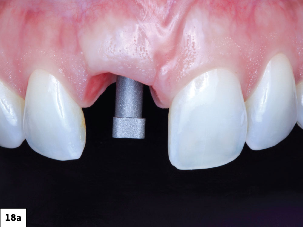
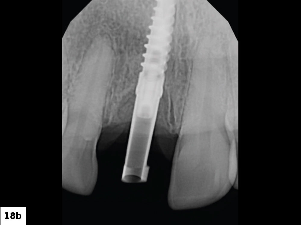
A Glidewell HT™ scan body was placed, and seating was verified with a vertical bitewing

Figures 18a, 18b: A Glidewell HTscan body was placed, and seating was verified with a vertical bitewing. 

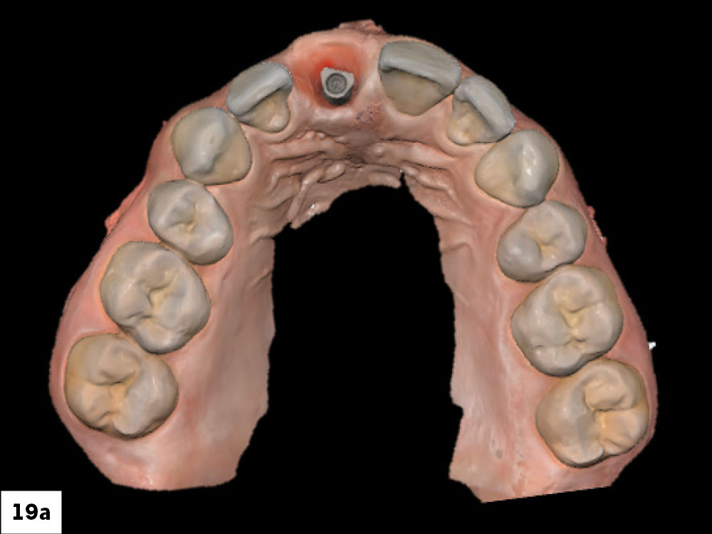
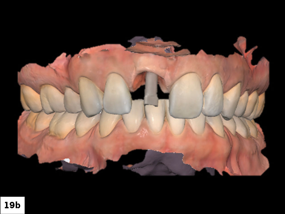
The scan was then completed, capturing both the scan body and peri-implant gingival contours to support accurate restorative design.

Figures 19a, 19b: The scan was then completed, capturing both the scan body and peri-implant gingival contours to support accurate restorative design.

The provisional was removed, revealing well-maintained soft tissue contours with a healthy gingival emergence profile

Figure 20: The provisional was removed, revealing well-maintained soft tissue contours with a healthy gingival emergence profile.  

DEFINITIVE RESTORATION AND OUTCOME

At the delivery appointment, the restoration was tried in to evaluate esthetics, function, and tissue response. Key parameters included shade integration with the adjacent dentition, incisal translucency, and the harmony of the gingival architecture shaped during the provisional phase. The emergence profile sculpted by the screw-retained provisional will seamlessly support the final crown contours, minimizing adjustments.
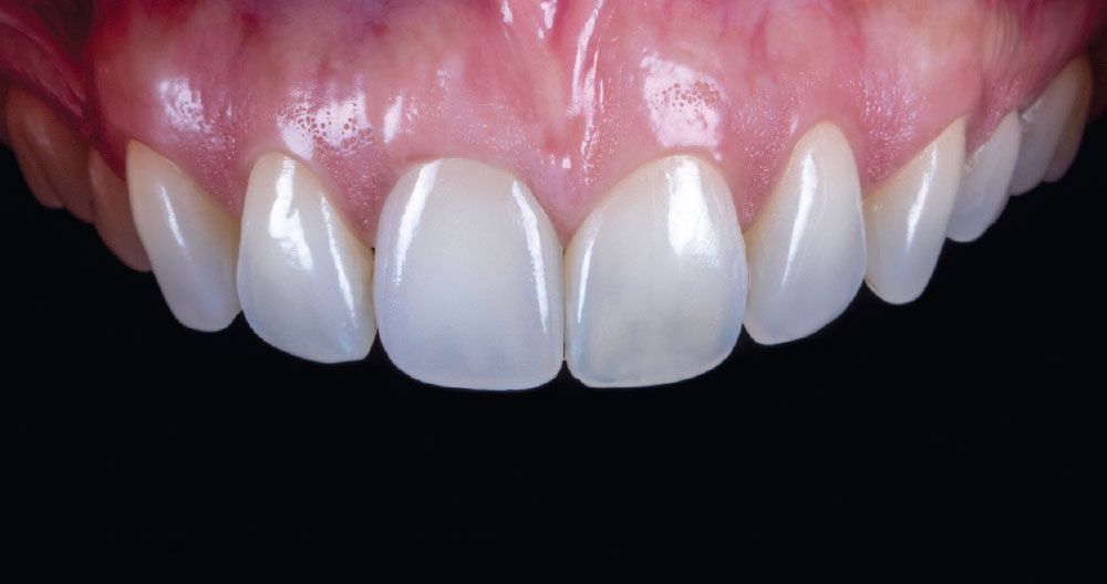
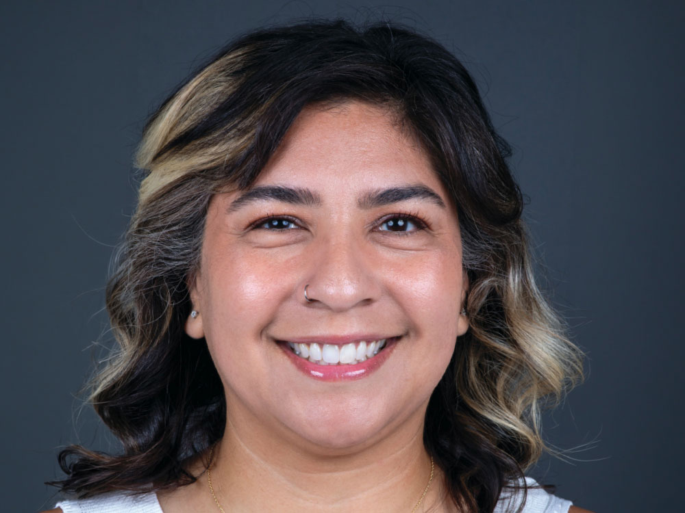
At the delivery appointment, the restoration was tried in to evaluate esthetics

Figure 21: For the definitive restoration, the material of choice was BruxZir® Fusion, which is a unique material developed by Glidewell’s Materials R&D department, that combines the advantages of two different BruxZir formulations. The restoration begins with an anatomically shaped core of BruxZir Full-Strength, providing strength, color, and depth. The core is then overlaid with a wraparound veneer of BruxZir Radiant, which lets light pass through incisal and interproximal surfaces.  The combination provides lifelike esthetics that are particularly compelling when used for single-tooth anterior restorations.

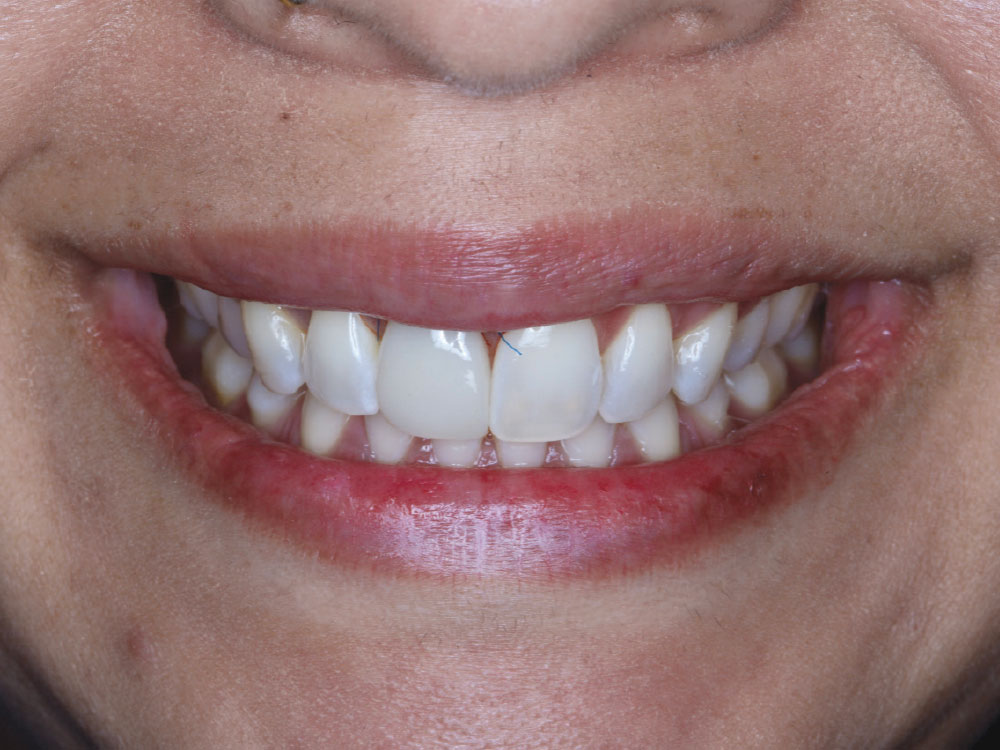
The patient was delighted with the transformation

Figure 22: The patient was delighted with the transformation.

Conclusion

In clinical scenarios where patients present with natural characterization, translucency, and high esthetic demands, BruxZir® Fusion provides an excellent balance between durability and lifelike appearance. In this case, the restoration looks so natural that the patient commented, “The old Melissa is back!” 

All third-party trademarks are property of their respective owners.
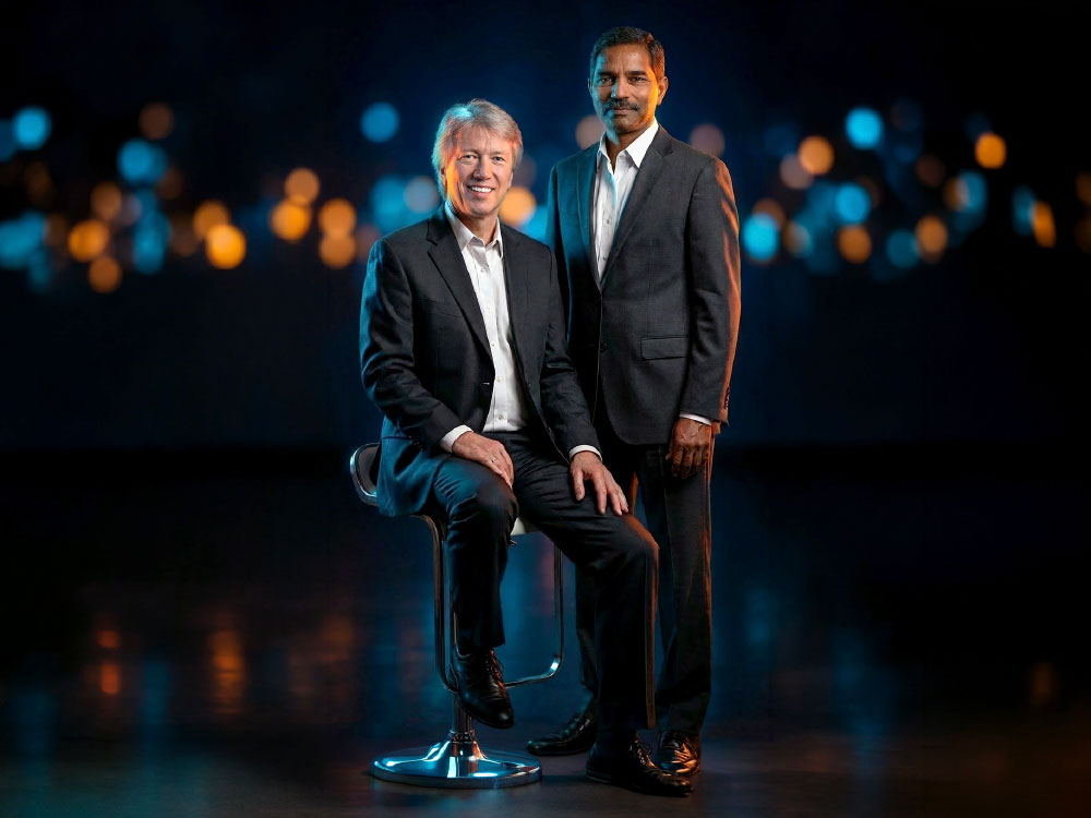
Raj Malyala VP of Materials R&D-Wolfgang Friebauer Director of R&D

The Innovators

Raj Malyala

VP of Materials R&D

With a Ph.D. in chemistry and three decades of experience in material science, Raj has built his career at the intersection of chemistry, innovation, and real‑world applications. Today, he leads a team of 60 scientists and engineers, whose collective expertise has delivered a wave of new products in ceramics, glass-ceramics, composites, and 3D-printed resins, while continuing to strengthen the legacy of flagship materials like BruxZir® zirconia.  “I’m fortunate to work with brilliant minds,” he says. “When you bring together true experts, innovation becomes the natural outcome.” 

Wolfgang Friebauer

Director of R&D

Wolfgang’s dental technology journey began in Germany’s apprenticeship system, combining education and real‑world experience. He expanded his expertise across orthodontics, dentures, ceramics, and combination techniques. Arriving at Glidewell in the 1990s with a German master dental technician degree, he focused on achieving greater uniformity and reliability with every case, regardless of the technician assigned, and laid out the groundwork for Glidewell’s first R&D efforts. “If something can be made better, we owe it to dentists, and their patients, to pursue it,” he says.