Implant Manufacturing

July 6, 2026
Implant Manufacturing

For several decades, implant dentistry has been accepted as a predictable, reliable method for the replacement of missing teeth. The ability to offer efficient, cost-effective treatment to our patients, however, has been a more recent innovation.

As implant dentistry expanded through the 1990s and early 2000s, laboratories faced growing operational challenges. Stock abutments, typically sourced from implant manufacturers, led to compromises in the design of the crucial interface between the implant restoration and the soft tissue. An improved, more biologically based understanding of that interface led to the increasing popularity of custom abutments, which allowed the development of a natural emergence profile and controlled placement of the restorative margin. In addition, concerns about the risks of cementation and the sequelae of retained excess cement led to a widespread shift from cemented to screw-retained restorations.

Glidewell developed a digital workflow that greatly improved the availability and pricing of custom abutments and implant restorations. Offering restorative products covering all common implant platforms, Glidewell’s CAD/CAM processes resulted in improved fit and faster turnaround, as well as lower, all-inclusive laboratory fees.

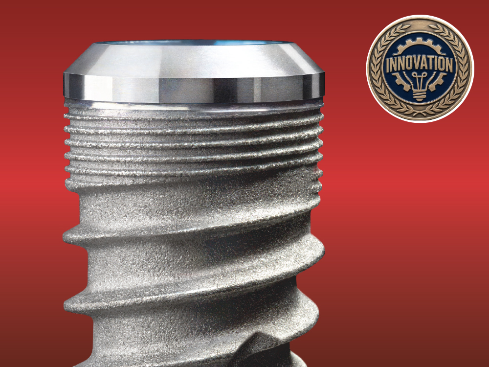
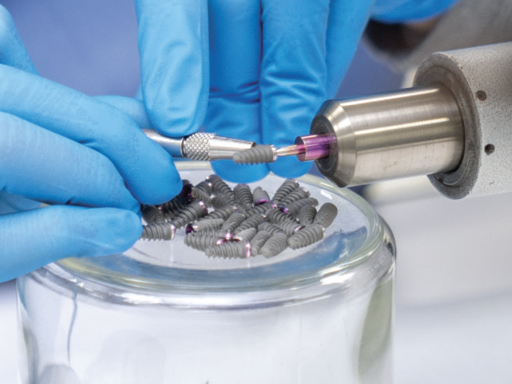
Anticipating the oncoming development of the digital ecosystem, in 2014, Glidewell partnered with Dr. Jack Hahn, a pioneer in the field of implant dentistry, to create the Hahn Tapered Implant System (now the Glidewell HT Implant System). Through the fabrication of many thousands of implant restorations for demanding patients and clinicians, Glidewell engineers understood the key characteristics of clinical success for both the surgical and restorative aspects of treatment.

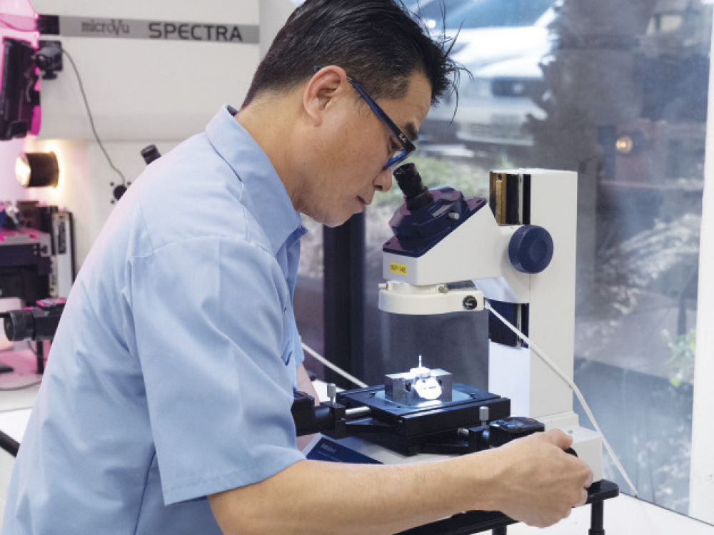
Implant manufacturing at Glidewell has now developed into a broader digital workflow that connects design, production, and clinical delivery. Glidewell’s investment in software and machining infrastructure to transition from basic component production to a fully integrated manufacturing operation provides key benefits for clinicians, including affordable pricing, reduced turnaround times, and improved workflow transparency between clinicians and the lab.
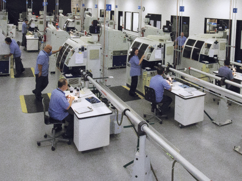
Glidewell developed a digital workflow that greatly improved the availability and pricing
Implant manufacturing at Glidewell has now developed into a broader digital workflow
Glidewell engineers understood the key characteristics of clinical success
From Extraction to Implant Placement to Screw-Retained Crown
From Extraction to Implant Placement to Screw-Retained Crown

Dr. Kosinski

by Timothy F. Kosinski, DDS, MAGD

Owner, Smile Creator of Bingham Farms

Bingham Farms, Michigan

CASE REPORT

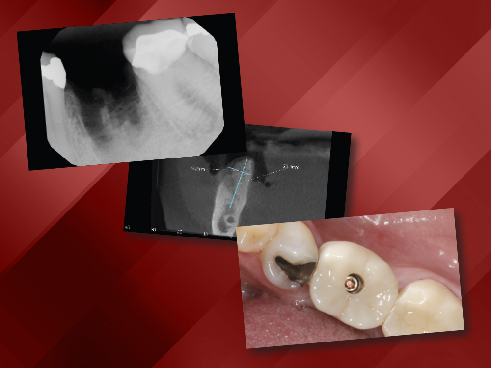
Denny is a 69-year-old who arrived at my practice with a symptomatic mandibular left first molar that had undergone silver point root canal therapy many years ago. The endodontist diagnosed a horizontal fracture along the mesial root and recommended extraction.
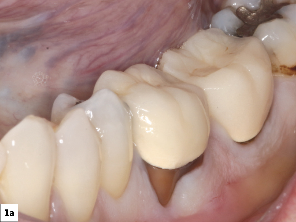
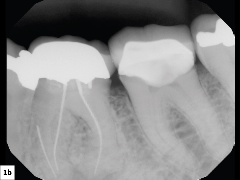
When presented with the treatment options, the patient chose a single-unit implant-supported restoration

Figures 1a, 1b: When presented with the treatment options, the patient chose a single-unit implant-supported restoration over the options of a conventional fixed bridge on #18–20 or a removable appliance.

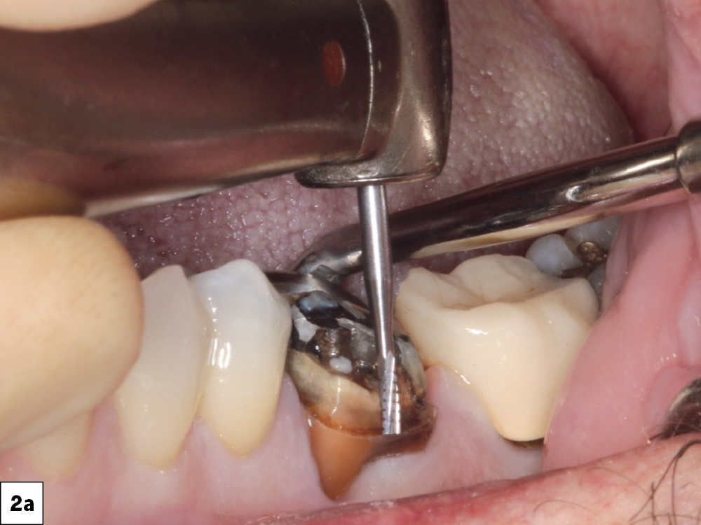
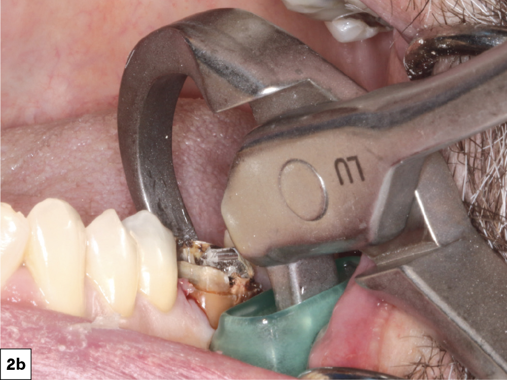
 After buccal vestibular infiltration and a lingual PDL injection, a mesio-distal root section was performed through the furcation
Each root was then removed individually, treating them as separate single-rooted teeth
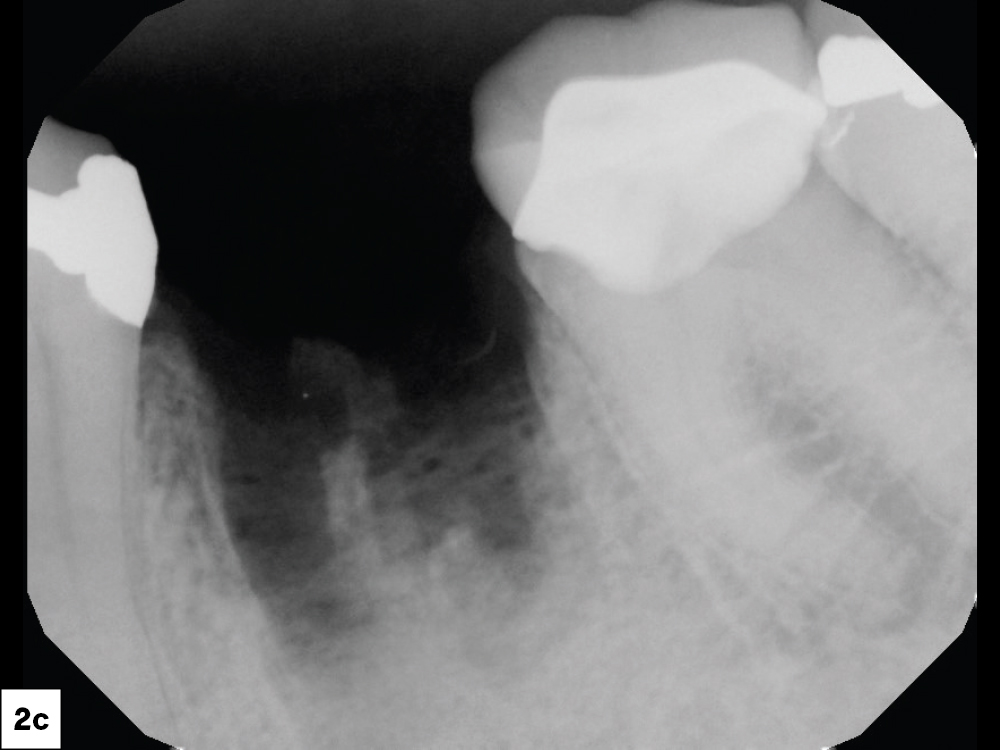
A periapical radiograph confirmed that all root structures had been removed

Figures 2a–2c: After buccal vestibular infiltration and a lingual PDL injection, a mesiodistal root section was performed through the furcation. Each root was then removed individually, treating them as separate single-rooted teeth. Following extraction, granulation tissue was curetted from the socket, and adequate bleeding was observed. A periapical radiograph confirmed that all root structures had been removed.

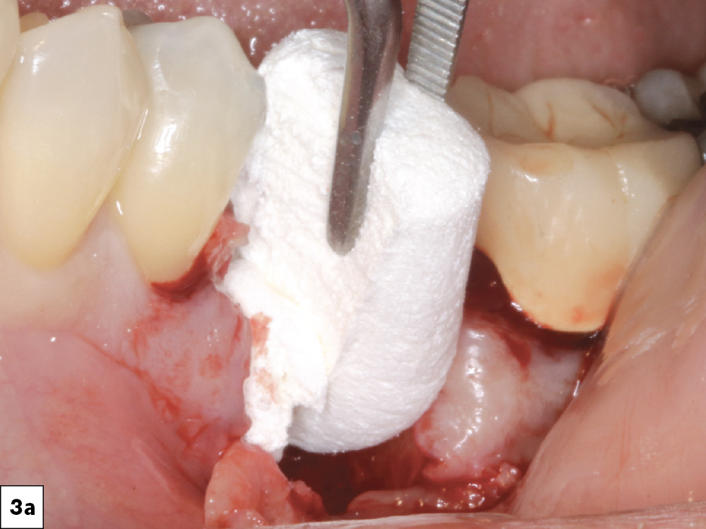
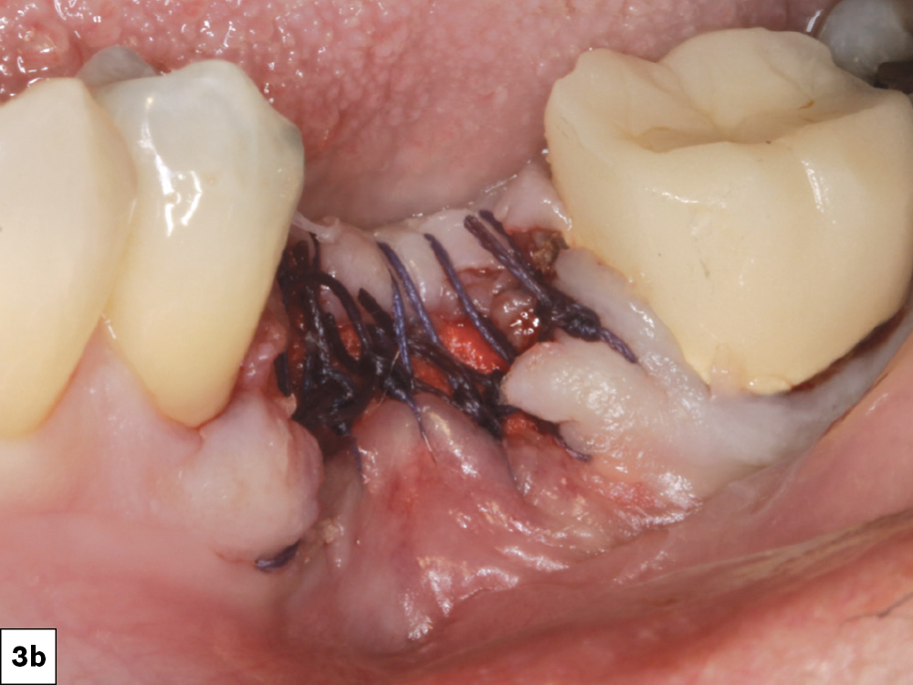
The socket was grafted using a large OsteoGen® plug
Primary closure was not required as the epithelium will grow over the top of the graft at a rate of 0.5 mm per day.

Figures 3a, 3b: The socket was grafted using a large OsteoGen® Plug (Glidewell Direct; Irvine, Calif.) cut in half and firmly condensed to the crest of the ridge, and the site was sutured using Newport Biologics PGA suture material (Glidewell Direct). Primary closure was not required as the epithelium will grow over the top of the graft at a rate of 0.5 mm per day.

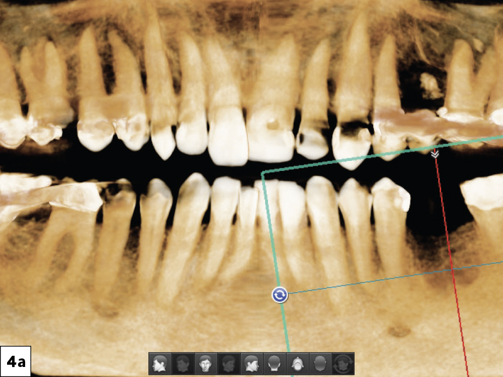
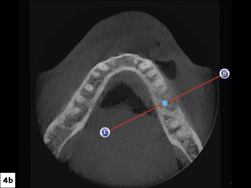
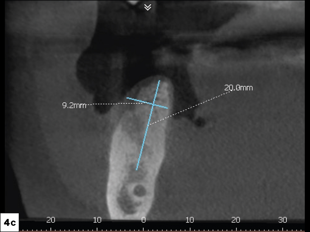
A CBCT analysis was completed, and the sagittal view helped determine that there was adequate bone for implant placement

Figures 4a–4c: A CBCT analysis was completed, and the sagittal view helped determine that there was adequate bone for implant placement.

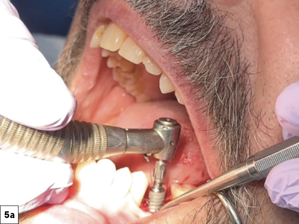
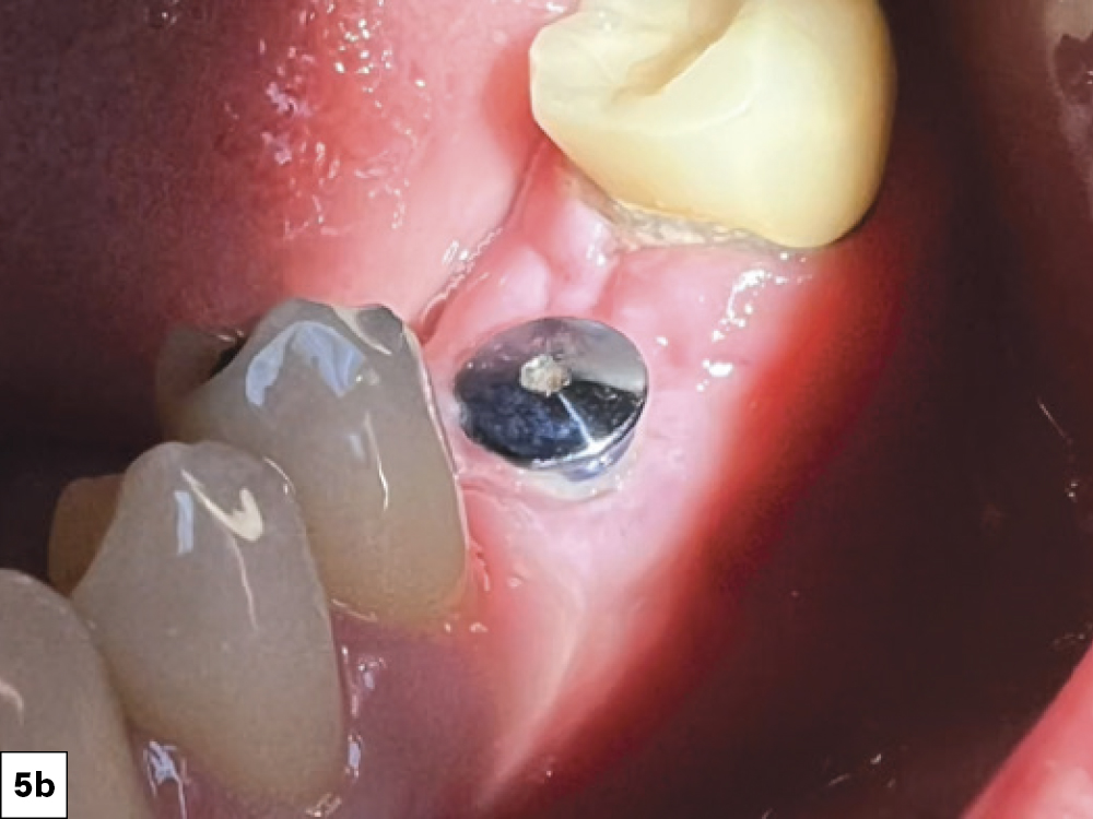
I performed a reflection on the facial of the edentulous space, demonstrating viable bone for implant placement
The facial reflection was then replaced and sutured, maintaining a band of attached gingiva on the facial aspect of the healing abutment

Figures 5a, 5b: A 5 mm x 10 mm Glidewell HT Implant (Glidewell Direct) was selected. I performed a reflection on the facial of the edentulous space, demonstrating viable bone for implant placement. I followed the Glidewell HT dental implant surgical protocol, including a 1.5 mm pilot drill and a 2.4 mm pilot drill, ideally positioning the implant both mesiodistally and facial-lingually. A 3-mm-tall healing abutment was threaded into the body of the implant and hand tightened. The facial reflection was then replaced and sutured, maintaining a band of attached gingiva on the facial aspect of the healing abutment.

THREE MONTHS AFTER IMPLANT PLACEMENT
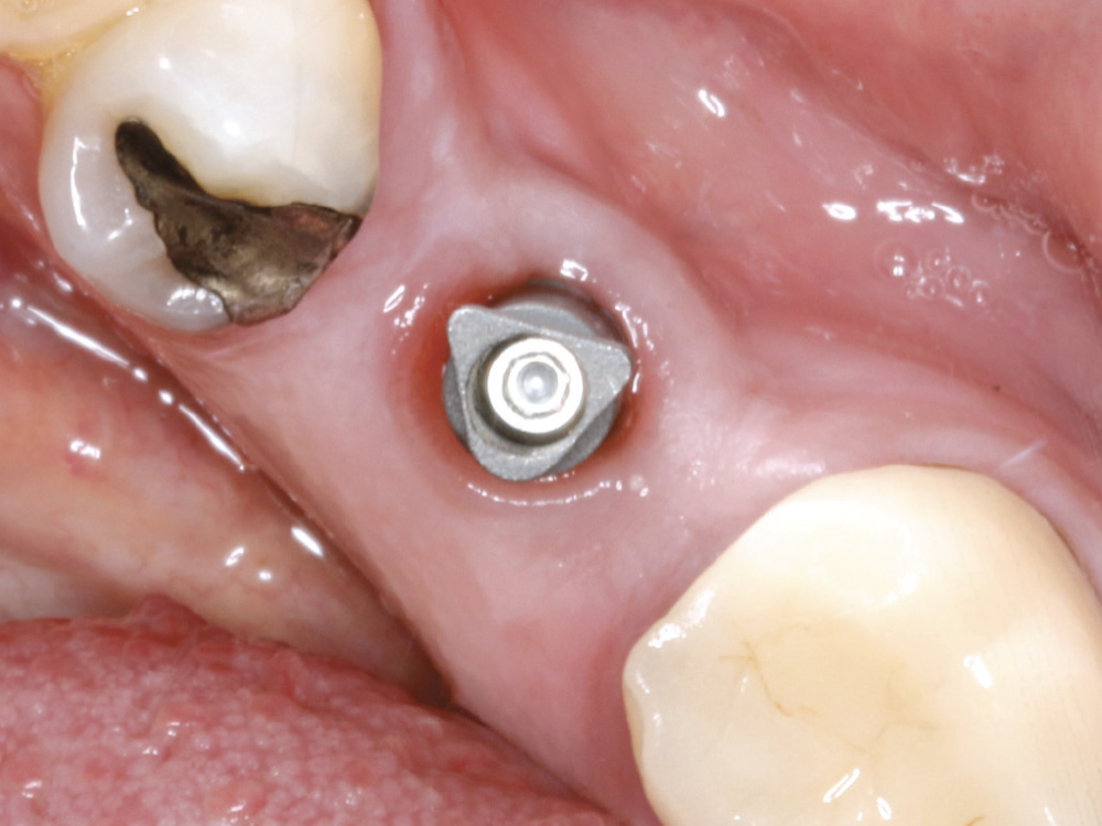
 I threaded the Glidewell scan body into the implant, and an intraoral scan was recorded with a fastscan.io™ Scanning Solution featuring Medit

Figure 6: I threaded the Glidewell scan body into the implant, and an intraoral scan was recorded with a fastscan.io Scanning Solution featuring Medit (Glidewell Direct). The scan was sent to the lab team at Glidewell, who fabricated a BruxZir® Full-Strength screw-retained crown.

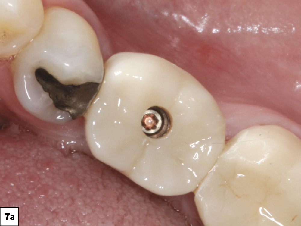
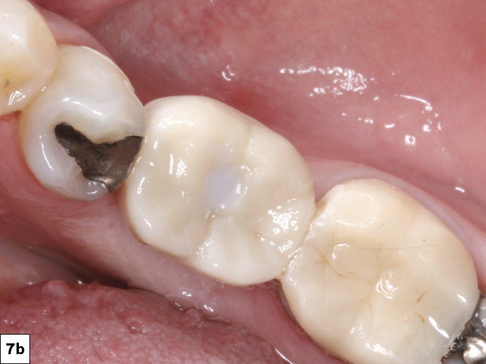
The crown was seated and the screw torqued to 35 Ncm after the occlusion and proximal contacts were verified
The scan was sent to the lab team at Glidewell, who fabricated a BruxZir® Full-Strength screw-retained crown

Figures 7a, 7b: The crown was seated and the screw torqued to 35 Ncm after the occlusion and proximal contacts were verified.

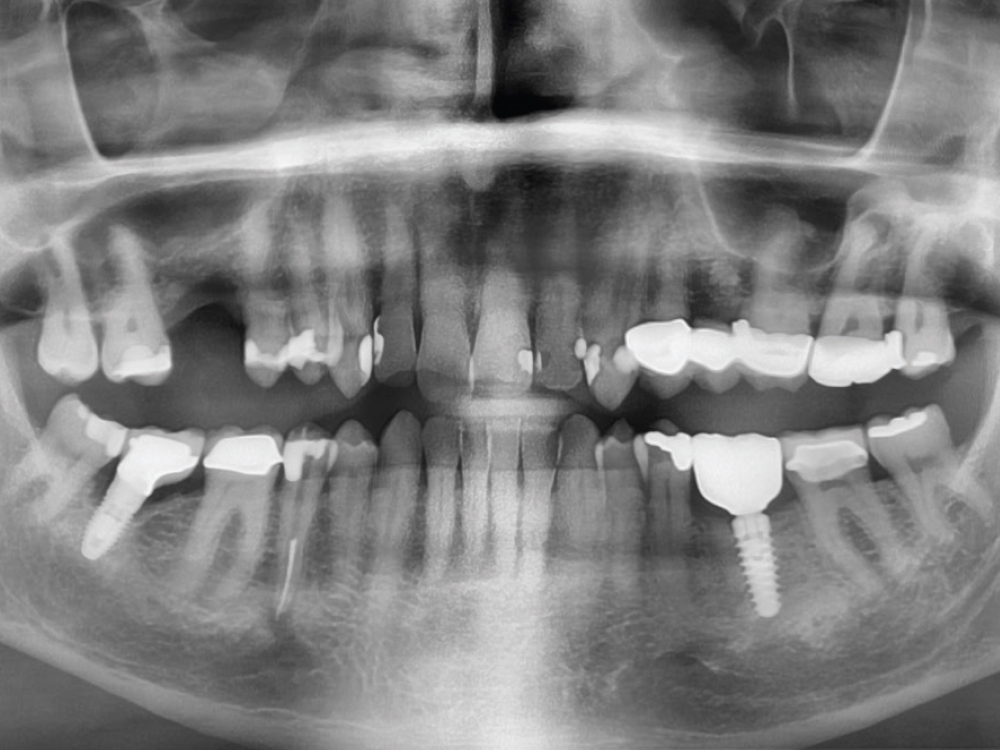
postoperative radiograph confirmed the complete seating of the screw-retained crown into the Glidewell HT implant

Figure 8: A postoperative radiograph confirmed the complete seating of the screw-retained crown into the Glidewell HT Implant.

CONCLUSION

The patient left with a fully functional, esthetic tooth replacement for his mandibular left first molar, and the Glidewell HT Implant System and laboratory helped make it a reality.

All third-party trademarks are property of their respective owners.
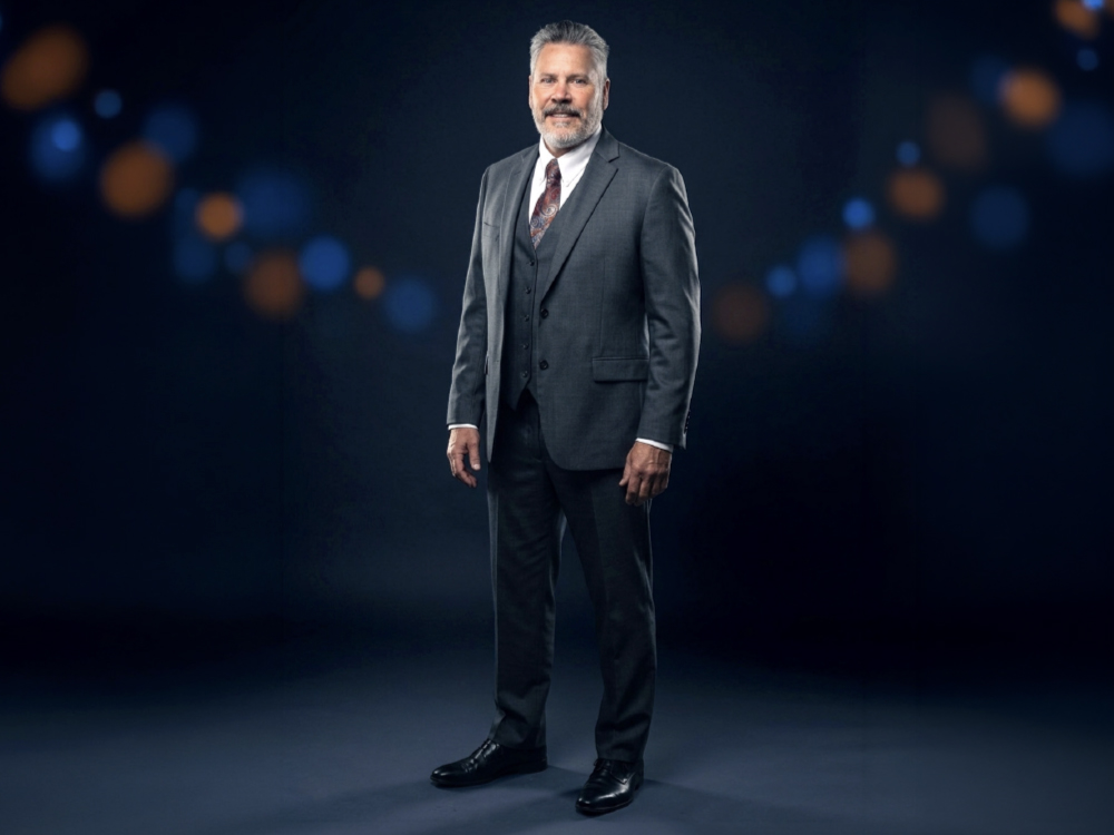
Grant Bullis

The Innovator

Grant Bullis

VP & General Manager
Implant Manufacturing

From cowboy life to Marine Corps service, Grant built his leadership style on discipline, accountability, and execution. After engineering studies at Irvine Valley College and Cal State Fullerton, followed by an MBA from the Kellogg School of Management, he worked in the aerospace, military, and dental manufacturing industries. Since joining Glidewell in 2007, he has focused on expanding the access and affordability of implants and implant restorations. Currently he leads a team that provides precision, reliability, and scale across Glidewell’s implant solutions.