Buccal Roll Technique for Augmentation of Keratinized Tissue (1 CEU)

December 27, 2023
 image
Paresh B. Patel, DDS
Buccal Roll Technique for Augmentation of Keratinized Tissue

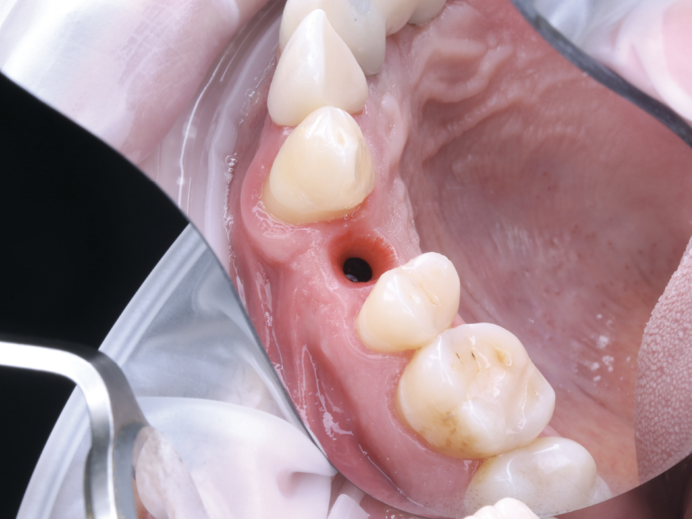
One of the most common questions asked by new implantologists is about when it is necessary to build keratinized tissue across the facial aspect of an implant. Generally, it’s recommended to have 3 mm of keratinized tissue occlusally (between the head of the implant and the top of the tissue), lingually, and buccally toward the mucogingival junction. This amount of tissue stability will ensure that the patient will not suffer loss of function around the implant site. If these conditions are not met after implant placement, surgical intervention is recommended.

An effective way to build keratinized tissue on the facial is by using a modification of Abrams’ roll technique for localized alveolar ridge augmentation, or the buccal roll.1 This simple technique uses deepithelialized palatal tissue to fold underneath buccal mucosa and create the necessary amount of keratinized tissue for an ideal functional and esthetic outcome.2 This case study will demonstrate how to use this technique by analyzing a mandibular premolar that required an implant restoration.

The patient came in with complaints of pain of tooth #4.

Figure 1: The patient came in with complaints of pain and presented with a vertical fracture of tooth #4. After discussing options with the patient, it was decided that an implant placement would offer the best long-term solution.

After extraction of the bifurcated tooth, the gingival architecture and structure of the buccal plate were shown to be well preserved

Figure 2: After extraction of the bifurcated tooth, the gingival architecture and structure of the buccal plate were shown to be well preserved, so a Hahn Tapered Implant (Glidewell Direct; Irvine, Calif.) was placed. When determining if an extraction site can handle immediate implant placement, it’s important to have at least 3 mm of bone apical from the tip of the root. Additionally, when planning for the possibility of using the buccal roll procedure, it’s necessary to keep a minimum of 2 mm of space between the implant and buccal plate. If the implant starts leaning toward the buccal after immediate placement, the best course of action is to dispose of that implant. To avoid long-term complications with stability, it’s best to graft the site and try again another day.

Newport Surgical Cortico/Cancellous Allograft Blend was used to graft the implant site.

Figure 3: Newport Surgical Cortico/Cancellous Allograft Blend (Glidewell Direct) was used to graft the implant site. Using a curette, the socket walls were scraped to facilitate bleeding before packing the allograft material. Not allowing enough blood to combine with the graft will detract from having it turn over into new bone.

After five months of healing, the buccal showed an insufficient amount of attached tissue

Figure 4: After five months of healing, the buccal showed an insufficient amount of attached tissue. At this stage, it’s advantageous to determine if a buccal roll is needed based on how much keratinized tissue there is around the implant site. With the presence of a frenum pull in this case, it was important to reinforce the thin, frail mucosa that could otherwise become functionally compromised with the final restoration in place. To ensure that a thick enough band of keratinized tissue could be formed, it was decided to implement the buccal roll technique.

For the buccal roll technique, a crestal incision should be used

Figures 5a, 5b: To expose the implant, a tissue punch is typically performed; however, doing so sacrifices existing keratinized tissue. For the buccal roll technique, a crestal incision should be used instead. Incisions for this procedure should be made closer to the palate as opposed to mid-crestal to create a thick enough layer of tissue to push toward the buccal. From there, the tissue can be lifted like a trapdoor as pictured above.

The epithelium was then removed from the dermis

Figures 6a, 6b: The epithelium was then removed from the dermis because it is not necessary for this technique. This thin layer of tissue is markedly colorless because it has no direct blood supply, making it easy to identify and scrape away with a blade.

The mucogingival junction and keratinized tissue were exposed

Figures 7a, 7b: The mucogingival junction and keratinized tissue were exposed after removal of the epithelium, which allowed the good-quality tissue to be rolled under itself to effectively double the thickness.

A cover screw was then connected to the Hahn implant

Figures 8a–8c: A cover screw was then connected to the Hahn implant to occupy the space created by the palatal incision. The roll of keratinized tissue was tucked on the mesial and distal sides of the cover screw and sutured to hold the tissue together.

After healing, the implant site showed an abundance of strong keratinized tissue

Figure 9: After healing, the implant site showed an abundance of strong keratinized tissue. The cover screw was removed, revealing how the buccal roll successfully created the desired 3 mm of tissue around the site, and from the occlusal aspect to the head of the implant.

A screwmentable BruxZir crown was selected as the final restoration

Figures 10a, 10b: A “screwmentable” BruxZir® crown was selected as the final restoration. This gave the patient the most esthetic result, as we were able to try in and remove the crown after the custom abutment was positioned, allowing for the contacts to be evaluated and adjusted before final seating of the crown.

The gingiva on the buccal side of the final restoration

Figure 11: The gingiva on the buccal side of the final restoration exhibited successful reconstruction of stable keratinized tissue, which provided an ideal esthetic and structural outcome.

CONCLUSION

Understanding how to perform the buccal roll technique to compensate for insufficient keratinized tissue is an important skill for doctors to have. Furthermore, being able to recognize the need for this procedure before delivery of the final crown will allow doctors to provide more efficient and cost-effective care.

References

  1. Scharf DR, Tarnow DP. Modified roll technique for localized alveolar ridge augmentation. Int J Periodontics Restorative Dent. 1992 Oct;12(5):415–25.

  2. Saquib SA, Bhat MYS, Javali MA, Shamsuddin SV, Kader MA. Modified roll technique for soft tissue augmentation in prosthetic rehabilitation: a case report. Clin Pract. 2019 Mar 14;9(1):1110.